Original Article
High expression of SLC6A10P contributes to poor
prognosis in lung adenocarcinoma
Kai Yuan1,2*, Zhao-Jia Gao1,2*, Wei-Dong Yuan1, Jun-Qiang Yuan1, Yong Wang1
1Division of Thoracic Surgery, The Affiliated Changzhou No. 2 People’s Hospital of Nanjing Medical University, Changzhou, Jiangsu Province, China; 2Heart and Lung Disease Laboratory, The Affiliated Changzhou No. 2
People’s Hospital of Nanjing Medical University, Changzhou, Jiangsu Province, China. *Equal contributors.
Received December 8, 2017; Accepted December 27, 2017; Epub February 1, 2018; Published February 15, 2018
Abstract: Purpose: To investigate the expression profile and prognostic value of SLC6A10P in patients with non-small cell lung cancer (NSCLC). Patients and methods: TCGA datasets were used to investigate the differential ex-pression of SLC6A10P in NSCLC, lung adenocarcinoma (LUAD), and lung squamous cell carcinoma (LUSC). Expres-sion of SLC6A10P was measured by in situ hybridization in tissue microarrays containing 136 NSCLC (51 LUAD and 85 LUSC) patients. The prognostic value of SLC6A10P was then evaluated. Results: SLC6A10P was highly expressed in tumor tissues compared with normal lung tissues. High SLC6A10P expression was associated with lymph node metastasis (NSCLC, P = 0.0054; LUAD, P = 0.0149), more advanced tumor stage (NSCLC, P = 0.0126; LUAD, P = 0.0416) and poor overall survival (NSCLC, P = 0.0248; LUAD, P = 0.0316) in NSCLC and LUAD. Multivariate analysis revealed that SLC6A10P was an independent prognostic factor in LUAD patients (P = 0.017). SLC6A10P showed no association with clinicopathological parameters and no prognostic value in LUSC. Conclusion: SLC6A10P is highly expressed in tumor tissues and its high expression predictspoor survival in patients with LUAD. SLC6A10P might serve as a novel therapeutic target and prognostic biomarker in LUAD patients in the future.
Keywords: SLC6A10P, lung adenocarcinoma, pseudogene, prognosis, bioinformatics
Introduction
Over the past few decades, the incidence and mortality of lung cancer has dramatically increased in China [1]. Currently, lung cancer is one of the most common malignancies and the leading cause of cancer-related deaths in China [2]. Non-small cell lung cancer (NSCLC), which contains two main histological types: lung ade-nocarcinoma (LUAD) and lung squamous cell carcinoma (LUSC), are the main histological types of lung cancer. Despite the development of diagnosis and treatment methods, the 5-year overall survival rate of lung cancer has not been significantly improved. Therefore, further investigation of the molecular mechanisms involved in carcinogenesis and progression of NSCLC is urgently needed.
In recent years, open data resources have been widely used for screening and identifying can-cer-related genes [3]. By analyzing the TCGA database and carefully reviewing the literature,
we found a novel lung cancerrelated candidate gene, SLC6A10P (Solute carrier family 6 mem-ber 10, pseudogene).
The SLC6A10P gene mapped to chromosome 16p11.2 [4, 5]. SLC6A10P belongs to the sol-ute carrier family 6 (SLC6) family, which con-tains nineteen functional members and four pseudogenes [6]. Aberrant expression of SLC6A10P has been observed in some malig-nancies [7]. However, to the best of our knowl-edge, the role of SLC6A10P in lung cancer has not been reported. In the present study, we aimed to investigate the gene’s expression pro-file and prognostic value in NSCLC.
Materials and methods
Bioinformatics
TCGA_LUSC_exp_HiSeq-V2-2015-02-24 were downloaded at the web-site of the UCSC Cancer Browser (https:// genome-cancer.ucsc.edu/). These datasets contained 94 paired NSCLC tissue samples, 46 paired LUAD tissue samples and 48 pair- ed LUSC tissue samples, respectively. Files named “genomic Matrix” in these datasets were used to obtain the mRNA expression lev-els of SLC6A10P (using Editplus® software).
Expression Atlas (http://www.ebi.ac.uk/gxa/ home/) was used to determine the expression profile of SLC6A10P in human tissues or cell lines.
Ethics, consent and permissions
This study was reviewed and approved by the Research Ethics Committee of Zhong- shan Hospital, Fudan University. Written informed consent was obtained from all of the patients. All specimens were handled and made anonymous based onethical and legal standards.
Patients and construction of tissue microarray (TMA)
A total of 140 tissue samples were obtained from patients with NSCLC who were operated on at the Thoracic Surgical Division of Zhongshan Hospital, Fudan University in the year 2005. A total of 10 normal lung tissues were obtained from patients, whose illness was diagnosed as benign lung tumor, undergoing operation at the Thoracic Surgical Division of Zhongshan Hospital, Fudan University in the year 2005. All patients were staged according to the international staging system and were graded according to the recent 2004 World Health Organization criteria. In the present study, no patients received adjuvant or neoad-juvant chemotherapy or radiation treatment prior to surgery, and no other malignancy within 5 years prior to diagnosis was present. As previ-ously described [8], tissue samples from 140 primary NSCLC cases and 10 normal lung tis-sues were arranged in rows and columns to construct a TMA.
In situ hybridization (ISH) and evaluation
Detection of SLC6A10P in FFPE TMA samples was performed using a SLC6A10P ISH Kit (Boster, Pleasanton, CA, USA). Antisense SLC- 6A10P probes were as follows: 5’-CCCAG- CACACTTGGCTCTCTAGGTAGGTCCTACTATTA- CT-3’, 5’-TCCATGGTGATCGTTTTCTACTGCAACAC- CTACTACATCA-3’ and 5’-TCTGCTGGACCTGGTC- TGGCCATCTGTTACCTGCCTATCT-3’. ISH was per-formed, essentially, as described by Nuovo et al. [9].
Briefly, 6 μm sections were deparaffinized, rehydrated and submitted to protease diges-tion followed by postfixadiges-tion for 10 minutes with 1% paraformaldehyde (PFA). Then, hybrid-ization was done at 37°C overnight with a mix-ture containing target probes to SLC6A10P. Posthybridization wash was done at 37°C in SCC, followed by incubation with blocking buf-fer at 37°C for 30 minutes. The blue color was developed by incubating the slides with DAB at 37°C.
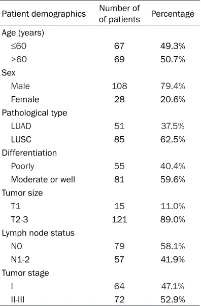
[image:2.612.91.289.96.398.2]SLC6A10P expression levels were determined as follows: The TMA was scanned with Aperio CS2 scanner (Leica Biosystems, San Diego, CA, USA), and representative images for each sam-ple were captured under ×200 magnification. Table 1. Clinical and histologic features of the
140 patients
Patient demographics Number of of patients Percentage Age (years)
≤60 67 49.3%
>60 69 50.7% Sex
Male 108 79.4%
Female 28 20.6%
Pathological type
LUAD 51 37.5%
LUSC 85 62.5%
Differentiation
Poorly 55 40.4%
Moderate or well 81 59.6% Tumor size
T1 15 11.0%
T2-3 121 89.0%
Lymph node status
N0 79 58.1%
N1-2 57 41.9%
Tumor stage
I 64 47.1%
II-III 72 52.9%
Then, by using Image-Pro Plus v6.0 software (Media Cybernetics, Bethesda, MD, USA), inte-grated optical density (IOD) of all the positive staining of SLC6A10P in each photograph was measured, and its ratio to total area of each photograph was calculated as SLC6A10P density.
[image:3.612.92.376.71.262.2]was 60 years, ranging from 26 to 79 years. Most NSCLC cases were in males and more than half of the tumors were LUSC. Clinical fol-lowups were recorded until July 2013. The median followup time was 42.5 months (range, 3~101 months). By the end of the followup, 89 patients had died.
Figure 1.In situ hybridization analysis of 140 lung cancer patients. A. Nega-tive SLC6A10P staining in normal lung tissues. B-D. PosiNega-tive SLC6A10P stain-ing in NSCLC tumor tissues, and the cytoplasmic expression varied.
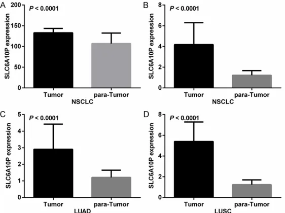
Figure 2. SLC6A10P is more highly-expressed in tumor tissues than in nor-mal lung tissues. (A) The expression level of SLC6A10P in NSCLC tissues is significantly higher than normal lung tissues (P < 0.0001). TCGA datasets further verified that SLC6A10P is an average 3.95-fold high-expressed in NSCLC tissue samples (P < 0.0001) (B), 2.80-fold higher-expressed in LUAD tissue samples (P < 0.0001) (C) and 5.05-fold higher-expressed in LUSC tis-sue samples (P < 0.0001) (D).
Statistical analysis
Data were presented as mean ± S.D. Overall survival (OS) times were defined as the period from primary surgery until the death of the patient or the latest followup. To cal-culate the optimum cutoff point for OS, the X-tile statisti-cal package (Version 3.6.1, Yale University School of Me- dicine, USA) was used. Stu- dent’s t-test, χ2 test,
Kaplan-Meier survival analysis,and Cox regression analysis were performed using SPSS 23.0 software (IBM Corp, Armonk, NY, USA). The graphs were made by GraphPad Prism 6.02 software (GraphPad Pri- sm Software, San Diego, CA, USA). P < 0.05 was consid-ered statistically significant. Results
The 136 patients’
character-istics
[image:3.612.93.374.325.535.2]The expression profile of SLC6A10P
Using the method previously described, we measured the in situ SLC6A10P mRNA in NSCLC patients. SLC6A10P was expressed in the cytoplasm of most tumor cells and to a lesser extent expressed in the cytoplasm of normal lung tissue cells. (Figure 1). The cyto-plasmic expression varied among tumors.
119.90 are regarded as low expression of SLC6A10P (Figure 3A).
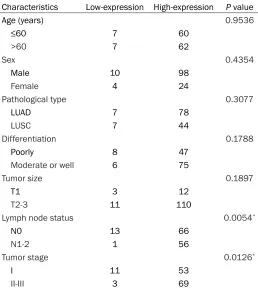
[image:4.612.95.523.74.208.2]We then investigated the association between SLC6A10P expression and clinicopathological variables. In NSCLC, SLC6A10P high expres-sion was significantly correlated with lymph node metastasis (P = 0.0054) and more advanced tumor stage (P = 0.0126) (Table 2). Figure 3. The optimum cutoff points are calculated by the X-tile statistical package. A. The optimal cutoff point of SLC6A10P for NSCLC is 119.90. B. The optimal cutoff point of SLC6A10P for LUAD is 128.60. C. The optimal cutoff point of SLC6A10P for LUSC is 145.30.
Table 2. Correlation between SLC6A10P expression and clinicopathological variables in NSCLC patients
Characteristics Low-expression High-expression P value
Age (years) 0.9536
≤60 7 60
>60 7 62
Sex 0.4354
Male 10 98
Female 4 24
Pathological type 0.3077
LUAD 7 78
LUSC 7 44
Differentiation 0.1788
Poorly 8 47
Moderate or well 6 75
Tumor size 0.1897
T1 3 12
T2-3 11 110
Lymph node status 0.0054*
N0 13 66
N1-2 1 56
Tumor stage 0.0126*
I 11 53
II-III 3 69
LUAD: lung adenocarcinoma; LUSC: lung squamous cell carcinoma; * Signifi-cant correlation.
The expression levels of SLC6A10P were measured as previously desc- ribed.
Compared with the 10 normal lung tissues, SLC6A10P is significantly higher expressed in the 136 NSCLC tissues (P < 0.0001) (Figure 2A). TCGA datasets were used to fur- ther verify this conclusion. By ana-lyzing the TCGA datasets, we found that SLC6A10P is an average 3.95-fold higher expressed in NSCLC tis-sue samples (P < 0.0001) (Figure 2B). Further analysis shows that SLC6A10P is an average 2.80-fold higher expressed in LUAD tissue samples (P < 0.0001) (Figure 2C) and 5.05-fold higher expressed in LUSC tissue samples (P < 0.0001) (Figure 2D).
SLC6A10P confers poor prognosis in NSCLC
[image:4.612.91.349.295.584.2]However, SLC6A10P expression showed no cor-relation with age (P = 0.9536), sex (P = 0.4354), pathological type (P = 0.3077), tumor differen-tiation (P = 0.1788) or tumor size (P = 0.1897) in NSCLC (Table 2). Further, the Kaplan-Meier survival curves were plotted to evaluate the prognostic value of SLC6A10P. The survival analysis reveals that high expressed SLC6A10P is significantly correlated with worse OS in NSCLC patients (P = 0.0248) (Figure 4A).
Further study shows that SLC6A10P confers poor prognosis in LUAD
We further explored the correlation between SLC6A10P expression and clinicopathological
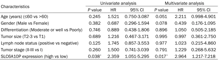
(Figure 4C). Univariate and multivariate analy-sis further revealed that high expression of SLC6A10P is an independent prognostic factor for worse OS in LUAD (P = 0.017) (Table 5). Discussion
[image:5.612.92.517.73.173.2]NSCLC is a highly malignant and aggressive tumor type and has shown a poor 5-year overall survival rate [2]. Therefore, it was urgent that we investigate the potential therapeutic targets of NSCLC. SLC6A10P (alias CT2) is located on 16p11.2 and isa pseudogene of the human creatine transporter gene, SLC6A8 (solute car-rier family 6 member 8, alias CT1) [4, 5, 10]. Historically, pseudogenes have been believed Figure 4. Kaplan-Meier survival curves according to SLC6A10P expression in NSCLC patients. Univariate survival analysis indicates that high SLC6A10P is associated with poorer prognosis in NSCLC (P = 0.0248) (A), as well as in LUAD patients (P = 0.0316) (B). (C) There wasno association between SLC6A10P and overall survival in patients with LUSC (P = 0.2740).
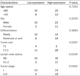
Table 3. Correlation between SLC6A10P expression and clini-copathological variables in LUAD patients
Characteristics Low-expression High-expression P value
Age (years) 0.7262
≤60 10 20
>60 8 13
Sex 0.2025
Male 15 22
Female 3 11
Differentiation 0.3693
Poorly 10 14
Moderate or well 8 19
Tumor size 0.5267
T1 4 5
T2-3 14 28
Lymph node status 0.0149*
N0 15 16
N1-2 3 17
Tumor stage 0.0416*
I 13 14
II-III 5 19
*Significant correlation.
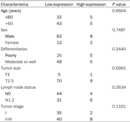
variables in LUAD and LUSC. For LUAD and LUSC patients, we used the grouping method described previously. The cutoff value for LUAD was 128.60, and for LUSC it was 145.30. Table 3 shows th- at similar with NSCLC, in LUAD, high SLC6A10P expression is con-cluded to be significantly correlat-ed with lymph node metastasis (P
[image:5.612.91.356.270.522.2]to represent nonfunctional genomic fossils, however, emerging evidence has suggested that many of them may be biologically active [11]. To the best of our knowledge, the role of SLC6A10P in lung cancer is still unclear. In our present study, we mainly investigated the expression profile and prognostic value of SLC6A10P in patients with NSCLC.
It was reported that SLC6A10P is detected in testis [4], brain [12] and ovaries [7]. By brows-ing the site (Expression Atlas, http://www.ebi. ac.uk/gxa/home/), we found that SLC6A10P is detected in many human tissues (heart, lung, prostate, etc.) and differential expressed in some cell lines (MCF-10A, etc.) (http://www.ebi. ac.uk/gxa/genes/ENSG00000214617?bs). In our present study, three TCGA datasets were downloaded and used to access the expression profile of SLC6A10P. Then, by analyzing the TCGA datasets, we found that SLC6A10P is sig-nificantly higher expressed in tumor tissues when compared with normal lung tissues. Based on the differential expression, we be- lieve that there are functional roles associated with SLC6A10P. This hypothesis has been sup-ported by some studies. A study done by Bayou et al. showed that SLC6A10P may be involved in the autistic phenotype in patients with
respectively. High SLC6A10P expression corre-lates with lymph node metastasis and more advanced tumor stage, leading to worse OS and is an independent prognostic factor for worse OS in LUAD patients, while SLC6A10P shows no correlation with any clinicopathologi-cal parameters and no prognostic value in LUSC. On the whole, SLC6A10P may be a use-ful marker for poor prognosis and a potential therapeutic target in NSCLC, especially in LUAD patients. However, it still takes time to explore the exact biological mechanisms of SLC6A10P in NSCLC.
In conclusion, our study indicates that SL- C6A10P is higher expressed in tumor tissues and its high expression predicts a poor survival in patients with LUAD. In the future, SLC6A10P may become a novel therapeutic target and prognostic biomarker in LUAD patients but that awaits further investigation.
Acknowledgements
[image:6.612.91.358.95.347.2]This research was supported by High-Level Medical Talents Training Project (Grant num- ber. 2016CZBJ042), Jiangsu Provincial Medical Youth Talent (Jiangsu Health Scientific Edu- cation [2017] No. 3), Postdoctoral Program of Nanjing Medical University, Basic Research Table 4. Correlation between SLC6A10P expression and
clini-copathological variables in LUSC patients
Characteristics Low-expression High-expression P value
Age (years) 0.6604
≤60 32 5
>60 43 5
Sex 0.7487
Male 63 8
Female 12 2
Differentiation 0.3440
Poorly 26 5
Moderate or well 49 5
Tumor size 0.6991
T1 5 1
T2-3 70 9
Lymph node status 0.2634
N0 44 4
N1-2 31 6
Tumor stage 0.1101
I 35 2
II-III 40 8
Project of Changzhou (Grant number. CJ2014- 0041).
Disclosure of conflict of interest
None.
Address correspondence to: Dr. Yong Wang, Division of Thoracic Surgery, The Affiliated Changzhou No. 2 People’s Hospital of Nanjing Medical University, 29 Xinglong Lane, Changzhou 213003, Jiangsu Province, China. Tel: +86 519 88123833; Fax: +86 0519 88123833; E-mail: doctor_wang1960@163. com
References
[1] Chen W, Zheng R, Zeng H and Zhang S. Epide-miology of lung cancer in China. Thorac Cancer 2015; 6: 209-215.
[2] Chen W, Zheng R, Baade PD, Zhang S, Zeng H, Bray F, Jemal A, Yu XQ and He J. Cancer statis-tics in China, 2015. CA Cancer J Clin 2016; 66: 115-132.
[3] Li CY, Xiong DD, Huang CQ, He RQ, Liang HW, Pan DH, Wang HL, Wang YW, Zhu HW and Chen G. Clinical value of miR-101-3p and bio-logical analysis of its prospective targets in breast cancer: a study based on the cancer genome atlas (TCGA) and bioinformatics. Med Sci Monit 2017; 23: 1857-1871.
[4] Iyer GS, Krahe R, Goodwin LA, Doggett NA, Si-ciliano MJ, Funanage VL and Proujansky R. Identification of a testis-expressed creatine transporter gene at 16p11.2 and confirmation of the X-linked locus to Xq28. Genomics 1996; 34: 143-146.
[5] Xu W, Liu L, Gorman PA, Sheer D and Emson PC. Assignment of the human creatine trans-porter type 2 (SLC6A10) to chromosome band 16p11.2 by in situ hybridization. Cytogenet Cell Genet 1997; 76: 19.
[6] Hoglund PJ, Adzic D, Scicluna SJ, Lindblom J and Fredriksson R. The repertoire of solute carriers of family 6: identification of new hu-man and rodent genes. Biochem Biophys Res Commun 2005; 336: 175-189.
[7] Ganapathi MK, Jones WD, Sehouli J, Michener CM, Braicu IE, Norris EJ, Biscotti CV, Vaziri SA and Ganapathi RN. Expression profile of CO-L2A1 and the pseudogene SLC6A10P predicts tumor recurrence in high-grade serous ovarian cancer. Int J Cancer 2016; 138: 679-688. [8] Gao ZJ, Wang Y, Yuan WD, Yuan JQ and Yuan K.
HIF-2alpha not HIF-1alpha overexpression confers poor prognosis in non-small cell lung cancer. Tumour Biol 2017; 39: 101042831- 7709637.
[9] Nuovo GJ, Elton TS, Nana-Sinkam P, Volinia S, Croce CM and Schmittgen TD. A methodology for the combined in situ analyses of the precur-sor and mature forms of microRNAs and cor-relation with their putative targets. Nat Protoc 2009; 4: 107-115.
[10] Eichler EE, Lu F, Shen Y, Antonacci R, Jurecic V, Doggett NA, Moyzis RK, Baldini A, Gibbs RA and Nelson DL. Duplication of a gene-rich clus-ter between 16p11.1 and Xq28: a novel peri-centromeric-directed mechanism for paralo-gous genome evolution. Hum Mol Genet 1996; 5: 899-912.
[11] Frankish A and Harrow J. GENCODE pseudo-genes. Methods Mol Biol 2014; 1167: 129-155.
[12] Bayou N, M’Rad R, Belhaj A, Daoud H, Zemni R, Briault S, Helayem MB, Ben Jemaa L and Chaabouni H. The creatine transporter gene paralogous at 16p11.2 is expressed in human brain. Comp Funct Genomics 2008: 609684. [13] Ndika JD, Lusink V, Beaubrun C, Kanhai W,
[image:7.612.91.524.87.206.2]Martinez-Munoz C, Jakobs C and Salomons GS. Cloning and characterization of the pro-moter regions from the parent and paralogous creatine transporter genes. Gene 2014; 533: 488-493.
Table 5. Cox regression analysis of overall survival in LUAD patients
Characteristics P valueUnivariate analysisHR 95% CI P valueMultivariate analysisHR 95% CI