THYROID DYSFUNCTION IN PATIENTS WITH HIV INFECTION
Dr. Sunanda Amonkar1, Dr. Vaibhav Dukle2, Dr. Lavita D’costa3*, Dr. Chitra Y. Dhume4
1
Associate Professor, Department of Internal Medicine, Goa Medical College, Bambolim-
Goa.
2
MD Medicine, Goa Medical College, Bambolim-Goa.
3
Assistant Lecturer, Goa Medical College, Bambolim-Goa.
4
Professor and Head, Department of Biochemistry, Goa Medical College, Bambolim-Goa.
ABSTRACT
The aims of the study were to investigate the prevalence of overt and
subclinical thyroid dysfunction of HIV-infected patients in Goa
Medical College, identify the patterns of thyroid dysfunction prevalent
in the study population, determine risk factors associated with the
development of thyroid dysfunction in HIV-infected patients and to
assess the applicability of thyroid function tests as biochemical
indicator of advanced HIV disease. The antiretroviral drug regimens
the patients in this study received comprised three of the following
antiretroviral drugs; zidovudine, stavudine, lamivudine, nevirapine and
efavirenz. In the present study, there was no statistically significant association between
receipt of the above mentioned antiretroviral drugs and thyroid dysfunction or
hypothyroidism. In the present study, the mean CD4 count was lower in patients with
abnormal thyroid function tests (205.6 cell/mm3) compared to those with normal thyroid
function (344.3 cell/mm3). The difference was found to be statistically significant. The study
range included subjects between 22-62 years. The mean age of the study population was 40.7
± 9.7 years. The patients with thyroid infection were older than the euthyroid patients (mean
age 43.8 ± 7.8 v/s 39.8 ± 10.1 months), however there was no statistically significant
association between age and development of thyroid dysfunction in HIV-infected individuals
(p value =0.143).
KEYWORDS: Thyroid dysfunction, HIV infection, T3 and FT3, CD4 cell counts, highly
Volume 5, Issue 12, 1144-1158. Research Article ISSN 2277– 7105
*Corresponding Author Dr. Lavita D’costa Assistant Lecturer, Goa
Medical College,
Bambolim-Goa. Article Received on 21 Oct. 2016,
Revised on 11 Nov. 2016, Accepted on 01 Dec. 2016
INTRODUCTION
Infection with the human immunodeficiency virus (HIV) results in a chronic systemic illness
with multi-organ involvement.[1] As acute complications of HIV decrease, the focus has to
shift to chronic complications of the decrease and chronic adverse effects of current treatment
regimes.[2]
A number of endocrine abnormalities develop in patients with HIV infection both during the
early and late stages of the disease. These abnormalities may contribute to the clinical
presentation and therefore appropriate treatment would theoretically improve the patient‟s
condition.[1,3] The endocrine dysfunction in these cases may be because of the possible
relationship between the immune system and the endocrine system, or through the direct
involvement of glands by HIV itself, opportunistic infections or malignancies.[3]
Increasing prevalence of thyroid function abnormalities has been reported in HIV-infected
patients in various cross-sectional studies.[4] These include overt hypothyroidism, subclinical
hypothyroidism, the low T3 syndrome, the sick euthyroid syndrome, hyperthyroidism
including Graves disease, subclinical hyperthyroidism, isolated low FT4 and isolated high
FT3. Subtle alteration of thyroid function tests are more common in HIV infection and are
sometimes already detectable in the early phase of the disease. Unique abnormalities of
thyroid function indices have also been recently documented. A progressive elevation in
serum TBG accompanying the decline of the CD4 lymphocyte count and a concomitant
increase in the serum T4 value has been reported.[5]
The exact mechanisms of thyroid dysfunction in HIV-infected patients are still unknown.[6]
The proposed mechanisms include thyroid gland destruction by opportunistic infections
(Pneumocystis jirovaci infection, cytomegalovirus infection, tuberculosis, cryptococcosis and
visceral leishmaniasis) or malignant diseases (Kaposi‟s sarcoma, lymphoma), drug-related
adverse effects especially with relation to HAART or side effects of the drugs used in the
course of HIV infection (e.g. rifampicin, ketoconazole, steroids etc.), effects of humoral
factors such as cytokines and direct infection of gland by HIV itself. The thyroid dysfunction
patterns consistent with non-thyroidal illness are less makred in HIV infection and are mainly
present in the final stages of the disease.[5-8]
The use of highly active antiretroviral therapy (HAART) in current clinical practice has been
HIV-related morbidity and mortality.[9-10] There is an increasing awareness of a growing
number of patients receiving HAART who present with symptoms of thyroid dysfunction.
[11-13]
Hypothyroidism is the most common thyroid function abnormality occurring in
HIV-infected patients, especially after treatment with HAART.[4,6]
Some studies have correlated worsening thyroid dysfunction with advancing HIV infection
and proposed that various thyroid function tests such as TBG (thyroglobulin), FT3, FT4, TSH
can be used as a surrogate markers, as these correlate with the progression of the disease.[14]
In view of the varying results of the previous studies concerning thyroid dysfunction in HIV
infected individuals and the absence of comparable data with respect to the Indian and Goan
population, it was decided to undertake a study of the prevalence and patterns of thyroid
dysfunction in HIV-infected patients in varied clinical settings in Goa Medical College.
MATERIALS AND METHODS
The study was conducted in Goa Medical College and Hospital, Bambolim, Goa in patients
attending the Outpatient Department (OPD) of the Department of Medicine and the
Anti-Retrovirus (ART) Centre during the period 2011 to 2012.
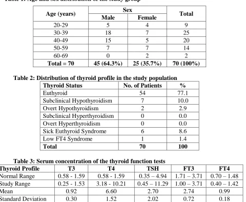
The study included a total number of 70 patients, 45 male and 25 female, with a mean age of
40 years and 42 respectively.
The study was approved by the Institutional Ethics Committee of the Goa Medical College.
All the participants were informed and explained about the study being undertaken and an
informed consent was obtained.
The study design was a single-centre observational cross-sectional study followed by a case
control study.
Inclusion Criteria
1. Diagnosed case of HIV infection.
2. Age between 18 years to 70 years.
Exclusion Criteria
2. Use of drugs known to interfere with thyroid hormone metabolism. (Phenytoin,
phenobarbitone, carbamazepine, carbimazole, warfarin, rifampicin, lithium, propranolol,
amiodarone, glucocorticoids).[15,16]
3. Abnormal renal function test.[17]
4. Abnormal liver function test.[18]
5. Pregnancy.[16]
6. Acute severe illness.
METHODOLOGY
A total of 70 HIV-infected subjects were included in the study. Data on age, gender, weight,
height, duration of HIV infection, WHO clinical stage, receipt of HAART including
treatment regimen and duration of HAART was collected in a pretested proforma meeting the
objectives of the study. A detailed history, clinical examination and necessary investigations
were recorded. Body mass index (BMI) was calculated as weight in kg/(height in
meters)[2].Thyroid dysfunction was clinically evaluated based on common symptoms and
clinical findings of hypothyroidism and hyperthyroidism. „Clinical features of overt thyroid disease‟ was defined as a Wayne score of ≥ 20 or a Billewicz score of > 30. Goitre was
graded according to the WHO/ UNICEF/ ICCIDD recommendations.[19] Treatment and
outcomes were not included in this study. Since the study was a cross-sectional study there
was no follow-up.
Detection of HIV infection: For the detection of HIV infection we employed the following three tests. The diagnosis of HIV infection was made when all the three tests were positive.
1. HIV ½ Triline Card Test: Immuno-chromatographic based assay for detection of antibodies to HIV-1 and HIV-2 in human serum or plasma. It demonstrates a sensivity
and specificity of 100%.
2. SD Bioline HIV ½ 3.0: It is an immune-chromatographic (Rapid) test for the qualitative detection of antibodies to HIV-1 and HIV-2. It demonstrates a sensitivity of 100% and
specificity of 99.8%.
3. HIV ½ Rapid Test Kit (Trispot): It is an immuno-concentration based assay which employs r- proteins for the detection of antibodies to HIV-1 and HIV-2 in human serum
or plasma. These proteins correspond to the highly antigenic structural and non structural
proteins of HIV and offer the advantage of high degree of sensitivity and specificity due
The patients diagnosed to have HIV were divided into 4 clinical stages using the „WHO
clinical staging for HIV/ AIDS for adults and adolescents (2006)‟.[20]
TSH: TSH was measured by the „Architect TSH assay‟ which is a Chemiluminescent Microparticle Immunoassay (CMIA) for the qualitative determination of human thyroid
stimulating hormone in human serum and plasma. A normal range for this test was
0.35µIU/ml to 4.94 µIU/ml. The assay had an analytical sensitivity of ≤ 0.0025 µIU/ml and
an analytical specificity of <10% cross-reactivity with FSH, LH and hCG at a concentration
of ≤500 µIU/ml, ≤500 µIU/ml and ≤200000 µIU/ml respectively. The ARCHITECT TSH
assay is designed to have a functional sensitivity of ≤0.01 µIU/ml, which meets the
requirement of a third generation TSH assay.
Free T3: Free T3 (FT3) was measured by the „ARCHITECT Free T3 assay‟ which is a two step immunoassay to determine the presence of free (unbound) T3 in human serum and
plasma using Chemiluminescent Microparticle Immunoassay (CMIA) technology. A normal
range for this test was 1.71pg/ml to 3.71pg/ml. The assay had an analytical sensitivity of ≤1.0 pg/ml and an analytical specificity of ≤0.001% cross-reactivity with T4 at a concentration of
1000000 pg/ml.
Free T4: Free T4 (FT4) was measured by the „ARCHITECT Free T4 assay‟ which is a two step immunoassay to determine the presence of free thyroxine in human serum and plasma
using Chemiluminescent Microparticle Immunoassay (CMIA) technology. A normal range
for this test was 0.70ng/dL to 1.48ng/dL. The assay had an analytical sensitivity of ≤0.4 ng/dL and an analytical specificity of ≤0.0035% cross-reactivity with T3 at a concentration of
12000 ng/dL in a sample containing 0.5 ng/dL of free T4.
Euthyroid state was defined as T3, FT3, T4, FT4 and TSH levels within the normal range.[16]
Overt hypothyroidism was defined as a high TSH level and low FT4 level;[21] subclinical
hypothyroidism was defined as a high TSH level and normal FT4 level.[21] and the category
“Low FT4 syndrome” as an FT4 level <0.70 ng/dL and a normal TSH level. Subclinical
hyperthyroidism was defined as a low TSH level and a normal FT4 level and overt
hypothyroidism was defined as a low TSH level and elevated FT4 level.[22] Sick euthyroid
syndrome/ Non-thyroidal illness was defined as low T3, FT3, T4 or FT4 in the presence of a
„Abnormal renal function test‟ was defined as a increased serum creatinine level and
estimated glomerular filtration rate (eGFR) less than 30 ml/min calculated using the
Cockroft-Gault formula. Serum creatinine was measured by Jaffe‟s method. Normal values
for males was 0.6-1.2mg/dl and females was 0.5-1.1mg/dl.
„Abnormal liver function test‟ was defined as SGOT and/ or SGPT levels greater than three
times the upper limit of the normal range or total serum bilirubin level greater than 1.2
mg/dL. SGOT and SGPT were measured by the NADH method and the normal values were
between 5-34 U/L and 0-55 U/L respectively. Total bilirubin was measured by the
„Diazonium salt‟ method and the normal values were between 0.2-1.2 mg/dL.
„Acute severe illness‟ was defined as any illness not included in the „WHO clinical staging for HIV/AIDS for adults and adolescents (2006).‟
A case-control study was conducted afterward in which patients with hypothyroidism (overt
hypothyroidism and subclinical hypothyroidism) were included as case patients and euthyroid
patients as control subjects. Patients with other patterns of thyroid dysfunction (i.e. Low FT4
level and sick euthyroid syndrome) were included in the prevalence study but excluded from
the case control study.
To study the usefulness of TFTs as a biochemical of advanced HIV disease, the cohort
population was divided into two groups. Group 1(n=34) were HIV infected patients in WHO
clinical stage 1 and 2, representing the early stages of HIV infection. Group 2 (n=36) were
HIV infected patients with WHO clinical stages 3 and 4, representing the advanced stages of
HIV infection.
RESULTS
Statistical analysis: Statistical analysis was performed using SPSS software 14.0 version (USA). Data with normal distribution were expressed as mean ± standard deviation. For
statistical analysis Pearson correlation test, Chi Square test, Student‟s t-test and
Mann-Whitney U test were used wherever appropriate. Pearson correlation test was applied to
determine correlation between CD4 cell counts and thyroid function tests. P<0.05 was
Demographic variables of the study population
The study group consisted of patients with HIV infection attending the Outpatient
Department (OPD) of the Department of Medicine and the Anti-Retroviral (ART) Centre at
Goa Medical College.
Table 1: Age and sex distribution of the study group
Age (years) Sex Total
Male Female
20-29 5 4 9
30-39 18 7 25
40-49 15 5 20
50-59 7 7 14
60-69 0 2 2
[image:7.595.64.545.178.572.2]Total = 70 45 (64.3%) 25 (35.7%) 70 (100%) Table 2: Distribution of thyroid profile in the study population
Thyroid Status No. of Patients %
Euthyroid 54 77.1
Subclinical Hypothyroidism 7 10.0
Overt Hypothyroidism 2 2.9
Subclinical Hyperthyroidism 0 0.0
Overt Hyperthyroidism 0 0.0
Sick Euthyroid Syndrome 6 8.6
Low FT4 Syndrome 1 1.4
Total 70 100
Table 3: Serum concentration of the thyroid function tests
Thyroid Profile T3 T4 TSH FT3 FT4
Normal Range 0.58 - 1.59 0.58 - 1.59 0.35 – 4.94 1.71 – 3.71 0.70 – 1.48
Study Range 0.25 - 1.53 3.18 - 10.21 0.45 – 11.29 1.00 – 3.71 0.40 – 1.42
Mean 0.92 6.60 2.70 2.74 0.99
Standard Deviation 0.30 1.52 2.02 0.72 0.18
Association between age and thyroid dysfunction: The study range included subjects between 22-62 years. The mean age of the study population was 40.7 ± 9.7 years. The
patients with thyroid infection were older than the euthyroid patients (mean age 43.8 ± 7.8 v/s
39.8 ± 10.1 months), however there was no statistically significant association between age
and development of thyroid dysfunction in HIV-infected.
Table 4: Association between BMI and thyroid dysfunction
Thyroid Function Test N Mean Std. Deviation Std. Error Mean
BMI Normal 54 20.346 2.8935 .3938
The participants with thyroid dysfunction had a lower BMI compared to euthyroid subjects
(mean BMI = 19.05 ± 3.06 v/s 20.34 ± 3.06 kg/m2).
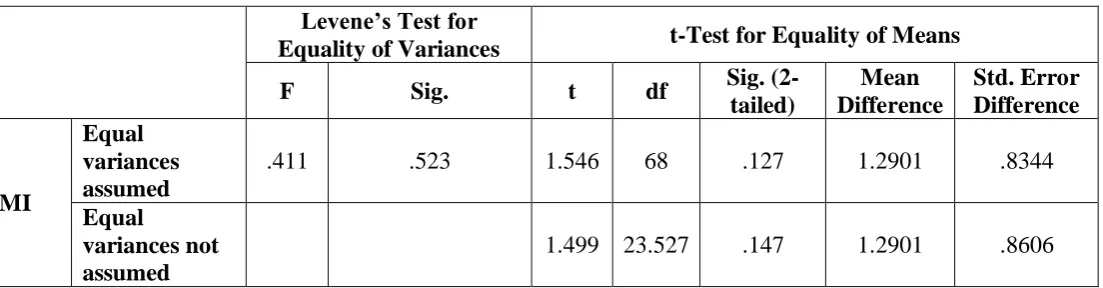
Table 5: Independent samples t-Test Levene’s Test for
Equality of Variances t-Test for Equality of Means
F Sig. t df Sig.
(2-tailed)
Mean Difference
Std. Error Difference
BMI
Equal variances assumed
.411 .523 1.546 68 .127 1.2901 .8344
Equal
variances not assumed
1.499 23.527 .147 1.2901 .8606
There was no statistically significant association between BMI and development of thyroid
dysfunction. (p value = 0.127).
Table 6: Association between receipt of HAART and thyroid dysfunction: Chi-Square Test
Receipt of HAART Thyroid Function Tests Total Normal Abnormal
Yes 39 12 51
No 15 4 19
Total 54 16 70
There was no significant association between the receipt of HAART and thyroid dysfunction
in HIV-infected patients (p value=0.826).
Association between receipt of specific antiretroviral drug and thyroid dysfunction The antiretroviral drugs evaluated in our study included zidovudine (AZT), stavudine (d4T),
lamivudine (3TC), nevirapine (NVP) and efavirenz (EFV). The study population was under
the following triple drug regimes: AZT + 3TC + NVP, d4T + 3TC + NVP, AZT + 3TC +
EFV, d4T + 3TC + EFV. The predominant drug regimen was AZT + 3TC + NVP in both the
euthyroid and hypothyroid group. All the participants received lamivudine and hence no
statistics were computed as it was a constant.
[image:8.595.30.582.145.289.2]Zidovudine
Table 7: Chi-Square Test
Receipt of Zidovudine Thyroid Function Tests Total
Normal Abnormal
No 29 7 36
Total 54 16 70
There was no significant association between the receipt of zidovudine and thyroid
dysfunction in HIV-infected patients (p value=0.484).
Stavudine
Table 8: Chi-Square Test
Receipt of Stavudine Thyroid Function Tests Total
Normal Abnormal
Yes 14 3 17
No 40 13 53
Total 54 16 70
There was no significant association between the receipt of stavudine and thyroid dysfunction
in HIV-infected patients (p value=0.557).
[image:9.595.78.504.195.271.2]Nevirapine
Table 9: Chi-Square Test
Receipt of Nevirapine Thyroid Function Tests Total Normal Abnormal
Yes 35 12 47
No 19 4 23
Total 54 16 70
There was no significant association between the receipt of nevirapine and thyroid
dysfunction in HIV-infected patients (p value=0.4446).
Efavirenz
Table 10: Chi-Square Test
Receipt of Stavudine Thyroid Function Tests Total Normal Abnormal
Yes 4 0 4
No 50 16 66
Total 54 16 70
There was no significant association between the receipt of efavirenz and thyroid dysfunction
in HIV-infected patients (p value=0.262). However, since only 4 patients in our study
population received efavirenz, statistical analysis of this variable was imprecise.
Table 11: Association between duration of HAART and Thyroid Dysfunction Thyroid
Function Test N Mean
Std. Deviation
Std. Error Mean
51 participants among our study population were receiving HAART.
The participants with thyroid dysfunction had a greater mean duration of treatment with
[image:10.595.26.583.182.338.2]HAART compared to euthyroid subjects.
Table 12: Independent samples t-Test Levene’s Test for
Equality of Variances t-Test for Equality of Means
F Sig. t df
Sig. (2-tailed)
Mean Difference
Std. Error Difference
Duration of HAART (months)
Equal variances assumed
0.03 0.953 -.506 49 .615 -3.929 7.770
Equal
variances not assumed
-.498 17.87 -.625 -3.929 7.895
There was no statistically significant association between duration of treatment with HAART
and development of thyroid dysfunction (p value=0.615).
[image:10.595.70.532.448.493.2]Association between duration of HAART and thyroid dysfunction Table 13: Mann-Whitney U Test
TFTs T3 CD4 Mean Std. Deviation CD4 Median Range
Normal 54 344.30 221.75 314 16 – 927
Abnormal 16 205.06 109.27 201 53 – 430
There was a significant difference in the CD4 count between euthyroid HIV-infected patients
and those with thyroid dysfunction (p-value = 0.027, <0.05).
To analyse the association between low CD4 count and thyroid dysfunction Table 14: Chi-Square Test
CD4 Count (cells/mm3) Thyroid Function Tests Total
Normal Abnormal
≤200(Low) 15 8 23
>200 39 8 47
Total 54 16 70
The association between thyroid dysfunction and low CD4 count did not attain statistical
[image:10.595.95.493.602.676.2]DISCUSSION
The present study included 70 patients with HIV infection, 45 males (64.3%) and 25 females
(35.7%) with a mean age of 40 years and 42 years respectively. The mean age of patients in
this study was 40.7 ± 9.7 years which was comparable to study groups Quirino et al,[23]
Beltran et al[6] and Grappin et al.[24]
Majority of patients in our study were found to have normal thyroid parameters. The
prevalence of thyroid dysfunction in our study was 22.9%. A similar prevalence of 24.5%
was documented by Madge et al.[25] in their study population. Madeddu et al.[13] and Grappin
et al[24] reported a lower prevalence of 12.6% and 12.3% respectively.
There is no uniform pattern of association between thyroid dysfunction and HIV infection.
Subclinical hypothyroidism has often been recognized in HIV-infected population with a
higher prevalence compared to HIV-negative individuals. Subclinical hypothyroidism was
the most common thyroid function abnormality in the present study. The prevalence of
subclinical hypothyroidism in our population was 10%. This observation was consistent with
several previous studies. Grappin et al.[24] found that 8.5% of their study populations had
subclinical hypothyroidism. Merenich et al.[8] reported an 8% prevalence of subclinical
hypothyroidism among HIV-infected patients receiving HAART, Calza et al.[26] reported a
high prevalence (12.2%) of subclinical hypothyroidism. Beltran et al.[6] reported a prevalence
of 6.6%, while Collazos et al.[27] and Madge et al.[25] reported a lower prevalence of 3.5% and
4% respectively. The prevalence of isolated low FT4 levels in our study was 1.4%. The
prevalence of isolated low FT4 has varied in each of the population studied. While generally
the mean FT4 is on the lower side of normal, isolated low FT4 incidence is reported in a
widely distributed spectrum of values. Collazos et al[27] found 1.3% of the determinations of
free thyroxine to be below the normal limits which is comparable with our population result.
It was observed that none of the patients in our study group had subclinical or overt
hyperthyroidism. This observation was consistent with the findings of Collazos et al.[27] and
Grappin et al.[24] Graves‟ disease is the leading cause of hyperthyroidism both in the general
population and in HIV-infected individuals. It is considered a late manifestation of immune
reconstitution inflammatory syndrome (IRIS) and is most commonly diagnosed 12-36 months
after initiation of HAART. The proposed mechanisms include thyroid gland destruction by
opportunistic infections or malignant diseases, drug-related adverse effects especially with
changes in thyroid hormone secretion or action due to direct infection of gland by HIV itself.
In our study, we evaluated the following risk factors for the development of thyroid
dysfunction; age, sex, BMI, duration of HIV infection, receipt of HAART, duration of
treatment with HAART, treatment with specific antiretroviral agent, low CD4 count and
WHO clinical stage of infection.
Age was not a risk factor for development of thyroid dysfunction or hypothyroidism in
HIV-infected patients. This observation was consistent with the previous studies of Beltran et al,[6]
Madge et al,[25] Quirino et al.[23] and Afhami et al.[4]
In our study population, the distribution of thyroid dysfunction between the sex groups
showed no statistical correlation. Similar results were observed in the studies by Madge et
al,[25] Nelson et al.[28] and Afhami et al.[4] Hypothyroidism has been reported to be more
prevalent in HIV-infected men than women, different from the gender distribution in women.
This finding was not replicated in our study. Quirino et al.[23] and Beltran et al.[6] documented
that the prevalence of subclinical hypothyroidism was higher in male patients with HIV
infection as compared with men in the general population. The prevalence of subclinical
hypothyroidism among the make subjects in our study was 8.9%, similar to that reported by
Beltran et al (8.1%).[6]
Longer duration of disease in HIV-infected patients treated with HAART might allow the
development of autoimmune thyroiditis as seen by Beltran et al.[6] There was no significant
association between the duration of illness and thyroid dysfunction or hypothyroidism in the
present study. This observation was concordant with the finding of Afhami et al.[4] and
Quirino et al.[23]
CONCLUSION
Abnormal thyroid function tests are common among HIV-infected individuals.The
prevalence of thyroid dysfunction in the study population was 22.9%. Subclinical
hypothyroidism was the most common abnormality identified in the study population,
followed by thyroid function test patterns consistent eith non-thyroidal illness (NTI). Overt
and subclinical hyperthyroidism is uncommon among HIV infected patients. Statistical
significance was not seen in the association between age, sex, BMI, duration of illness,
receipt of HAART, receipt of specific antiretroviral drug, duration of HAART, low CD4
count (≤ 200 cell/mm3
significant association between „WHO clinical stage of HIV disease‟ and the development of
thyroid dysfunction and hypothyroidism in HIV-infected patients. T3 and FT3 can be used as
biochemical indicators of advanced HIV disease as they correlate with CD4 cell counts and
the progression of disease. The majority of the thyroid abnormalities detected in our cohort
would not be routinely treated and hence the role of screening for thyroid dysfunction in
HIV-infected patients is debated.
REFERENCES
1. Hinz S, McCormack D, Van Der Spuy ZM. Endocrine function in HIV-infected women.
Gynecol Endocrinol, 2002; 16: 33-8.
2. Kalra S, Sleim H, Kotwal N. Human immunodeficiency virus and the endocrine system.
Indian J Endocrinol Metab, 2011; 15: 231-3.
3. Koskuba J, Kreze A, Kosák Metal. Endocrine complications in HIV/AIDS patients. Klin
Mikrobiol Infekc Lek, 2010; 16: 108-10.
4. Afhami S, Haghpanah V, Heshmat R et al. Assessment of the factors involving in the
development of hypothyroidism in HIV-infected patients: a case-control study. Infection,
2007; 35: 334-8.
5. Lambert M. Thyroid dysfunction in HIV infection. Baillieres Clin Endocrinol Metab,
1994; 8: 825-35.
6. Beltran S, Lescure F-X, Desailloud R et al. Increased prevalence of hypothyroidism
among HIV infected patients: a need for screening. Clin Infect Dis, 2003; 37: 579-83.
7. Bongiovanni M, Adorni F, Casana M et al. Subclinical hypothyroidism is HIV-infected
subjects. J Antimicrob Chemother, 2006; 58: 1086-9.
8. Merenich J, McDermott T, Asp A, Harrison M, Kidd G. Evidence of endocrine
involvement early in the course of human immunodeficiency vrus infection. J Clin
Endocrinol Metab, 1990; 70: 566-71.
9. Palella FJ, Delaney KM, Moorman AC et al. Declining Morbidity and Mortality among
Patients with Advanced Human Immunodeficiency Virus Infection. N Engl J Med, 1998;
338: 853-60
10.Mocroft A, Vella S, Benfield TL et al. Changing patterns of mortality across Europe in
patients infected with HIV-1. EuroSIDA Study Group. Lancet, 1998; 352: 1725-30.
11.Lima MK, Freitas LL, Montandon C et al. The thyroid in acquired immunodeficiency
12.Danoff A. HIV and the thyroid – what every practicing endocrinologist needs to know.
Nat Clin Endocrinol Metab, 2006; 2: 602-3.
13.Maddedu G, Spanu A, Chessa F et al. Thyroid function in human immunodeficiency virus
patients treated with highly active anti-retroviral therapy (HAART): a longitudinal study.
Clin Endocrinol (Oxf), 2006; 64: 375-83.
14.Jain G, Devpura G, Gupta BS. Abnormalities in the Thyroid Function Tests as Surrogate
Marker of Advancing HIV Infection in Infected Adults. J Assoc Physicians India, 2009;
57: 508-10.
15.Larsen PR, Davies TF, Schlumberger M-J, Hay ID. Thyroid Physiology and Diagnostic
Evaluation of Patientswith Thyroid Diease. In, Kronenberg HM, Melmed S, Polonsky
KS, Larsen PR (ed). Williams Textbook of Endocrinology, 11th edition. Philadelphia,
Saunders Elsevier, 2008; 299-332.
16.Jameson JL, Weetman AP. Disorders of the Thyroid Gland. In, Longo DL, Kasper DL,
Jameson JL, Fauci AS, Hauser SL, Loscalzo J (ed). Harrisons Principles of Internal
Medicine, 18th edition. USA, Mc-Graw Hill, 2012; 2911-39.
17.Iglesias P, Díez JJ. Thyroid dysfunction and kidney disease. Eur J Endocrinol, 2009; 160:
503-15.
18.Hepner GW, Chopra IJ. Serum thyroid hormone levels in patients with liver disease. Arch
Intern Med. 1979; 139: 1117-20.
19.WHO/ UNICEF/ ICCIDD Chapter 2: Selecting target groups and chapter 5: Selecting
appropriate indicators. In, Indicators for assessing Iodine Deficiency disorders and their
control through salt iodination. Geneva. WHO/NUT/94. 6: 1994.
20.Guidelines for Prevention and Management of Common Opportunistic Infections/
Malignancies among HIV-infected Adult and Adolescent, Annex. 1. WHO Clinical
Staging of HIV/AIDS for Adults and Adolescents (2006).
21.Garber JR, Cobin RH, Gharib H et al. Clinical Practice Guidelines for Hypothyroidism in
Adults: Co-sponsored by American Association of Clinical Endocrinologists and the
American Thyroid Association. Endocr Pract, 2012; 11: 1-207.
22.Bahn RS, Burch HB, Cooper DS et al. Hyperthyroidism and other cuases of
thyrotozicosis: management guidelines of the American Thyroid Association and
American Association of Clinical Endocrinologists. Endocr Pract, 2011; 17: 456-520.
23.Grappin M, Piroth L, Verges B et al. Increased prevalence of subclinical hypothyroidism
24.Quirino T, Bongiovanni M, Ricci E et al. Hypothyroidism inHIV-infected patients who
have or have not received HAART. Clin Infect Dis, 2004; 38: 596-7.
25.Madge S, Smith CJ, Lampe FC et al. No association between HIV disease and its
treatment and thyroid function. HIV Med, 2007; 8: 22-7.
26.Calza L, Mandredi R, Chiodo F. Subclinical hypothyroidism in HIV infected patients
receiving highly active antiretroviral therapy. J Acquir Immun Defic Syndr, 2002; 31:
361-3.
27.Collazos J, Ibarra S, Verges B et al. Thyroid hormones in HIV infected patients in the
highly active anti-retroviral therapy era. AIDS, 2003; 17: 763-5.
28.Nelson M, Powles T, Zeitlin A et al. Thyroid dysfunction and relationship to therapy in
HIV-positive individuals in the HAART era. J Acquir Immun Defic Syndr, 2009; 50: