Crackles in the Early Detection of Asbestosis
1
’
2
RAYMOND L. H. MURPHY, JR, EDWARD A. GAENSLER, STEPHEN K. HOLFORD, ELIZABETH A. DEL BONO, and GARY EPLER
Introduction
M
inimal interstitial pulmonary fibrosis caused by the inhalation of asbestos can be difficult to detect (1, 2). Sympto-matology is subjective and pulmonary function studies are often nonspecific. Roentgenographic manifestations of early interstitial disease are often difficult to distinguish from normal shadows (1-3), and observer variability in interpretation of radiographs is also a problem. Discontinuous adventitious lung sounds (crackles, rales) have been recognized as prominent features of pulmonary asbestosis (4—6) and are thought to be an early finding (7—9). Although chest auscultation is regarded as highly subjective, recently developed technology has brought the promise of objective quantification of lung sounds. One such method, called time-expanded waveform (TEW) analysis, consists of tape recordings of sounds for analysis by a computer. Amplitude is then plotted versus time, with a time axis of 800mm/s or more, allowing visualization of minute details of pulmonary sounds (10). Because TEW analysis provides documentation of the presence of crackles, it can be used to train technicians in auscultation (11). Moni-toring of exposed workers by ausculta-tion is attractive because it is noninvasive and inexpensive. The purpose of this investigation was to quantify the relationship between the prevalence of crackles assessed by a technician, whose performance had been objectively validated, to other criteria for asbestosis in exposed workers.
Methods
The 386 workers included in this study were exposed to asbestos during the manufacture of paper and insulation materials and in a shipyard. Lung sounds were recorded at 4 preselected basilar sites during breathing with the mouth open while the worker was in a sitting position. Workers were asked to breathe slightly more deeply than the usual tidal breathing as in standard clinical practice. These sites were the right and left bases in the midscapsular line, the right midcla- vicular line, and the left anterior axillary line. The number of crackles was estimated at each auscultation site using a
scale ranging from 0 (none) to 3.0 (indicating many), and a weighted crackle score (WCS) was then calculated. Scores
from the right and left bases posteriorly were doubled and added to the scores from the anterior sites. The resulting WCS ranged from 0 to 18. Lung sounds were recorded at the time of auscultation, and selected tape recordings were analyzed for frequency content and TEW analysis with and without 800-Hz filtered sound. These were compared with the independent observations of a trained technician.
For this assessment, worker identification was masked by use of code numbers. Comparison of the WCS was made by 2 observers who interpreted the TEW analyses while simultaneously listening to the sounds. The technician’s results were also compared with the pulmonary physician’s listening during the surveys. The Kappa statistic was calculated according to the method of Fleiss (12). Kappa is a numerical method of separating agreement from chance association, and it varies from 0 (no agreement) to 1.0 (complete agreement). For interpretation of the intermediate values, we employed the criteria of Landis and Koch (13): a Kappa value of 0.81 to 1.0 indicates almost perfect agreement, 0.61 to 0.80 indicates substantial agreement, 0.41 to 0.60 indicates moderate agreement, and 0.4 and below indicates slight to poor agreement.
Other data were collected using a questionnaire on respiratory history and occupational exposure, a physical examination including independent observations on lung sounds by 2 or more chest physicians, chest roentgenographic readings according to the 1W scheme, and detailed pulmonary func-
tion testing (14). Asbestosis in these exposed workers was considered to be present when 3 or 4 of the following
criteria were present:
SUMMARY We studied 386 workers exposed to asbestos to assess the value of chest ausculta. tion by a trained technician in detecting asbestosis as defined by previously reported clinical, physiologic, and roentgenoiogic criteria. The presence and degree of crackles were assessed at preselected basilar lung sites by a technician whose performance was validated by comparison with computer.generated time.expanded waveforms of tape recordings of lung sounds. Asbestosis was present in only 2.8% of the total population, but it was present in 8.6% of those with over 25 yr or more of employment. The technician correctly identified all the workers in whom the diagnosis was most certain, that is, those with all criteria positive. The overall true positive rate was 55%. The majority (94.8%) of those with no abnormal criteria were correctly classified. Auscuitation by an objectively validated technician can be a useful noninvasive method for screening industrial populations exposed to asbestos.
AM REV RESPIR DIS 1984; 129:375-379
(1) radiograph showing irregular opacification of a profusion of 1/2 or more, (2) single-breath diffusing capacity
less than 80% predicted, (3) vital capacity less than
80% predicted, and (4) crackles at 2 or more basilar sites.
Results
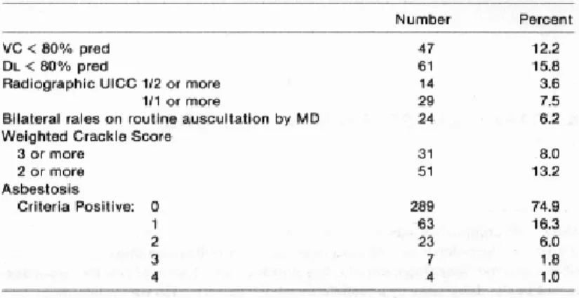
Asbestosis by our criteria was present in only 2.3% of the workers and did not occur in workers with less than 11 yr of exposure. In the 93 workers with over 25 yr of employment, 8.6% were considered to have asbestosis. The prevalence of abnormal findings in each of these criteria is shown in table 1. In general, the prevalence of these criteria increased both with exposure and with age, as is expected in populations wherein age and duration of exposure are closely correlated (figure 1). Pleural abnormalities also showed a similar re-lationship to duration of exposure and age (figure 2).
The relationship between the tech-
(Received in original form May 26, 1982 and in
revised form October 18, 1983)
From the Tufts University School of Medicine, the Boston University School of Medicine, and the Pulmonary Service, Faulkner Hospital, Boston, Massachusetts.
2 Requests for reprints should be addressed to
Raymond L. H. Murphy, Jr., M.D., Director, Pul-monary Service, Faulkner Hospital, 1153 Centre Street, Boston MA 02130.
nician’s auscultatory findings and the number of asbestos criteria found in individual workers is presented in table 2. The technician correctly identified all the workers in whom the diagnosis was most certain, that is, those with 4 of 4 positive criteria. The majority of those (94.8%) with no abnormal criteria were also correctly identified. If the crackles heard by physicians were omitted from the assessment to avoid the possibility of circular reasoning, the results were similar.
The technician identified 80% of those with all 3 of the remaining criteria. The observations of the technician while recording at the industrial site correlated well with interpretations of waveforms (table 3). The epidemiologic diagnosis of asbestosis related poorly to symptomatic and roentgenologic evidence of obstructive disease (table 4). The weighted crackle score (WCS) was associated with pack-years of cigarette smoking (table 5) but related poorly to the diagnosis of bronchitis with obstruction, as defined by the combination of a history of productive
cough plus a coexistent forced expiratory volume in one second of 70%. The interaction of smoking history, years of exposure, and crackle index was statistically evaluated using logistics regression, using both a trichotomous and dichotomous response variable (15, 16). Using the trichotomous response variable, age appeared to be the most significant effect, and the smoking-asbestos interaction was nonsignificant. The effect of asbestos exposure was present only when comparing a WCS of 3+ with a WCS of 0,
Fig. 1. Relationship of the weighted crackle score and of abnormalities consistent with pulmonary asbestosis and our diagnosis of asbestosis to duration of exposure and age in 386 asbestos-exposed workers. In general, an increasing prevalence is seen with increasing exposure for each of the criteria and for asbestosis. The weighted crackle score (WCS 3+) also shows a similar
ex-posure and age relationship. For definition of abbreviations, see table 1.
Fig. 2. Pleural thickening and/or calcification shows an increase in prevalence with increasing exposure and age, as expected in a population with exposure to asbestos.
suggesting that there was no asbestos-related differences between a WCS or 1 or 2 and a WCS of 0. The effect of smoking was more difficult to interpret. The smoking effect was present only when comparing a WCS of 1 or 2 with a WCS of 0. The results suggest the WCS of 1 or 2 may be the result of smoking, but a WCS of 3+ is associated with asbestosis.
A dichotomous model was then examined. When comparing a WCS of 0, 1, or 2 with a WCS of 3+, an asbestos effect was seen (p = 0.06) but no smoking
effect was found. Age was shown to be highly significant (p = 0.0099) when there
is no smoking-asbestos interaction (p =
.85). In comparing a WCS of 0 versus 1 +, a smoking effect is seen (p = 0.024), no
asbestos effect is found (p = 0.85), the age
effect is again highly significant (p
= 0.00026), and no smoking-asbestos
interaction is found (p = 0.65). The results
of the dichotomous model confirm those of the tnichotomous model.
Discussion
Prior to discussing the possibility of our false positives reflecting slight forms of asbestosis we will briefly review the history of crackles in asbestosis. Fifty years ago Wood and Gloyne (17) pointed out that crackles were a common feature of pulmonary asbestosis. Since that time, reports have varied as to the importance and specificity of auscultation findings. Hunter (18) said crackles occurred “sometimes” and Wyers (19) said they were “generally” present. Wyers believed that the adventitious sounds tend to be “evanescent,” and that the dry crackling sounds could disappear altogether. Smither (9) believed that the crackles of asbestosis were characteristic in their sound and distribution, present first at the bases in the midaxillary lines, and tending to spread to the posterior bases. As the disease advanced, the
crackles were distributed upwards from the base at the scapular level.
The belief that crackles are an early sign is also consistent with many pub-lished reports. Crackles have been re-garded as a feature of pulmonary asbestosis for over 50 yr, and even in early reports were considered to be present with minimal disease (17). Clinical features of their nature and distribution on the chest were described in some detail by Smither (9). Mitchell and coworkers (20) found crackles more closely related to duration of exposure to heat-resistant and friction composites than was vital capacity, and they recommended that chest auscultation be included as a biologic monitor of the work environment. Reported rates vary, but about half of the persons considered to have asbestosis on clinical grounds are reported to have crackles (21—23). These represent selected cases. In order to understand the strength of an association between an exposure and a health effect, it is important to know the status of all persons exposed. If only patients are studied, the effects may appear more striking than if asymptomatic persons are also included. Population-based studies, therefore, allow more precise examination of the frequency of crackles in asbestos-exposed persons. These show prevalences ranging from about 10 to 20%. Such prevalences depend on a variety of factors including the method of auscultation, the severity and duration of exposure, the age of population, the prevalence of the diseases causing crackles, etc. Neverthe-less, the association between crackles and asbestos exposure is clearly established by: (1) high frequency in diagnosed cases, (2) common occurrence in exposed populations, and (3) increased frequency with increased duration of exposure (24, 25). Furthermore, the-prevalence in control populations is low
(24—27).
The correct detection of those in whom the epidemiologic diagnosis of asbestosis was most certain suggests that a trained technician can screen a group of exposed workers to estimate the prevalence of asbestosis. Despite the high true positive rate there are errors in the method. In our study, 5 of the 7 workers with 3 of 4 positive criteria were missed (false negative). Although this may represent technician error, it is known that not all those with asbestosis have crackles. Indeed, Epler and coworkers (7) reported crackles in
only 60% in their pathologically proved cases of usual interstitial pneumonia. The magnitude of the false negative rate for each asbestos criteria was assessed by comparing the number of positives by the test with those workers positive by each of the remaining tests. The diffusing capacity was abnormal in 4 of the 5 workers positive for each of the remaining tests. Corresponding rates were 4/7 for vital capacity, 4/6 for radiograph, 4/5 for physician-crackles, and 4/5 for technician crackles. The false negative rate of crackles for the identification of asbestosis was, therefore, similar to the false negative rate for each of the other criteria, when considered separately. In any case, a false negative rate would have to be accepted for auscultation, as is true for any of the other available screening methods.
Some of the false negative results may have been due to the method we employed. Mitchell and coworkers (8) pointed out that inhalation to total lung capacity can abolish or decrease the number of fine rales heard in a sub-sequent inspiration. Conversely, Shirai and associates (26) have shown very high prevalences by breathholding volumes prior to deep breathing. Presumably this maneuver accentuates collapse of dependent lung regions, which reexpand on the subsequent inspiration. This is consistent with the theory that the opening of collapsed airways is the mechanism of crackle generation (28). We have also found that crackles are more common after this maneuver, particularly if the initial inspiration is performed slowly. Unfortunately, observer variability and artifacts appear to be somewhat greater, and more careful investigations will be required before this becomes an acceptable method.
The “false positives,” that is, those with a WCS of 3+ but otherwise not meeting our criteria for asbestosis, are of particular interest. Possible explanations for this include: (1) technician error, (2) crackles caused by conditions unrelated to exposure, or (3) crackles indicating lesser degrees of disease than do the other methods employed.
Technician Error
Such error is likely small in circum-stances such as this study wherein the technician is highly trained and focuses on a specific task. We evaluated the accuracy of the technician in assessing the presence, degree, and quality of crackles,
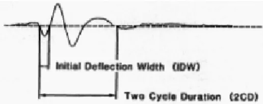
Fig. 3. A single crackle from time-expanded wave-form analysis. On the basis of 2 wavewave-form using objective methods of features, Holford (30) devised a classifier for separating fine from coarse crackles. The first measurement (the Initial deflection width or IDW) is the time in milliseconds of the first deflection of an identifiable crackle above or below the baseline. The two-cycle duration is the time in milliseconds for 2 S-shaped waves or cycles to occur. A discriminant curve on the classifier then separates fine from coarse crackles based upon both IDW and 2CD measurements for each crackle.
detecting crackles by tape recordings and time-expanded waveform analysis. Indeed, the association between the observations of the technician and the waveform interpretation was over 90%; the Kappa statistic likewise was high, indicating that this was due to agreement rather than to mere association, as it was in a more extensive study of observer variation in chest auscultation (29). The accuracy of the technician in this study in classifying crackles as fine or coarse compared with the computer classifier devised by Holford (30) was 84.4% (Kappa, 0.68). Crackle measurements made on TEW analyses are illustrated in figure 3.
Other Causes of Crackles
Numerous conditions other than in-terstitial fibrosis, such as chronic bronchitis, cause crackles. Our technician was carefully trained by listening to tape recordings of crackles from patients with asbestosis to facilitate recognition of these sounds. Indeed, the prevalence of crackles did not correlate significantly with the symptoms of chronic bronchitis, pulmonary function showing obstruction, or with roentgenographic evidence of emphysema. There was, however, a significant correlation between WCS 3+ and pack-years of smoking. The degree to which crackles reflect the industrial exposure rather than the effects of cigarette smoking is difficult to determine in these workers. Weiss (31) has suggested that it is important, but not all studies confirm this opinion (32). The study of Rossiter and Berry (21) is interesting in this regard. These inves-tigators showed an interaction between
smoking and asbestos exposure in the production of crepitations. In an asbestos textile factory, those who did not
smoke or smoked less than 5 cigarettes per day were less likely to develop crepi-tations or be certified for asbestosis than those who smoked more, although the amount of asbestos exposure was similar. Clarification of this point awaits more thorough investigation of the relationship of smoking to interstitial fibrosis and more careful delineation of the differentiating characteristics of crackles in bronchitis and asbestosis. As cigarette smoking and resulting chronic obstructive lung diseases are common in industrial workers, this is an important considera-tion. The effect of removing persons with a ratio of forced expiratory volume in one second to vital capacity of less than 70% on the age and duration of exposure plots is shown in figure 1. These tend to strengthen the relationships.
The fine crackles of interstitial fibrosis are similar in character to those caused by congestive heart failure. The latter diagnosis can usually be readily distinguished on clinical grounds. In this regard, it is important to mention that our criteria were designed for epidemiologic screening and in no way were intended to obviate the need for careful clinical evaluation of suspected positives.
Crackles as an Early Diagnostic Sign of Asbestosis
Whether our false positives reflected lesser degrees of asbestosis than other tests is unknown. However, the average duration of exposure of the workers with high crackle scores and no other criteria was longer than that of the entire group (19.6 and 13.8 yr, respectively).
Most investigators consider crackles to be an early finding (7, 26, 33). Not all investigators have concurred. In the study of 386 ship repair workers by Selikoff and associates (34), radiologic changes were considered to be a far more sensitive index than were crackles. Some of this variation in opinion is likely related to the criteria employed. We have discussed the rationale for our choice previously (10, 35, 36). Selikoff and associates apparently considered a small opacity of 0/1 or greater or any
worker in the course of developing asbestosis, it is likely that microscopic changes of alveolar thickening and peribronchial fibrosis precede any roentgenologic abnormality. It is also pleural change to asbestosis. Indeed, be evidence of in an asbestos
probable that those who develop severe asbestosis have roentgenographic pro-gression from UICC grade 0/0 to 3/4 in a relatively orderly fashion. Our reluctance to regard the lesser UICC grades of small opacifications as prima facie evidence of an effect of asbestos is based on the reliability of such evidence. First, the early diagnosis of interstitial fibrosis has long been regarded as one of the most subjective in radiology (2, 3) (other causes for roentgenologically similar shadows include soft tissues, inadequate inspiration, etc.). Second, it is likely that a variety of other illnesses and exposures produce similar findings (previous pul-monary infection, other silicates, drugs, congestive heart failure, etc.). Third, in our studies, lesser grades of opacifica-tions in asbestos workers could not be distinguished reliably from shadows seen on roentgenograms of the control subjects (10, 36). Decreasing the degree of disease required to classify an individual as a positive case in epidemiologic studies frequently leads to an increase in sensitivity. Unfortunately, this also often decreases the specificity or accuracy of the observation (13). The relative “earliness” of pulmonary function abnormalities, roentgenographic findings, and auscultation are all subject to this phenomenon. The relevant issue is which test or set of tests provides reliable monitoring of workers to assure, as far as practicable, their safety. In this regard, a significant advantage of auscultation is its noninvasiveness, as worker acceptance must also be considered. Roentgenograms offered by employers are frequently refused by workers because of the fear of exposure to radiation or personal considerations.
The results of these studies indicate that auscultation, by a properly trained and objectively validated technician, can be a useful method for screening industrial populations with exposure to asbestos. Although true positives may be underestimated, the false negative rate is low and the method is inexpensive. With the use of modern recording and analysis techniques, chest auscultation can be verified and permanent records can be made. Further studies of the sensitivity and specificity are warranted in view of the noninvasive nature of auscultation.
Acknowledgment
The writers wish to thank Drs. J. Glassroth,
J. O’Brien, M. L. Petusevsky, L. Sicilian, C. P. Wall, and D. Woodford who
participated in the industrial surveys, and Drs. R. J. McCunney and B. Raff for their participation in observer variability studies. We also
thank R. C. Bloom who performed the logistics regression analysis, J. G. Berera for tech-nical assistance, and L. M. Starr for secretarial help in the manuscript preparation.
References
1. Parkes WR. Occupational lung disorders. London: Butterworth & Co., Ltd, 1974:305. 2. Mackenzie FAF. The radiological investigation of the early manifestations of exposure to asbestos dust. Proc R Soc Med 1971; 64:34.
3. Fletcher DE, Edge JR. The early radiological changes in pulmonary and pleural asbestosis. Clinical Radiol 1970; 21:355.
4. Heard BE, Williams R. The pathology of as-bestosis with reference to lung function. Thorax 1961; 16:264.
5. Mitchell CA. Asbestos and health today. Mod Med Aust 1976; 14-6.
6. Wedler HW. Klinik der lungen asbestose, Arbeit und Gesundheit. In: Martineck G, ed, Sozialmedizi nische Schriftenreihe aus dem Gebiete des Reichsarbeitsministeriums. Vol 34. Leipzig: Georg Thieme, 1939.
7. Epler GR, Gaensler EA, Carrington CB. Crackles (rales) in the interstitial pulmonary diseases. Chest 1978; 73:333—9.
8. Mitchell CA, Charney M, Schoenberg JB. Early lung disease in asbestos-product workers. Lung 1978; 154:261—72.
9. Smither WJ. Secular changes in asbestosis in an asbestos factory. Biological effects of asbestosis. Ann NY Acad Sci 1965; 132:166—81.
10. Murphy RLH, Holford SK, Knowler WC. Visual lung sound characterization by time-expanded wave-form analysis. N Engl J Med 1977; 296:968—71.
11. Workum P, Del Bono E, Shirai S. et a!.
Observer variability in breath sound interpretation. Presented at the Fourth International Conference on Lung Sounds, Chicago, Illinois, Sept.
1980.
12. Fleiss JL. Statistical methods for rates and proportions. New York: John Wiley and Sons, 1973.
13. Landis Ri, Koch CG. The measurement of observer agreement for categorical data. Biometrics 1977; 33:159.
14. Epler GR, Saber FA, Gaensler EA. Determi-nation of severe impairment (disability in interstitial lung disease). Am Rev Respir Dis 1980;
121 :647-59.
15. Pagano M, Trichler D. An algorithm for logistic regression with polych otomous response. Technical Report. Dept. of Biostatistics and Epi-demiology. Sidney Faber Institute, Boston, MA. 16. Cox DR. Analysis of binary data. London:
Chapman & Hall, 1970.
17. Wood WB, Cloyne SR. Pulmonary asbestos. Lancet 1930; 1:447.
18. Hunter D. The diseases of occupations. London: English University Press, 1955:877-8. 19. Wyers H. Asbestosis. Postgrad Med J 1949; 25:631—8.
20. Mitchell C, Charney M, Schoenberg J. A survey of pulmonary disease in the friction materials industry. Am Rev Respir Dis 1974; 109: 728.
21. Rossiter CE, Berry G. The interaction of asbestos exposure and smoking on respiratory health. Bull Eur Physiopathol Respir l978;l4: 197-204.
22. Huuskonen MS. Clinical features, mortality and survival of patients with asbestosis. Scand J Work Environ Health 1978; 4:265—74.
23. Segarra F, Baselga MM, Lopes IP, Perez NJ. Asbestosis in a Barcelona fibrocement factory. Environ Res 1980; 23:292-300.
24. Dreesen WC, Dallavale JM, Edwards TI, et a!.
A study of asbestos in the asbestos textile industry. Public Health Bulletin No. 241. Washington, D.C.: Government Printing Office, 1938.
25. Murphy RLH, Ferris BG, Burgess WA, Worcester J, Gaensler EA. Effects of low concen-trations of asbestos. Clinical, environmental, radiologic and epidemiologic observations in shipyard pipe coverers and controls. N Engl J Med 1974; 285:1271—8
26. Shirai F, Kudoh 5, Shubuya A, Sada K, Mikami R. Crackles in asbestos workers. Auscul-tation and lung sound analysis. Br J Dis Chest 1981; 75:386—96.
27. Kleinfeld M, Massitte J, Kooyman 0, et aL
Effects of asbestos dust inhalation of lung function. Arch Environ Health 1966; 12:741.
28. Forgacs P. Applied cardiopulmonary physi-ology. The functional basis of pulmonary sounds. Chest 1978; 73:399—405.
29. Davidson F, Murphy RLH. Gravity depen-dence of crackles. In: Nair 5, ed, Computers in critical care and pulmonary medicine. New York: Plenum Press, 1983:279-86.
30. Holford SK, Discontinuous adventitious lung sounds: measurement, classification and modeling. Sc.D. Thesis. Massachusetts Institute of Technology, 1982.
31. Weiss W. Cigarette smoking and diffuse pulmonary fibrosis. Am Rev Respir Dis 1969; 99: 67—72.
32. Chew PK, Chia M, Chew SF. Asbestos workers in Singapore. Arch Environ Health 1973; 26:290—3.
33. Elmes PC. Health parameters other than mesothelioma. Proceedings of Asbestos Sym-posium, Johannesburg, S. A., Oct 3-7, 1977. Randburg: National Institute for Metallurgy, 1978:9— 16.
34. Selikoff IJ, Nicholson Wi, Lilis R. Radio-logical evidence of asbestos disease among ship repair workers. Am J Ind Med 1980; 1:9—22.
35. Murphy RLH, Gaensler EA, Ferris BG. Diagnosis of “asbestosis.” Observations from a longitudinal survey of shipyard pipe coverers. Am J Med 1978; 65:488-98.
36. Murphy RLH. Asbestosis in pipe coverers engaged in new ship construction (thesis). Harvard School of Public Health, Boston, MA, April, 1968.