17 Page 17-23 © MAT Journals 2019. All Rights Reserved
Brain Tumor Detection and Classification of MR Images Using
SVM Algorithm
Dr. C. Poongodi1, M. Sandhiya2, I. K. Surya Vikashini2
1
Associate Professor, Department of Electronics and Communication Engineering, Bannari Amman Institute of Technology, Erode, Tamil Nadu, India
2
PG Student, Department of Electronics and Communication Engineering, Bannari Amman Institute of Technology, Erode, Tamil Nadu, India
Email: [email protected]
DOI: http://doi.org/10.5281/zenodo.2604967
Abstract
Brain tumor is a mass of tissue and it occurs an abnormal growth of cells, then it form within the brain. To identifying tumor detection and classification using brain MRI image. There are several algorithms are developed for brain tumor detection and classifications in the field of medical image processing.In this undertaking we have proposed a crossover calculation for recognition cerebrum tumor in Magnetic Resonance pictures utilizing measurable highlights and Support Vector Machine (SVM) classifier. The proposed procedure comprises of four phases specifically, Noise decrease, Feature extraction, Feature decrease and Classification. In the first stage anisotropic filter is applied for noise reduction and to make the image suitable for extracting features. In the second stage, acquires the surface highlights identified with MRI pictures. In the third stage, the highlights of attractive reverberation pictures have been decreased utilizing Principles Component Analysis (PCA) to the most fundamental highlights. At the last stage, the Supervisor classifier based SVM has been utilized to group subjects as ordinary and strange mind MR pictures.
Keywords: Brain tumor, Segmentation, GLCM, PCA, SVM Classification
INTRODUTION
Brain tumor is an intracranial solid neoplasm which is characterized by an cluster of abnormal growth cells within the brain or the central spinal canal[1]Human have extraordinarily large and complex brains. The anatomy of the brain is complex due its complicate structure and function. The brain is the part of the central nervous system. It is the centre to control the mental processes and physical action of a human being. Brain abnormality is a symptom where motor impairment and neuropsychological problems affect the central nervous system. It is an abnormal growth of cells within the brain, which can be cancerous or non-cancerous. Brain tumors present at any location and different types of shapes and sizes in image, then it occurring any person almost any age. Brain tumors can be malignant (cancerous) or benign
(non-cancerous). Low grade gliomas and meningiomas are non-cancerous and glioblastoma multiforme is a cancerous tumor which represents the common primary brain neoplasm. To date, numerous researches of brain abnormality detection had been conducted due to its important roles in identifying anatomical areas of interest for diagnosis, treatment, or surgery planning paradigms. Brain tumor are normally situated in the posterior fossa in children and in the foremost of the cerebral hemispheres, in fact it canaffect any part of the brain.
The main common symptoms of brain tumors are headaches; numbness or tingling in the arms or legs; seizures; memory problems; mood and personality changes; balance and walking problems; nausea and vomiting; or changes in speech, vision, or hearing.
18 Page 17-23 © MAT Journals 2019. All Rights Reserved In this paper, first initially pre-processing
process using MRI brain image in anisotropic filter for removing noise and stripping the skull from image, after pre-processed image it can be segmented by using an interpolation algorithms. For feature extraction proposed system uses the
Gray Level Co-occurrence
Matrix(GLCM)for feature vector and then feature reduction can be used for the Principle Component Analysis(PCA). Finally, for tumour classification, Support Vector Machines (SVM) classification algorithm is used. Support Vector Machines were used to solve the pattern classification and regression problems. While using this classification process for overall performance and accuracy also increased compare other classification techniques.
RELATED ARTICLES
BRAIN TUMOR DETECTION AND CLASSIFICATION OF MR IMAGES USING TEXTURE FEATURES AND FUZZY SVM CLASSIFIER
The authors Jayachandran and Dhanasekaran propose a half and half calculation for recognition mind tumor in Magnetic Resonance pictures utilizing Fuzzy Support Vector Machine (FSVM) classifier [1]. For fruitful treatment, tumor position and size are essentially characterized and exactness to gauge. In by and large have a few calculations for cerebrum tumor recognition and groupings in the field of medicinal picture preparing. In this paper they proposed different approach consists of four stages such as: Noise reduction, Feature extraction, Feature reduction and Classification. In thisproposed a hybrid algorithm for detection brain tumor in Magnetic Resonance images using statistical features and Fuzzy Support Vector Machine (FSVM) classifier.
In the first stage Pre-processing process involves removing their low-frequency
background noise, smoothing smudges then removing reflections and masks etc. Then pre-processing process used some different types of filters such as Median filter, Anisotropic filter, Mean filter, Frequency filters, Gaussian smoothing, Laplacian filters etc.
Anisotropic filter is used to remove the background noise and thus preserving the edge points in the image. Anisotropic filter is applied for noise reduction and to make the image suitable for extracting features. After pre-processing, segmentation can be used for an noise reduction and edge reduction to be used.The result of image segmentation is a set of segments that collectively cover the entire image, or a set of contours extracted from the image. In the second stage, obtains the texture features related to MRI images. In that feature extraction can be used for the GLCM (Gray Level Co-occurrence Matrix). GLCM is an estimate of the second-order statistical information of neighboring pixels of an image.
In the third stage, the features of magnetic resonance images have been reduced using principles component analysis to the most essential features. The PCA is an existing input features into a new lower dimensional feature space. PCA can be used for reduce the dimensionality of features and it results are more accurate and efficient.
At the last stage, the SVM (Support Vector Machines) are supervisor classifier based has been used to classify subjects as normal and abnormal brain MR images. SVMs are set of related administered learning strategies utilized for classification and regression. SVM did not depend on any dimension or feature set. It changes the higher dimensional information into nonlinear map function.
19 Page 17-23 © MAT Journals 2019. All Rights Reserved
AUTOMATIC CLASSIFICATION OF MR BRAIN TUMOR IMAGES USING DECISION TREE
The authors Hemarajini and Narmatha proposed an automatically classifies brain tumor using decision tree [5]. It has five different types of tumors such as glioblastoma multiforme, astrocytoma, metastatic, glioma and pituitary macro. For tumor grouping utilized MR pictures and its considered a component extraction for T1-weighted pictures are differentiate for each hub cut through the brain.A double choice tree is an established and coordinated tree with two sorts of tree vertices are, for example:
A binary decision tree is a rooted and directed tree with two types of tree vertices are such as: Terminal vertices andNon-terminal vertices.
TERMINAL VERTICES
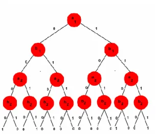
Terminal Vertices v has no children and is labelled by value (v) Є {0, 1}.A binary decision tree for the two-bit comparator, given by the formula
F(a1,a2,b1,b2)=(a1←→a2)(b1←→b2) (1) NON - TERMINAL VERTICES:
Each non- terminal vertex v is a labelled by an variable vertex i.e. var(v) and has two succesors:
Low (v) similarly to the case where var(v) is assigned 0 and
High (v) similarly to the case where var(v) is assigned 1.
The proposed strategy has three phases. They are pre-preparing, include extraction and order. In the main stage, the clamor is expelled utilizing a wiener channel. In the second stage, six surface highlights are extricated utilizing dim dimension co-event network.
The features extracted are angular second moment, contrast, inverse difference moment, entrophy, correlation and variance. Finally, a decision tree classifier is used to classify the type of tumor image.
Figure 1: General Binary Decision Tree. The extracted features are compared with the stored features in the knowledge base to classify the type of tumors.
The proposed method has three stages. They are pre-processing, feature extraction and classification.
Figure 2: Block Diagram for Decision Tree Algorithm.
Recognizing a consequently order the five distinct sorts of tumors like glioblastoma multiforme, astrocytoma, metastatic, glioma and pituitary large scale in mind MR pictures utilizing choice tree and exactness likewise estimated.
AN OVERVIEW ON SUPPORT VECTOR MACHINES (SVM) AND CLASSIFICATION USING
INTERSECTION KERNEL SUPPORT The authors Dussa Harsha Praneeth proposed an overview of SVM classification and kernel support process are also explained.
First initial process is done a pre-processing after segmenting after doing a feature reduction or feature extraction can be used. And then finally classification it’s
Pre-processing: Noise removal Feature extraction using GLCM Classifier: Decision tree
20 Page 17-23 © MAT Journals 2019. All Rights Reserved very important one of the process.
Classification analyses the different numerical properties of various image features and also organises the data and that classification types have differently splitted such as: Supervised Learning and Unsupervised Learning.
The classification have different types of analysed such as:
1. Artificial Neural Network(ANN) 2. Support Vector Machines(SVM) 3. Fuzzy Based Support Vector
Machines(FSVM) 4. Decision Tree.
The ANN is a type of artificial intelligence that imitates some functions of the person mind. ANN has a normal tendency for storing experiential knowledge. An ANN consists of a sequence of layers; each layer consists of a set of neurones. All neurones of every layer are linked by weighted connections to all neurones on the preceding and succeeding layers.
In ANN characterizes are uses an Non-parametric approach. Performance and accuracy depends upon the network structure and number of inputs
Decision Tree (DT) calculates class membership by repeatedly partitioning a dataset into uniform subsets Hierarchical classifier permits the acceptations and rejection of class labels at each intermediary stage. This method consists of 3 parts: Partitioning the nodes, find the terminal nodes and allocation of class label to terminal nodes.
DT characterized is DT are based on hierarchical rule based method and use nonparametric approach.
SVM is a help vector machines. SVM tackles the matched arrangement. SVM is used to aggregate the class name by disconnecting the information point with a straight line. In any case, in some datasets, it is preposterous to hope to seclude the
information point by one straight line. SVM all the while limit the experimental classification mistake and boost the geometric edge. So SVM called Maximum Margin Classifiers. SVM depends on the Structural hazard Minimization (SRM). SVM outline vector to a higher dimensional space where a maximal isolating hyperplane is developed. Two parallel hyperplanes are built on each side of the hyperplane that different the information.
KERNELS IN SVM
The kernel capacity may change the information into a higher dimensional space to make it conceivable to play out the division. Kernel capacities are a class of calculations for example examination or acknowledgment, whose best realized component is the support vector machine (SVM). Training vectors xi is mapped into a higher (might be endless) dimensional space by the capacity.
The decision of a Kernel relies upon the current issue since it relies upon what we are endeavouring to demonstrate. A polynomial kernel, for instance, enables us to demonstrate highlight conjunctions up to the request of the polynomial. Spiral premise capacities permits to choose hovers - interestingly with the Linear kernel, which enables just to select lines (or hyperplanes).
A NOVEL BRAIN TUMOR
DETECTION METHOD USING DWT AND CLUSTERING TECHNIQUES FROM T2-WEIGHTED BRAIN MRI IMAGES
The authors T. Lakshmi Narayana and T. Sreenivasulu Reddy proposed a mind tumor recognition utilizing DWT strategies. In that framework utilized T2-weighted cerebrum MRI pictures utilized for bunching strategies.This paper describes a novel method for brain tumor
21 Page 17-23 © MAT Journals 2019. All Rights Reserved detection from MRI images using hybrid
DWT and fuzzy C means technique and support vector machine classifier.
In this paper a proposed system consists of four stages. Such as,
Stage 1 is pre-processing, Stage 2 is segmentation,
Stage 3 is feature extraction and Stage 4 is classification.
In the first stage the images are acquired and enhanced the quality by removing the noise by median filtering technique.The median filtering is widely used denoising technique used to improve the quality of a brain MRI image by removing highfrequency components without disturbing the edges. T2-weighted MR image is chosen as the reference image. In the second stage the discrete wavelet transform ad fuzzy c means techniques partitioned the images to identify the abnormal part. It is the most important task in medical imaging. Here the DWT and fuzzy C means are the segmentation techniques used to segment the MRI images of human brain.
The identified tumor part is extracted in stage three. The GLCM has used to determine the spatial information and distribution of neighbour pixels. The GLCM has powerful features, though time consuming. Contrast, correlation, homogeneity, entropy and energy are the features determined by using this GLCM technique.
In Fourth stage is a classification technique which is used to classify the output obtained from the feature extraction stage. The classification classifies the features into the proper class. The support vector machine (SVM) is the best binary classifier for braintumor detection. SVM did not depend on any dimension or feature set.
And at final, calculated the performance
parameters of the brain MRI images and compared with existing techniques. The proposed method is robust, efficient and fast computing technique comparing with existing methods.
BRAIN TUMOR DETECTION USING K-MEAN CLUSTERING AND SVM The authors Anushree A. wankhade, Dr. A. V.Malviya proposes a Brain tumor detected by K-Mean clustering and SVM approach.
In this project firststage are image is enhanced in the way that finer details are improved and noise is removed from the image. Most commonly used enhancement and noise reduction techniques are implemented that can give best possible results. Enhancement will result in more prominent edges and a sharpened image is obtained.
Segmentation is an essential process to extract information from complex medical images. The main objective of segmentation is to simplify or change the representation of an image.
In third stage k-means algorithm is to cluster the data. Kmeans algorithm is one of the simplest partitions clustering method. K-Means is the one of the unsupervised learning algorithm for clusters. Clustering the image is grouping the pixels according to the some characteristics. In the k-means algorithm initially we have to define the number of clusters k. Then k-cluster center are chosen randomly.
And finally last stage is Support vector machine (SVM) is the linear learning algorithm used for classification. The process of classification forward through training and testing. Support vector machine (SVM) is the linear learning algorithm used for classification. SVM will classify the image into normal and Abnormal. SVM is a powerful supervised
22 Page 17-23 © MAT Journals 2019. All Rights Reserved classifier and accurate learning technique.
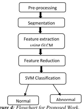
METHODOLOGY AND ANALYSIS In this paper proposed an SVM algorithm for classification method. SVM is an (Support Vector Machines). And feature extraction using an GLCM matrix. Then the Feature reduction using an PCA method and ICA analysis their images. And it steps to analysis the proposed work.
Figure 3 (a): Input Image (b) Segmented Image (c).Output Image.
Steps occupied in the development of the proposed work:
1. Read the input brain MRI image. 2. Resize the input image into 256x256
sizes.
3. If the image is in color, then convert it to gray scale.
4. The image has filtered by using Aniostropic filter which is used to eliminate the noise from the gray scale image so as to make image is smoother and preserve the edges.
5. Segmentation is a significant process to extract edge detection. Each of the pixels in a region is similar with respect to some characteristic or computed property, such as color, intensity, or texture.
6. After segmenting process in the proposed technique, skull stripping is used for the segmentation of brain tissues
7. Apply the gray level co-occurrence matrix to find different features which are used for the performance evaluation. It is estimated of a joint Probability Density Function (PDF) of gray level pairs in an image.
8. Apply the Principle Component Analysis (PCA) and Independent component analysis (ICA) are used to
feature reduction.This two tools are used for transforming the existing input features into a new lower dimensional feature space.
9. Classifies the image is having tumor or not. It used an SVM algorithm.
10.Calculate the area (size) of the tumor in terms of mm2 and number of pixels occupied by tumor. And specified the tumor is benign (noncancerous) or malignant (cancerous).
Figure 4: Flowchart for Proposed Work.
CONCLUSION
This paper proposes discovery of cerebrum tumor and picture division utilizing a SVM calculation. After segmentation, the feature extracted image using GLCM technique and feature reduction used a PCA method. The performance of two-tier classifier system in terms of statistical measures such as sensitivity, specificity and accuracy of tumor will be analyzed. REFERENCES
1. Jayachandran A, Dhanasekaran R “Brain tumour detection and classification of MR images using texture features and fuzzy svm classifier”.,Res.J.Eng.Tech,June2013,p p 2264-2269.
2. Rajini, N.H., Narmatha, T., Bhavani, Pre-processing Segmentation Feature extraction using GLCM SVM Classification Abnormal Feature Reduction Normal
23 Page 17-23 © MAT Journals 2019. All Rights Reserved R.: “Automatic classification of MR
brain tumor images using decision tree”. Special Issue of Int. J. of Computer Applications, March2012, pp. 10–13.
3. Dussa Harsha Praneeth “An Overview On Support Vector Machines (Svm) And Classification Using Intersection Kernel Support”. International Journal of Management, Technology and Engineering, ISSN NO: 2249-7455. 4. T. Lakshmi Narayana,T. Sreenivasulu
Reddy, “A Novel Brain Tumor Detection Method using DWT and Clustering Techniques from T2-Weighted Brain MRI Images”, International Journal of Management, Technology And Engineering, ISSN NO : 2249-7455.
5. Anushree A. wankhade, Dr. A. V.Malviya, “Brain Tumor Detection Using K-Mean Clustering And Svm”,International Research Journal of Engineering and Technology, ISSN: 2395-0072.
6. Clark, M.C., Hall, L.O., Goldgof, D.B., Velthuzien, R., Muztagh, F.R., Silbiger, M. “Automatic tumor segmentation using knowledge based techniques”, IEEE Trans. Med. Imaging,vol.17,Feb.2012,pp. 187–192.
7. Ramteke, R.J., Monali,
Y.K.:“Automatic medical image classification and abnormality detection using K-nearest neighbour”, International Journal Advanced Computer. Res.s, vol.2,March 2012, pp. 190–196.
8. Barakbah Ridho, A., Kiyoki, Y.: “A pillar algorithm for k-means optimization by distance maximization for initial centroid designation”,Proc. of IEEE Symp. On Computational Intelligence and Data Mining, Feb.2011, pp. 61–68.
9. Pal, N.R., Pal, S.K.:“A review on image segmentation techniques”, Pattern Recognition .pp. 1277–1294. 10.M.S.B PhridviRaja, C.V GuruRaob
(2014). Data mining – past, present and future – a typical survey on data streams. Procedia Technology 12, 255 – 263.
Cite this article as: Dr. C. Poongodi, M. Sandhiya, & I. K. Surya Vikashini. (2019). Brain Tumor Detection and Classification of MR Images Using SVM Algorithm. Journal of Signal
Processing, 5(1), 17–23.
http://doi.org/10.5281/zenodo.260496 7