Article
1
Efficient Shielding of Polyplexes using
2
Heterotelechelic Polysarcosines
3
Philipp Michael Klein 1,*, Kristina Klinker 2,4, Wei Zhang 1, Sarah Kern 1, Eva Kessel 1,
4
Ernst Wagner 1,3, Matthias Barz 2,*
5
1 Ludwig-Maximilians-Universität (LMU) Munich, Pharmaceutical Biotechnology, Department of
6
Pharmacy, Butenandtstrasse 5-13, D-81377 Munich, Germany; [email protected]
7
2 Institute of Organic Chemistry, Johannes Gutenberg University, Duesbergweg 10-14, D-55128 Mainz,
8
Germany Affiliation 2; [email protected]
9
3 Nanosystems Initiative Munich, Schellingstraße 4, D-80799 Munich, Germany
10
4 Graduate School Materials Science in Mainz, Staudinger Weg 9, 55128 Mainz, Germany
11
12
* Correspondences: [email protected]; Tel.: +49-89-2180-77794
13
[email protected]; Tel.: +49-6131-39-26256
14
15
Abstract: Shielding agents are commonly used to shield polyelectrolyte complexes, e.g. polyplexes,
16
from agglomeration, precipitation in complex media, like blood, and thus enhance their circulation
17
times in vivo. Since up to now primarily poly(ethylene glycol) (PEG) has been investigated to shield
18
non-viral carriers for systemic delivery, we report on the use of polysarcosine (pSar) as a potential
19
alternative for steric stabilization. A redox-sensitive, cationizable lipo-oligomer structure
20
(containing two cholanic acids attached via a bioreducible disulfide linker to an oligoaminoamide
21
backbone in T-shape configuration) was equipped with azide-functionality by solid phase
22
supported synthesis. After mixing with small interfering RNA (siRNA), lipopolyplexes formed
23
spontaneously and were further surface-functionalized with polysarcosines. Polysarcosine was
24
synthesized by living controlled ring-opening polymerization using an azide-reactive
dibenzo-aza-25
cyclooctyne-amine as an initiator. The shielding ability of the resulting formulations was
26
investigated with biophysical assays and by near-infrared fluorescence bioimaging in mice. The
27
modification of ~100 nm lipopolyplexes was only slightly increased upon functionalization. Cellular
28
uptake into cells was strongly reduced by the pSar shielding. Moreover, polysarcosine-shielded
29
polyplexes showed enhanced blood circulation times in bioimaging studies compared to unshielded
30
polyplexes and similar to PEG-shielded polyplexes. Therefore, polysarcosine is a promising
31
alternative for the shielding of non-viral, lipo-cationic polyplexes.
32
Keywords: shielding agent, polysarcosine, biodistribution, click-chemistry, lipopolyplex, nucleic
33
acid carrier
34
35
1. Introduction
36
Therapeutic nucleic acids are powerful tools, which can be used to specifically control gene
37
expression inside cells [1-5]. For several diseases, including severe metastatic tumors, systemic
38
delivery is required to achieve therapeutic effect. Naked oligonucleotides have limited stability in
39
biological fluids because they are actively targeted and degraded by nucleases. Although this issue
40
might be addressed by the incorporation of chemical modifications [6,7], the renal clearance of small
41
oligonucleotides or siRNA usually occurs within a few minutes, which limits the time to reach their
42
desired site of action [8,9]. Liposome-based formulations, lipo-polymer micelles, and polymer-based
43
complexes increase the size usually beyond the renal cut-off and thus enhance circulation times,
44
whenever a stealth-like corona protects the systems from unspecific aggregation [6,10-21]. Various
45
polycations, cationic lipids and combinations with helper lipids are used to form polyplexes
46
[4,14,16,17,22-24], lipoplexes or lipid nanoparticles (LNPs) [1,11,25-30]. Precise editing of the
47
components' chemical structure enables the fine-tuning of a carrier's stability and size, but also other
48
properties, which are important for the delivery process like cellular uptake, endosomal escape
49
ability, and cell tolerability [31-34].
50
Solid phase-supported synthesis (SPS) is a very convenient and precise way to optimize a
51
delivery system in a step-wise manner [18,35]. Recently, we developed our own customized amino
52
acids, such as succinoyl tetraethylene pentamine (Stp), which contains of short defined repeats of the
53
diaminoethane motif prepared in boc/fmoc protected form. With artificial building blocks, natural α
-54
amino acids and fatty acids we sequentially synthesized monodisperse cationic oligomers via SPS,
55
which are highly adaptable to different demands in the field of gene delivery [8,12,18,31-33,36-39].
56
By precise incorporation of a bioreducible cleavage site between the cationic and a lipophilic block,
57
for instance, it was possible to destabilize polyplexes only after reaching the cytoplasm of the cell
58
[39]. Thereby the carrier system remained stable in serum and transfection efficiency as well as cell
59
viability could be increased in certain cell lines.
60
Besides size and stability, the surface character of a nanoparticle is of utmost importance for its
61
systemic delivery. Shielding agents attached to the surface prevent interactions with neighboring
62
particles and/or blood components, which usually leads to extended circulation in the body's
63
bloodstream [40-42]. Already in 1990 it could be demonstrated that polyethylene glycol (PEG) could
64
extend the blood circulation half-life of systemically administered liposomes from <30 min to several
65
hours [40]. Its hydrophilic character enables PEG to generate a hydrated shell covering the
66
nanoparticles and thereby sterically reduce unwanted interactions with biomolecules or other poly-
67
or lipoplexes [43]. PEG is the most prominent shielding agent and has often been used to shield
68
cationic polyplexes in numerous applications [44-49]. A major drawback, however, is that more and
69
more researchers in academia or industry observe immune responses towards PEGylated
70
nanoparticles [43,50-55]. For this reason several new alternatives were evaluated for shielding, such
71
as natural proteins [56], oligosaccharides [57,58], poly(N-(2-hydroxypropyl)methacrylamide)
72
(pHPMA) [58-60], hydroxyethyl starch (HES) [61] or polypeptides (poly(glutamic acid) [62],
73
poly(hydroxyethyl-L-asparagine) [63], poly(hydroxyethyl-L-glutamine) [63], prolin-alanin-serin
74
motif (PAS) [64,65]). Nevertheless, according to the Whiteside’s rules for protein resistant surfaces an
75
ideal alternative to PEG should mimic its chemical properties, being a hydrophilic, non-charged
76
polymer and a weak hydrogen acceptor without donor properties, which is not the case for all
above-77
mentioned polymers. In contrast, polysarcosine fulfills all the described criteria and has already
78
demonstrated protein resistant properties on various surfaces [66-68]. In addition, it can be also
79
synthesized by living controlled ring opening polymerization of the corresponding N
-80
carboxyanhydrides (NCA) [69,70]. However, in vivo data on polysarcosine is rarely reported in
81
literature [71]. In contrast to polypeptides, the side chain of polypeptoids is situated at the nitrogen
82
rather than the α-carbon, in the case of pSar the nitrogen is methylated. As a result, polysarcosine
83
adopts a random coil conformation in aqueous solution and possesses a comparable second virial
84
coefficient and molecular weight dependency like PEG [72]. All these properties provide a high
85
resistance against protein adsorption [73] and make it in theory an ideal material for shielding
86
electrostatic complexes in vivo [74]. Importantly, it has been reported that polysarcosine has so far
87
demonstrated neglectable complement activation or immunogenicity in mouse, rat and rabbit animal
88
models [75,76]. And pSar-shielded polyplexes, micelles, colloids and nanohydrogels demonstrated
89
the absence of aggregation in human serum [77-80].
90
In the current work, we have incorporated azide domains into a previously described
redox-91
sensitive T-shaped bis-(cholanic acid amido) oligoaminoamide siRNA carrier system [39] and used
92
strain-promoted azide-alkyne cycloaddition (SPAAC) reaction to equip the surface of lipopolyplexes
93
with ~8 kDa polysarcosine (DP=119) chains. We report on the ability of polysarcosine to shield siRNA
94
lipoplexes and analyzed the in vivo stability and biodistribution after intravenous administration into
95
mice. In a second approach, we modified the system with a folate ligand to target the folate receptor
96
2. Materials and Methods
98
2.1 Materials
99
Protected Fmoc-α-amino acids, 2-chlorotrityl chloride resin, N,N-dimethylformamide (DMF),
100
N,N-diisopropylethylamine (DIPEA) and trifluoroacetic acid (TFA) for solid-phase syntheses were
101
purchased from Iris Biotech (Marktredewitz, Germany). Triisopropylsilane (TIS),
1-102
hydroxybenzotriazole (HOBt), 5β cholanic acid. Dimethylformamide (DMF) for DBCO-PSar
103
syntheses was purchased from Acros Organics (99.8% Extra), further dried over CaH2 and
104
fractionally distilled in vacuo. Folic acid (FolA) was purchased from Acros Organics (96–102% pure).
105
Triethylamine (TEA) and N,N-diisopropylethylamine (DIPEA) were dried over NaOH and
106
fractionally distilled in vacuo. (Benzotriazol-1-yloxy) tripyrrolidino phosphonium
107
hexafluorophosphate (PyBOP),
2-(1H-Benzotriazol-1-yl)-1,1,3,3-tetramethyluronium-108
hexafluorphosphat (HBTU) and microreactors were obtained from MultiSynTech (Witten, Germany).
109
Cell culture media, antibiotics and fetal calf serum (FCS) were purchased from Invitrogen (Karlsruhe,
110
Germany), HEPES from Biomol GmbH (Hamburg, Germany), glucose from Merck (Darmstadt,
111
Germany), agarose (NEEO Ultra-quality) from Carl Roth GmbH (Karlsruhe, Germany), and
112
GelRed™ from VWR (Darmstadt, Germany). Cell culture 5 × lysis buffer andD-luciferin sodium salt
113
were obtained from Promega (Mannheim, Germany). Ready-to-use siRNA duplexes were obtained
114
from Axolabs GmbH (Kulmbach, Germany): eGFP-targeting siRNA (siGFP) (sense: 5′
-115
AuAucAuGGccGAcAAGcAdTsdT-3′; antisense: 5′-UGCUUGUCGGCcAUGAuAUdTsdT-3′) for
116
silencing of eGFPLuc; control siRNA (siCtrl) (sense: 5′-AuGuAuuGGccuGuAuuAGdTsdT-3′;
117
antisense: 5′-CuAAuAcAGGCcAAuAcAUdTsdT-3′); Cy5-labled siRNA (Cy5-siAHA1) (sense:
5’-118
(Cy5)(NHC6)GGAuGAAGuGGAGAuuAGudTsdT-3’; antisense:
5’-119
ACuAAUCUCcACUUcAUCCdTsdT-3’); Cy7-labled siRNA (Cy7-siAHA1) (sense:
5’-120
(Cy7)(NHC6)GGAuGAAGuGGAGAuuAGudTsdT-3’; antisense:
5’-121
ACuAAUCUCcACUUcAUCCdTsdT-3’) small letters: 2′methoxy; s: phosphorothioate. All other
122
chemicals were purchased from Sigma (Munich, Germany), Iris Biotech (Marktredwitz, Germany),
123
Merck (Darmstadt, Germany) or AppliChem (Darmstadt, Germany), Acros Organics, Alfa Aesar, or
124
Fluka.
125
2.2 Synthesis of oligomers and DBCO shielding agents
126
2.2.1 Synthesis of oligomers
127
See supporting information for detailed description on syntheses of oligomers.
128
2.2.2 Synthesis of sarcosine-N-carboxyanhydride
129
The synthesis was performed as described in Klinker et al. [71] Sarcosine (15.16 g, 170.2 mmol,
130
1 eq) was weighed into a pre-dried three-necked flask and dried under vacuum for 1 hour. 300 mL
131
absolute (abs) THF was added under a steady flow of nitrogen. The apparatus was connected to two
132
gas washing bottles filled with aqueous sodium hydroxide solution. Diphosgene (16.26 ml,
133
134 mmol, 0.8 eq) was added slowly via syringe. The colorless suspension was heated to 70 °C
134
yielding a clear solution after 3 hours of stirring. The solvent was evaporated under reduced pressure
135
yielding a brown oil as crude reaction product. The oil was heated to 50 °C and dried under reduced
136
pressure to obtain an amorphous solid. The crude reaction product was redissolved in 60 mL THF
137
and precipitated with 300 mL abs n-hexane. The precipitate was filtered off under N2-atmosphere
138
and dried with a stream of dry nitrogen for 60 - 90 minutes to remove residual traces of solvents. The
139
next day, the product was dried in high vacuum for 2 hours in the sublimation apparatus and
140
subsequently sublimated at 80 – 85 °C and < 1x10⁻² mbar. The product was collected from the
141
sublimation apparatus in a glove box on the same day. Colorless crystals were obtained (50 – 67 %).
142
mp = 104.3 °C; 1HNMR (300 MHz, CDCl3) δ[ppm] = 2.86 (s, 3H, NH-CH3), 4.22 (s, 2H, NH-CH2-CO).
143
2.2.3 Synthesis of DBCO-PSar
146
DBCO-PSar was synthesized using ring-opening polymerization of SarNCA as described in
147
Klinker et al. [71] In a typical experiment, 461.8 mg of SarNCA (4.012 mmol) were transferred under
148
nitrogen counter flow into a pre-dried Schlenk-tube, equipped with a stir bar and again dried in
149
vacuum for 30 minutes. The NCA was then dissolved in 3.5 ml of dry DMF. A stock solution of
150
DBCO-amine (0.074 mmol, 1/110 eq, M/I = 110) in 2 mL DMF was prepared and 1 mL of this stock
151
solution were added to the monomer solution via syringe. The solution was stirred at 40 °C and kept
152
at a constant pressure of 1.25 bar of dry nitrogen via the Schlenk-line to prevent impurities from
153
entering the reaction vessel while allowing CO2 to escape. Completion of the reaction was confirmed
154
by IR spectroscopy (disappearance of the NCA peaks (1853 and 1786 cm-1)). Directly after completion
155
of the reaction, the polymer was precipitated in cold diethyl ether and centrifuged (4500 rpm at 4 °C
156
for 15 min). After discarding the liquid fraction, new ether was added and the polymer was
157
resuspended using sonication. The suspension was centrifuged again and the procedure was
158
repeated. The polymer was then dissolved in H2O and lyophilized to obtain a fluffy powder (279.7
159
mg, 98 %). 1H-NMR: (400 MHz, DMSO−d6): δ [ppm] = 0.88 – 0.79 (m, ini, 9H, -C(CH3)3), 2.58 - 3.11 (br,
160
3nH, N-CH3), 3.73 - 4.57 (br, 2nH, -CO-CH2-N), = 7.86 – 7.10 (m, 8H, benzylic protons).
161
2.2.4 Synthesis of DBCO-PSar-Ac
162
DBCO-PSar119 (Mn =8735 g mol-1) (36 mg, 0.004 mmol), acetic anhydride (4.2 mg, 3.9 µL, 0.04
163
mmol), and triethylamine (10.7 mg, 14.6 µL, 0.08 mmol) were dissolved in absolute DMF (1 mL) and
164
stirred at 25 °C for 24 h under an argon atmosphere. Subsequently, the polymer was precipitated in
165
diethyl ether, extensively dialyzed against water (MWCO = 3500 g mol-1), and lyophilized. Yield after
166
dialysis: 25 mg (69%).
167
2.2.5 Synthesis of DBCO-PSar-FolA
168
DBCO-PSar110 (Mn =8095 g mol-1) (55.8 mg, 0.007 mmol) was separately dissolved in absolute
169
DMSO. Folic acid (30.4 mg, 0.068 mmol), HBTU (26.1 mg, 0.068 mmol), and HOBt (9.31 mg, 0.068
170
mmol) were dissolved in DMSO and cooled to 0 °C. DIPEA (17.8,mg, 24.0 µL, 0.138 mmol) was added
171
and the mixture was left to react for 30 minutes at 0 °C. The in situ formed activated ester was added
172
to the predissolved polymer and the reaction mixture was stirred at 25 °C for 24 h under an argon
173
atmosphere. The crude reaction product was purified by size exclusion chromatography in DMSO
174
using a Sephadex LH-20-packed column. The purified conjugate was lyophilized from water. Yield
175
after SEC: 31 mg (55%).
176
2.2.6 Gel permeation chromatography
177
Polymer molecular weight and dispersity index were determined by gel permeation
178
chromatography (GPC). GPC in hexafluoro-2-propanol (HFIP) was performed with 3 g L-1 potassium
179
trifluoroacetate (KTFA) at 40 °C. The columns were packed with modified silica (PFG columns,
180
particle size: 7 µm; porosity: 100 and 1000 Å). A refractive index detector (G 1362A RID, Jasco) and a
181
UV/vis detector (UV-2075 Plus, JASCO, λ=230 nm; λ=330nm for folic acid detection) were used to
182
detect the polymer. Molecular weights were calculated using calibration performed with PMMA
183
standards (Polymer Standards Services GmbH). Toluene was used as the internal standard.
184
185
2.2.7 UV-vis spectroscopy
186
UV-vis absorbance spectra were recorded using a spectrophotometer V-630 (Jasco) with water
187
being the solvent.
188
2.3 Formation of siRNA polyplexes
192
siRNA was dissolved in 20 mM HEPES buffered 5% glucose pH 7.4 (HBG) at a concentration of
193
50 ng/µL for in vitro experiments and 500 ng/µL for in vivo experiments. According to the indicated
194
nitrogen/phosphate (N/P) ratio, the oligomer solution was prepared in a separate tube. Only
195
protonatable nitrogens were considered in the N/P calculation. The same volume of siRNA solution
196
was added to the oligomer. The mixture was rapidly pipetted at least five times and incubated for
197
40 min at RT resulting in a polyplex solution with 25 or 250 ng of siRNA/µL respectively.
198
2.4 Functionalization of polyplexes with DBCO reagents
199
For functionalization of siRNA polyplexes with DBCO click agents, solutions with reagents were
200
prepared in ¼ of the volume of polyplex solutions prepared before. The concentration of the solution
201
was calculated according to the respective equivalents (eq). Equivalents represent the molar ratio of
202
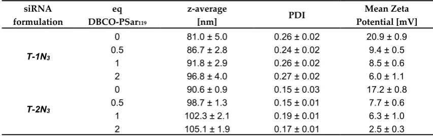
shielding agent to oligomer in the polyplex solution. The reaction time was 16 h for biophysical and
203
in vitro assays and 4 h for in vivo experiment respectively.
204
2.5 siRNA binding assays
205
siRNA binding assays were performed analogously as described in Klein et al. [86]. A 1%
206
agarose gel was prepared by dissolving agarose in TBE buffer (10.8 g of trizma base, 5.5 g of boric
207
acid, 0.75 g of disodium EDTA, and 1 L of water) and subsequent boiling. After cooling down to
208
about 50 °C, GelRedTM was added. Formulations were prepared with 50 ng of siRNA. Samples were
209
placed into the pockets after 4 µl of loading buffer (prepared from 6 mL of glycerine, 1.2 mL of 0.5 M
210
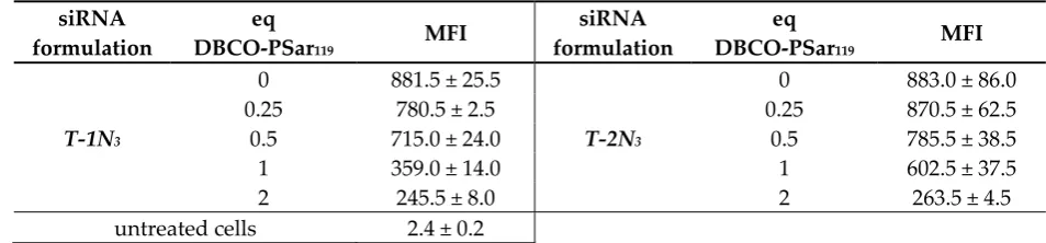
EDTA, 2.8 mL of H2O, 0.02 g of bromophenol blue) was added. Electrophoresis was performed at
211
70 V for 60 min.
212
2.6 Particle size and zeta potential measurements
213
Dynamic light scattering (DLS) measurements of polyplex solutions were performed in a folded
214
capillary cell (DTS 1070) using a Zetasizer Nano ZS with backscatter detection (Malvern Instruments,
215
Worcestershire, UK). Polyplexes were formed using 2 µg siRNA in a total volume of 80 µL. For size
216
measurements, the equilibration time was 0 min, the temperature was 25 °C and an automatic
217
attenuator was used. The refractive index of the solvent was 1.330 and the viscosity was 0.8872 mPa·s.
218
Each sample was measured 3 times. For detection of the zeta potential, the sample was diluted to
219
800 µL volume with 10 mM NaCl solution. Measurements with at least 6 runs were performed. Zeta
220
potentials were calculated by the Smoluchowski equation. Ten to fifteen sub runs lasting 10 s each at
221
25 °C (n = 3) were measured.
222
2.7 Cell culture
223
The mouse neuroblastoma cells (Neuro2a) were cultured in DMEM low glucose medium
224
(Sigma, Munich, Germany). As FR-expressing cell lines, human cervix carcinoma cells (KB), and
225
human cervix carcinoma cells stably transfected with the eGFPLuc (enhanced green fluorescent
226
protein/luciferase) gene (KB/eGFPLuc) were cultured in folate-free RPMI 1640 medium (Invitrogen,
227
Karlsruhe, Germany). All media were supplemented with 10% FBS, 100 U/mL penicillin, and 100
228
µg/mL streptomycin. The cells were maintained in ventilated flasks in the cell incubator at 37 °C with
229
5% CO2 in a humidified atmosphere. Cell lines were grown to 80-90% confluency and harvested.
230
2.8 Cell association and internalization of siRNA polyplexes measured with flow cytometry
231
For untargeted polyplexes, Neuro2a cells were seeded in 24-well plates with 5 × 10⁴ cells/well at
232
24 h before the experiment, and fresh growth medium was provided before the experiment.
233
Polyplexes containing 1.5 µg of siRNA (including 20% Cy5-labeled siRNA) were added into each
234
well incubated for four hours at 37 °C in 5% CO2. Cells were then incubated with 500 I.U. heparin to
235
For folate-targeted polyplexes, KB cells were seeded in 24-well plates with 5 × 10⁴ cells/well at
237
24 h before the experiment, and fresh growth medium was provided before the experiment.
238
Polyplexes containing 1.5 µg of siRNA (including 20% Cy5-labeled siRNA) were added into each
239
well incubated 30 min on ice for cell association or 45 min at 37 °C in 5% CO2 for cellular
240
internalization, respectively. Cells were washed with PBS to remove free polyplexes. For cellular
241
internalization, cells were then incubated with 500 I.U. heparin to remove polyplexes non-specifically
242
associated to the cell surface.
243
Finally, cells were collected and resuspended in PBS buffer with 10% FBS. All samples were
244
measured by flow cytometry with CyanTM ADP (Dako, Hamburg, Germany) through excitation at
245
635 nm, and detection of emission at 665 nm. Dead cells were differentiated by DAPI fluorescence
246
and removed by gating in order to analyze cellular uptake of polyplexes in living cells. Data were
247
analyzed by FlowJo 7.6.5 flow cytometric analysis software.
248
2.9 Confocal laser scanning microscopy (CLSM)
249
Neuro2a cells were seeded into an 8-well Lab-Tek chamber slide (Nunc) at a density of 3 × 10⁴
250
cells/well in 300 µL of growth medium 24 h prior to treatment. Polyplexes were formed as described
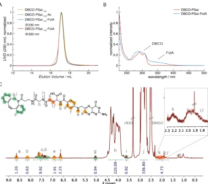
251
using a 1.5µg of a mixture of 80% of siCtrl and 20% Cy5-labeled siRNA and oligomer at N/P 12 in 60
252
µL of HBG followed by the indicated agent in 20µL. Cells were incubated with 220 µL of fresh growth
253
medium and polyplex solution was applied. For the uptake study, the incubation with polyplexes
254
was at 37 °C for 4 h. The growth medium was removed, cells were washed twice with 300 µL of PBS
255
and fixed with 4% PFA solution for 30 min at room temperature. Cell nuclei were stained with DAPI.
256
A Leica TCS SP8 confocal microscope was used for image acquisition.
257
2.10 Mouse tumor model
258
Female six- to seven-week-old nude mice, Rj: NMRI-nu (nu/nu) (Janvier, Le Genest-Saint-Isle,
259
France), were housed in isolated ventilated cages under specific pathogen-free condition with a 12 h
260
light/dark interval and were acclimated for at least 7 days prior to experiments. Food and water were
261
provided ad libitum. Animals were injected subcutaneously with 5 × 10⁶ Neuro2a cells. The body
262
weight was recorded, and the tumor volume was measured by caliper and calculated as
263
[0.5 × (longest diameter) × (shortest diameter)2]. All animal experiments were performed according
264
to guidelines of the German Animal Welfare Act and were approved by the local animal ethics
265
committee.
266
2.11 Biodistribution study
267
For near infrared (NIR) in vivo imaging, unlabeled control siRNA (siCtrl) was spiked with 50%
268
of Cy7-labeled siRNA (Cy7-siAHA1) in HBG. When tumors reached the size of 500 - 1000 mm3, the
269
mice (n = 2/per group) were anesthetized with 3% isoflurane in oxygen. siRNA polyplexes containing
270
50 µg of Cy7-labeled siRNA (N/P 10) in 250 µL (containing 100 µL of siRNA solution, 100 µL of
271
oligomer solution, 50 µL of agent solution or buffer) of HBG were injected intravenously (i.v.), and
272
fluorescence was measured with a CCD camera at different time points. For evaluation of images, the
273
efficiency of fluorescence signals was analyzed after color bar scales were equalized using the IVIS
274
Lumina system with Living Image software 3.2 (Caliper Life Sciences, Hopkinton, MA, USA).
275
3. Results and Discussion
276
3.1 Design and synthesis of a lipo-oligomer for click chemistry
277
In previous work, we have established a new class of redox-sensitive lipo-oligomers prepared
278
by solid-phase supported synthesis (SPSS) to serve as carriers for siRNA delivery [39]. Beside
279
beneficial effects of lipid-based delivery systems, such as enhanced nanoparticle stability and
280
endosomal escape capability, the in vivo distribution is often limited to certain tissues, primarily liver,
281
ones that were synthesized in this approach demonstrate the strongest retention in liver tissue [89,90].
283
Those effects might be related to a high stickiness of cationic particles, but it is also possible that
284
certain serum proteins, which incorporate onto nanoparticle surfaces may impair tissue specificity
285
[88,91-93]. An efficient shielding should reduce both types of interactions and should enable a better
286
distribution in the body. For this reason, one of the best performing candidates from redox-sensitive
287
lipo-oligomers, T-shape structure T-0N3 (published as ID 992 in [39]) was chosen and extended by
288
click-reactive azide functionality. After the formation of siRNA lipopolyplexes, the particle surface
289
was further modified with the shielding agents.
290
291
292
Figure 1. Overview of chemical compounds. Table, top: schematic illustration of sequence-defined
293
oligomers with T-shape topology; T-0N3 (ID: 992 published [39]), T-1N3 (ID: 1073) and T-2N3 (ID:
294
1086) with no, one or two terminal azidolysines K(N3). Other units of the oligomers: Y: tyrosine, K:
295
lysine, G: glycine, Stp: succinoyl-tetraethylene-pentamine, ssbb: succinoyl-cystamine, CholA: 5β
-296
Cholanic acid. The broken lines represent amide linkages, the triangle (▲) is the starting point of the
297
synthesis. IDs are unique database identification numbers. Table, bottom: structure of the shielding
298
agents DBCO-PSar119, DBCO-PSar119-Ac and DBCO-PSar110-FolA. Scheme of the formulation of a
299
shielded polyplex.
300
T-0N3 was chosen as starting point for further modifications, because it forms stable siRNA
301
polyplexes with sizes below 200 nm hydrodynamic diameter, which show high transfection efficiency
302
in mouse neuroblastoma (Neuro2a) cells. This structure combines natural amino acids and artificial
303
building blocks (Figure 1 - top). It consists of four repeats of the cationic polyamino acid
succinoyl-304
tetraethylene-pentamine (Stp) used for complexation of nucleic acid and for endosomal buffering.
305
Two tyrosine trimer units flanking the cationic domain stabilize the polyplex due to their
306
hydrophobicity and π–π stacking ability. In the center of the cationic Stp units, two hydrophobic
307
cholanic acids branch off the cationic backbone (T-shape) for lipopolyplex stabilization. The lipid and
308
cationic domains are connected via a bioreducible linking unit (ssbb) [39]. In this approach, the azide
309
function was incorporated into the oligomer during standard Fmoc solid-supported synthesis via an
310
azidolysine residue at the N- and/or C-terminus of the backbone (structures T-1N3 with one azide
311
and T-2N3 with two azides, Figure 1 - top).Consequently, the structure can be subsequently further
312
3.2 Synthesis of DBCO-modified polysarcosine
314
Polysarcosine is a hydrophilic, nonionic peptoidic structure with exclusively weak hydrogen
315
bond acceptor properties. As shown empirically by Whitesides and co-workers for protein-resistant
316
surfaces, these properties are essential to achieve "stealth"-like properties in a material [71,74].
317
Polysarcosine can be functionalized at its N-terminal (via post-polymerization modification) and C
-318
terminal (via functional initiators) end. It is conveniently synthesized by controlled living
ring-319
opening polymerization of α-amino acid N-carboxyanhydrides (NCA) with low dispersity index
320
(ÐGPC ≤ 1.1; see Table S1) [71]. Initiating the reaction with dibenzo-aza-cyclooctyne-amine
(DBCO-321
amine) leads to a C-terminal DBCO end group (Figure S1A) [94]. To ensure end group accessibility
322
and steric stabilization at once we aimed for a degree of polymerization of around 115, which
323
correlates with the PEG5k used as reference material. Therefore, a theoretical degree of
324
polymerization of 115 was set by the monomer to initiator ratio. The polymerization was carried out
325
under the conditions recently reported by Klinker et al. [95], NMR end group and SEC analysis (using
326
pSar standards as described by Weber et al. [72]) revealed a number average degree of polymerization
327
(DP) of 119 and a weight average DP of 8735 g mol-1 (Table S1), which is within the experimental
328
error. The accuracy molecular weight determination is perfectly in line with the calculated one. The
329
synthesized polymer displays a symmetrical SEC elugram indicating a Poisson-like molecular weight
330
distribution, as expected for the amine initiated NNCA polymerization. The terminal DBCO can be
331
clearly detected in the 1H-NMR spectra and thus can be employed for the strain promoted
azide-332
alkyne cycloadditon (SPAAC) with azides. For SPAAC no catalyst is needed, no side reactions with
333
other functional domains of the oligomer can occur, and no toxic by-products are generated [96].
334
Mixing azide-modified cationic oligomers with siRNA leads to spontaneous assembly of polyplexes.
335
Due to oligomer excess, several azide functionalities are accessible on the polyplex surface and can
336
serve as attachment points for functionalization with DBCO-modified pSar. The N-terminal free
337
amine group can further be modified with carboxylic acid-bearing molecules to introduce a second
338
functionality, e.g. targeting ligands such as folic acid or alternatively may be capped by acetylation
339
to remove the terminal amine, which is positively charged in aqueous solution of neutral pH. All
340
synthesized agents are presented in Figure 1.
341
3.3 Polyplex formation and pSar-shielding
342
For polyplex formation, the structures T-1N3 and T-2N3 were incubated with siRNA for 40 min
343
with a final concentration of 25 ng siRNA/µL. The electrophoretic mobility of incorporated siRNA
344
was measured with an agarose gel shift assay to test the oligomers' abilities to bind nucleic acid.
345
Different N/P values depict the ratio of amines (N) of the oligomers that can be protonated to the
346
phosphates (P) of the siRNA. Like its azide-free analogue T-0N3 [39], the oligomers showed complete
347
retention of siRNA in the pockets of the gel at an N/P ratio of 12 (Figure S2). No changes in binding
348
ability were observed for the incorporation of one or two azides.
349
Next, the heterotelechelic polysarcosine with DBCO-end group (DBCO-PSar119; Figure 1,
350
bottom) was used to react with the azides on the preformed polyplexes (N/P ratio 12) to introduce a
351
shielding layer. The SPAAC was allowed to proceed until full conversion for 16 hours (see scheme in
352
Figure 1, bottom).
353
Afterwards, the influence of the pSar shielding agent on electrophoretic mobility was evaluated
354
with respect to the number of azide functions incorporated into the polyplex-forming core structures
355
(N3 = 0, 1, 2). The amount of DBCO-PSar119 added to the polyplexes was kept constant (Figure 2A).
356
In this experiment, only the azide-bearing polyplexes migrated in the gel towards the cathode,
357
whereas the azide-free polyplex remained in the loading pocket. This demonstrates that a covalent
358
bond connecting the shielding agent to the nanoparticle is crucial to provide this migratory effect.
359
The migration of the integrated siRNA against its own negative charge shows that it is fully shielded
360
against the force of the electric field. With increasing equivalents of DBCO-PSar119, stronger
361
migration could be observed. This effect can be explained by the degree of polyplex surface
362
modification (Figure 2B). For the oligomer with only one azide functionality (T-1N3), maximum
363
PSar119 (2 eq) did not increase the effect. T-2N3 siRNA polyplexes with two azide functionalities
365
within the carrier also showed the maximum migration for equimolar ratios of azide to DBCO (2 eq
366
DBCO-PSar119). These findings are in line with covalent modification with PEG5k [86].
367
368
369
Figure 2. Electrophoretic mobility of siRNA polyplex formulations analyzed with an agarose gel shift
370
assay A) siRNA polyplexes formed with oligomers bearing no (T-0N3), one (T-1N3) and two (T-2N3)
371
azide functions incubated with 0.75 equivalents of DBCO-PSar119 1 % agarose gel, 70 V, 80 min
372
runtime. B) Formulations with increasing equivalents (eq mol/mol) of DBCO-PSar119. 0.75 % agarose
373
gel, 100V, 80 min runtime. All polyplexes were incubated for 40 min at N/P 12, followed by
DBCO-374
PSar119 addition for 16 h at room temperature. The right lane shows the running distance of free
375
siRNA not complexed by lipo-oligomers.
376
A second indirect measure for the efficiency of polyplex shielding is the zeta potential or
377
electrochemical mobility. The latter can be determined by measuring a particle's mobility in an
378
electric field with light scattering. In this respect, we observed that the positive zeta potential of an
379
unshielded particle can be strongly reduced from 21 mV to 6 mV in case of T-1N3 polyplexes and
380
from 17 mV to 3 mV in case of T-2N3 polyplexes, when the particle is shielded with an excess of
381
DBCO-PSar119 (Table 1). By using 0.5 eq DBCO-PSar119 / oligomer, the zeta potential can already be
382
reduced to 50%. It should be noted here that due to the N-terminal cationic tail group, the zeta
383
potential always remained slightly positive.
384
As determined by single-angle dynamic light scattering (DLS), hydrodynamic diameters of the
385
polyplexes were approximately 100 nm. With increasing amounts of DBCO-PSar119, the nanoparticle
386
size increased by up to 16 nm in diameter. pSar covering the particle surface seems to be the most
387
plausible explanation for the increase in size.
388
Table 1. Particle size (z-average) and zeta potential of pSar-shielded siRNA formulations determined
389
by a dynamic light scattering (DLS) zetasizer. siRNA polyplexes were prepared at N/P 12
390
siRNA formulation
eq DBCO-PSar119
z-average
[nm] PDI
Mean Zeta Potential [mV]
T-1N3
0 81.0 ± 5.0 0.26 ± 0.02 20.9 ± 0.9
0.5 86.7 ± 2.8 0.24 ± 0.02 9.4 ± 0.5
1 91.8 ± 2.9 0.26 ± 0.02 8.5 ± 0.6
2 96.8 ± 4.0 0.27 ± 0.02 6.0 ± 1.1
T-2N3
0 90.6 ± 0.9 0.15 ± 0.03 17.2 ± 0.8
0.5 98.7 ± 1.3 0.15 ± 0.01 7.7 ± 0.6
1 102.3 ± 2.1 0.19 ± 0.01 6.3 ± 1.0
2 105.1 ± 1.9 0.17 ± 0.01 2.5 ± 0.3
3.4 Evaluation of pSar-shielding agents in vitro
392
Through the incorporation of pSar the unspecific interaction of polyplexes with cell membranes
393
should be efficiently reduced as already demonstrated for other stealth-like polymers, e.g. PEG. To
394
prove our assumption we performed uptake studies with pSar-shielded polyplexes. Polyplex
395
formulations were prepared with Cy5-labled siRNA for this assay to follow the fluorescent cargo,
396
incubated with neuroblastoma Neuro2a cells for 4 h at standard culture conditions and analyzed by
397
flow cytometry. The signal intensity of cells labeled with fluorescent dye correlates with the amount
398
of polyplexes being internalized (Table 2 and Figure S3). Unshielded material and material shielded
399
with low equivalents of DBCO-PSar119 showed significant uptake into cells already after 4 h
400
incubation time for both polyplex formulations prepared with one and two azide-bearing backbones
401
(T-1N3 and T-2N3). For T-1N3 formulations, a significant reduction in fluorescence intensity of more
402
than 50% was observed for 1 eq of DBCO-PSar119 per oligomer, whereas 2 eq of DBCO-PSar119 were
403
needed for T-2N3 formulations to reduce cell uptake (Table 2).
404
Table 2. Mean fluorescence intensity (MFI) for cellular internalization of Cy5-labeled siRNA
405
formulations (left: T-1N3; right: T-2N3) shielded with increasing equivalents (eq mol/mol) of
DBCO-406
PSar119 determined by flow cytometry.
407
siRNA formulation
eq
DBCO-PSar119 MFI
siRNA formulation
eq
DBCO-PSar119 MFI
T-1N3
0 881.5 ± 25.5
T-2N3
0 883.0 ± 86.0
0.25 780.5 ± 2.5 0.25 870.5 ± 62.5
0.5 715.0 ± 24.0 0.5 785.5 ± 38.5
1 359.0 ± 14.0 1 602.5 ± 37.5
2 245.5 ± 8.0 2 263.5 ± 4.5
untreated cells 2.4 ± 0.2
408
The effect on internalization can be visualized by confocal laser scanning microscopy (CLSM).
409
Cells were incubated with T-1N3 siRNA formulations for 4 h and the Cy5-labeled siRNA (red)
410
representing the localization of the polyplex was detected (Figure 3). Compared to the unshielded
411
material, which was avidly taken up by cells, 0.5 eq of DBCO-PSar119 showed a slight reduction in
412
cellular internalization. For 1 eq of DBCO-PSar119, only a few polyplexes were taken up by cells,
413
indicating a strong shielding ability. This experiment confirmed the observations made in the
414
previously described flow cytometry studies.
415
420
Figure 3. Intracellular distribution of T-1N3 siRNA formulations in Neuro2a-eGFP-Luc cells with
421
increasing equivalents (eq mol/mol) of DBCO-PSar119 acquired by confocal laser scanning
422
microscopy. Cells were incubated with the formulations for 4 h and washed with PBS buffer. Nuclei
423
were stained with DAPI (blue) and siRNA was spiked with 20 % Cy5-labeled siRNA (red). The
424
overlay image shows the merged channels and the light microscope image. Scale bar: 10 µm
425
In conclusion, covalent surface modification of polyplexes by SPAAC reduced cell binding and
426
uptake substantially. Interestingly, the increase of azide functionalities in the T-2N3 backbone did not
427
lead to a better surface passivation of the formed polyplex. As depicted in Table 2, both polyplexes
428
behave comparably and differ only slightly at full polysarcosinylation levels. This observation may
429
relate to differences in microstructure between T-1N3 and T-2N3 based polyplexes, which seems to
430
influencethe accessibility of azide on the polyplex surface.
431
3.5 Distribution of pSar- functionalized polyplexes in vivo
432
After the shielding ability of pSar-functionalized polyplexes could be demonstrated in
433
biophysical and in vitro assays, we aim to explore the in vivo behavior of polysarcosinylated
434
polyplexes. For in vivo biodistribution studies, the unshielded T-1N3 siRNA polyplex, which showed
435
the lowest interaction with cells, was used to prepare a formulation to which either DBCO-PEG5k
436
and a formulation using acetylated polysarcosine (DBCO-PSar119-Ac; Figure 1, bottom) was
437
covalently linked by SPAAC. The acetyl end group of DBCO-PSar119-Ac seems to be better
438
comparable to the commercial methoxylated PEG agent in terms of surface polarity. The cap of the
439
N-terminal sarcosine slightly reduced migration distance of polyplexes in the gel in comparison to
440
the non-acetylated pSar (Table S2, Figure S4). T-1N3 siRNA polyplexes were prepared with 50%
Cy7-441
labeled siRNA and incubated with 1 eq of the respective shielding agent per oligomer for 4 h. A final
442
concentration of 200 ng siRNA / µL was used for this experiment. 50 µg of siRNA and oligomers at
443
an N/P ratio of 10 were used.
444
446
Figure 4. Biodistribution of T-1N3 siRNA formulations (50 µg siRNA; 50% Cy7-labeled) in
NMRI-447
nude mice bearing Neuro2a tumors after i.v. administration. NIR fluorescence bioimages show
448
formulations with 1 eq DBCO-PEG5k, 1 eq acetylated DBCO-PSar119-Ac or HBG buffer
(non-449
shielded). Experiments were performed with two animals per group for time points until 60 min and
450
one animal per group for later time points; a representative animal of each group is shown. Animals
451
are presented in the dorsal, ventral and lateral view.
452
The formulations were injected into Neuro2a tumor-bearing mice via i.v. tail-vain injection and
453
the distribution of the near infrared (NIR) fluorescent dye attached to the siRNA was monitored at
454
various time points over 24h by bioimaging in mice (Figure 4, Figure S5). The unshielded polyplexes
455
started accumulating in the liver after 15 min. Such a finding could also be observed for other
456
unmodified T-shape backbone structures in previous work [89,90]. In contrast to the unshielded
457
polyplexes, both shielded formulations showed much-extended circulation times and tumor
458
accumulation. 60 minutes after injection of the material, the shielded formulations were still
459
detectable in all areas of the body including the tumor site. After 4 h the intensity of the signals
460
decreased, indicating a slow removal of polyplexes from circulation. The strongest signals remained
461
in liver and bladder. In mice injected with shielded polyplexes, a strong signal was detected in the
462
exposed periphery, such as the paws after more than 4 hours (Figure S5). In direct comparison,
463
polyplexes with the DBCO-PSar119-Ac and the DBCO-PEG5k displayed negligible differences in
464
biodistribution, circulation time or tumor accumulation. A pronounced accumulation at the tumor
465
site, however, could not be observed and may require further strategies to enhance tumor
466
accessibility and retention.
467
3.6 Attachment of the targeting ligand folate to polysarcosine
468
The inhibition of unspecific cell binding is an important requirement for any specific interaction
469
with cell surface receptors or proteins in solution. Thus, attaching a targeting ligand onto shielded
470
polyplexes is expected to enable targeting of specific cell surface receptors. Since pSar offers the
471
possibility to be further functionalized at its free secondary amino function, we chose folic acid (FolA)
472
as a ligand to be conjugated to the N-terminus of DBCO-PSar110 by peptide bond formation. Folic
473
acid is an interesting ligand, because it is a commercially available small molecule with carboxyl
474
groups for conjugation and it is the natural ligand to the folic acid receptor (FR) overexpressed on
475
several tumor types, e.g. prostate cancer [82-84,97-100]. The applied coupling conditions using
476
equimolar amounts of folic acid, DBCO-PSar polymer and coupling reagents (2-(1H
-benzotriazole-1-477
yl)-1,1,3,3-tetramethyluronium hexafluorophosphate (HBTU), 1-hydroxybenzotriazole (HOBt)) and
478
the steric hindrance of the polymer avoid the formation of divalent folic acid conjugates. Concerning
479
regioselectivity, it has been reported that both isoforms result for N,N-dicyclohexylcarbodiimide
480
(DCC)-mediated amidation in DMSO or DMSO/DMF, but with an observed regioselectivity of 80%
481
for the γ- conjugate (DBCO-PSar110-FolA; Figure 1, Figure 5, Table S1) [101].
483
Figure 5. Characterization of DBCO-PSar ligands for post-shielding of polyplexes. A) GPC elugrams
484
of PSar in HFIP with different end groups. B) UV-vis spectrum of PSar and
DBCO-485
PSar-FolA, respectively. C) 1H NMR spectrum of DBCOPSar-FolA in DMSO-d6 (400 MHz).
486
The properties of T-1N3 siRNA polyplexes equipped with this negatively charged ligand
487
changed in an unexpected way. For increasing equivalents, aggregates with high polydispersity were
488
found by DLS (Table S1). An interesting finding was the change in size from around 80 to 25 nm of
489
nanoparticles when folic acid was involved, which indicates strong compactation of the polyion
490
complex. For small degrees of particle modification with DBCO-PSar110-FolA, the size of polyplexes
491
did not significantly change compared to unshielded polyplexes. For higher amounts, aggregates
492
were found in DLS measurements. Similar findings were observed for folic acid-targeted
493
lipopolyplexes [102]. In the latter case, aggregation could be avoided by incorporation of
tetra-494
glutamylated folic acid into the shielding agent. We observed that for further increase of
DBCO-495
PSar110-FolA to equimolar amounts, small defined particles of ~25 nm were found. This can only be
496
explained with DBCO-PSar110-FolA-induced instability following a complete rearrangement of
497
particles into a uniform population. The folic acid's chemical properties - its hydrophobic character
498
and negative charge – seem to play a major role in this reassembly process, since it was not observed
499
for untargeted polysarcosine-shielded particles.
500
When testing the folate targeted formulation with DBCO-PSar110-FolA on a FR-overexpressing
501
KB/eGFPLuc cell line, we found that targeted polyplexes showed increased binding to the cell
502
surface, which could be blocked by folic acid competition (Figure S6A) ensuring FR mediated
503
binding. Much to our surprise, the internalization of the polyplexes into the cell was extremely low
504
(Figure S6A+B). As a consequence, no gene silencing activity was achieved with such systems (Figure
505
efficiencies [103-107], can be excluded, since co-incubation with the lysosomotropic agent
507
chloroquine did not improve gene silencing activity (Figure S6D). The trafficking of the vitamin folate
508
via FR is reported to occur by a non-clathrin, non-caveolar pathway also known as CLIC/GEEC
509
endocytosis pathway [104,108]. For folate-targeted nanoparticles however, it could be demonstrated
510
that pathways like caveolae- and clathrin-mediated endocytosis occur [105,107,109]. The size and the
511
ligand density on their surface were reported to influence the cellular uptake pathway. The well
512
shielded, ~25 nm siRNA polyplexes do not seem to trigger any of the pathways in HeLa-derived KB
513
cells efficiently. Further, the bioreducible carrier T-1N3 within the liopolyplex might be an easy prey
514
for disulfide cleavage, which was reported to occur distinctly in the extracellular environment of
515
HeLa cells [110]. The consequence of insufficient cellular uptake was a lack of gene silencing activity.
516
This effect has been observed for folate-targeted polyplexes with 3.5 kDa PEG chains before [86]. At
517
this point, we cannot provide an explanation for the observed findings and further studies need to
518
be conducted to understand the fact that specific receptor binding was achieved, while receptor
519
mediated endocytosis seems to be inhibited. In light of these in vitro data, a transfer of targeted
520
polyplexes to in vivo studies was not performed based on ethical considerations.
521
4. Conclusions
522
To investigate the use of ability of pSar to shield polyplexes and enhance their circulation times
523
and reduce unspecific interactions, we synthesized a polyplex formulation based on sequence
524
defined lipo-oligomers and applied PEG and pSar based polymers for shielding of the preformed
525
polyplexes. In previous work, a new class of redox-sensitive lipo-oligomers was successfully
526
established for siRNA delivery. [39] For this reason, one of the best performing candidates from
527
redox-sensitive lipo-oligomers was chosen and extended by a click-reactive azide functionality,
528
resulting in carrier T-1N3. After the formation of siRNA lipopolyplexes, the particle surface was
529
further modified with the shielding agent polysarcosine. The SPAAC could be performed between
530
the DBCO-PSar polymer and the azide-containing lipo-oligomers within the polyplex. In addition, it
531
was demonstrated that the grafting could be controlled stoichiometrically introducing a shielding
532
layer. The shielding of the formed pSar corona has been observed in vitro in gel retardation assays
533
and cell studies. In contrast to unmodified polyplexes, binding and cellular uptake was substantially
534
reduced for all pSar-modified systems.
535
Furthermore, biodistribution in mice revealed that 8 kDa polysarcosine can strongly expand the
536
circulation of the siRNA lipopolyplexes from several minutes to hours. The difference in
non-537
shielded and shielded formulations is most pronounced at the 60 minutes time point, where in case
538
of non-shielded polyplexes, most of the polyplexes have accumulated in the liver, but stable
539
circulation is still observed for pSar-shielded polyplexes. While the biodistribution between
non-540
modified polyplexes and polysarcosinylated systems differ substantially, such systems behaved
541
similar to PEGylated polyplexes in vivo. Therefore, we can conclude that in terms of polyplex
542
shielding pSar and PEG behave identically and can be both applied to reduce unspecific interactions
543
of lipo-oligomer polyplexes and thus enhance blood circulation substantially from minutes to hours.
544
When DBCO-PSar was, however, modified with folic acid to target cell surface receptors, not only
545
the size of polyplexes was reduced from 80 to 25 nm, but also specific binding to FR-positive KB cell
546
membranes did not boost cellular internalization. Therefore, we need to conclude that further
547
investigations are necessary to combine favorable in vivo shielding with efficient receptor-targeted
548
gene silencing for pSar-functionalized lipo-oligomer polyplexes.
549
550
Supplementary Materials: The following are available online, Figure S1: Synthesis of heterotelechelic
DBCO-551
PSar polyplex shielding agents by NCA polymerization and subsequent amidation for further introduction of
552
functionalities, Figure S2: siRNA binding ability of T-shape structures analyzed with an agarose gel shift assay,
553
Figure S3: Cellular internalization of siRNA formulations shielded with increasing equivalents of DBCO-PSar119
554
determined by flow cytometry, Figure S4: Electrophoretic mobility of formulations, Figure S5: Biodistribution of
555
siRNA formulations in NMRI-nude mice bearing Neuro2A tumors after i.v. administration, Figure S6: Cellular
556
analogues, Table S1: Analytical data of synthesized heterotelechelic DBCO-PSar ligands, Table S2: Particle size
558
and zeta potential of siRNA formulations determined with a DLS zetasizer.
559
Author Contributions: Philipp Klein performed chemistry of oligomers, preparation of the formulations and
560
biophysical and cell-free in vitro experiments. Kristina Klinker synthesized and analyzed the DBCO agents. Wei
561
Zhang performed transfections and FACS studies. Sarah Kern and Eva Kessel performed the in vivo experiment.
562
Ernst Wagner and Matthias Barz supervised the experimental work and contributed with scientific discussions.
563
Philipp Klein wrote the draft manuscript, Kristina Klinker, Matthias Barz and Ernst Wagner edited the
564
manuscript, all other authors checked and contributed to the finalization of the manuscript.
565
Funding: This work was supported by German Research Foundation (DFG) CRC1066-1/-2 Projects A6, B5 and
566
B12 as well as the Excellence Cluster Nanosystems Initiative Munich (NIM). KK would like to thank "Materials
567
Science in Mainz" (MAINZ) and HaVo-foundation. WZ appreciates receiving a Bayerischen
568
Gleichstellungsförderung fellowship as support for her postdoctoral research at LMU Munich.
569
Acknowledgments: We thank Miriam Höhn for generating the CLSM images.
570
References
![Figure 1. Overview of chemical compounds. Table, top: schematic illustration of sequence-defined oligomers with T-shape topology; T-0N3 (ID: 992 published [39]), T-1N3 (ID: 1073) and T-2N3 (ID: 1086) with no, one or two terminal azidolysines K(N3)](https://thumb-us.123doks.com/thumbv2/123dok_us/7954267.1319894/7.595.131.487.192.488/overview-chemical-compounds-schematic-illustration-oligomers-published-azidolysines.webp)