International Journal of Nanomedicine
O R I G I N A L R E S E A R C H
open access to scientific and medical research
Open Access Full Text Article
Dovepress
A highly sensitive and selective viral protein
detection method based on RNA
oligonucleotide nanoparticle
Changhyun Roh1
Ho-Young Lee2
Sang-Eun Kim2
Sung-Kee Jo1
1Radiation Research Division for
Biotechnology, Advanced Radiation Technology Institute (ARTI), Korea Atomic Energy Research Institute (KAERI), Sinjeong-dong, Jeongeup, Jeonbuk, South Korea; 2Department
of Nuclear Medicine, College of Medicine, Seoul National University, South Korea
Correspondence: Changhyun Roh Radiation Research Division for Biotechnology, Advanced Radiation Technology Institute (ARTI), Korea Atomic Energy Research Institute (KAERI), 1266, Sinjeong-dong, Jeongeup, Jeonbuk 580-185, South Korea Tel +82 63 570 3227 Fax +82 63 570 3229 Email [email protected]
Sung-Kee Jo
Radiation Research Division for Biotechnology, Advanced Radiation Technology Institute (ARTI), Korea Atomic Energy Research Institute (KAERI), 1266, Sinjeong-dong, Jeongeup, Jeonbuk 580-185, South Korea Tel +82 63 570 3200 Fax +82 63 570 3229 Email [email protected]
Abstract: Globally, approximately 170 million people (representing approximately 3% of the population worldwide), are infected with hepatitis C virus (HCV) and at risk of serious liver disease, including chronic hepatitis. We propose a new quantum dots (QDs)-supported RNA oligonucleotide approach for the specific and sensitive detection of viral protein using a biochip. This method was developed by immobilizing a HCV nonstructural protein 5B (NS5B) on the surface of a glass chip via the formation of a covalent bond between an amine protein group and a ProLinkerTM glass chip. The QDs-supported RNA oligonucleotide was conjugated via an
amide formation reaction from coupling of a 5′-end-amine-modified RNA oligonucleotide on the surface of QDs displaying carboxyl groups via standard EDC coupling. The QDs-conjugated RNA oligonucleotide was interacted to immobilized viral protein NS5B on the biochip. The detection is based on the variation of signal of QDs-supported RNA oligonucleotide bound on an immobilized biochip. It was demonstrated that the value of the signal has a linear relationship with concentrations of the HCV NS5B viral protein in the 1 µg mL-1 to 1 ng mL-1 range with a
detection limit of 1 ng mL-1. The major advantages of this RNA-oligonucleotide nanoparticle
assay are its good specificity, ease of performance, and ability to perform one-spot monitoring. The proposed method could be used as a general method of HCV detection and is expected to be applicable to other types of diseases as well.
Keywords: hepatitis C virus, viral protein, RNA oligonucleotide, quantum dots, biochip
Introduction
Hepatitis C virus (HCV) is a major human health problem worldwide which can result in acute and chronic hepatitis, cirrhosis, and/or development of hepatocellular carci-noma.1,2 HCV is a single-stranded positive sense RNA virus, the 9.6 kb genome of which
is organized to contain a single, large translational open-reading frame that encodes a large polyprotein precursor (3010–3030 amino acids).3,4 The genome is replicated via
a negative-stranded intermediate by the nonstructural protein 5B (NS5B), that is viral RNA-dependent RNA polymerase, a crucial and unique component of viral replication machinery.5–15 Because of its essential role in viral replication, HCV NS5B viral protein
is regarded as a prime target for antiviral therapy.
There has been considerable interest in the development of simple and reliable methods for detection of HCV for applications in diagnostic medicine. Presently, the most widely used method for diagnosing HCV is the detection of anti-HCV antibodies using a screening enzyme-linked immunosorbent assay (ELISA), based on recombinant proteins from the genome of HCV.16,17 Although it is highly specific,
this assay has limitations. For example, it cannot detect viruses during the early stage
International Journal of Nanomedicine downloaded from https://www.dovepress.com/ by 118.70.13.36 on 23-Aug-2020
For personal use only.
Number of times this article has been viewed
This article was published in the following Dove Press journal: International Journal of Nanomedicine
Roh et al Dovepress
of infection, at a time when antibodies against HCV antigens are not yet produced. The ELISA method sometimes gener-ates false-positive or false-negative results.18 In addition,
antibody use is temperature-sensitive, has specific reaction conditions, and requires a secondary antibody conjugated with enzyme and fluorescent dyes.
Quantum dots (QDs), which are colloidal nanoparticles of semiconductor materials with a CdSe/ZnS core shell, have attracted considerable attention in the fields of nanotechnol-ogy and biotechnolnanotechnol-ogy, especially influorescence-based biological imaging applications.19–23 Semiconductor QDs
possess remarkable optical characteristics compared with conventional organic fluorophores in terms of being bright, tunable, and having narrow fluorescence emission, as well as broad absorption spectra.24–26
Recently, there have been many studies on the devel-opment of fluorescence-based assays for HCV RNA detection.27,28 Furthermore, a variety of assay methods have
been developed for the qualitative and quantitative detec-tion of HCV. Among the developed methods, biosensors based on RNA-oligonucleotide nanoparticles have attracted significant attention and are a promising method for specific detection of oligonucleotide because of their high sensitiv-ity, low cost, rapid response, portability (compatibility for miniaturization), and low labor requirement. Several papers have been published on RNA biosensors for HCV detection,29,30 however, to date, little has been reported on
HCV viral protein detection using QDs-supported RNA oligonucleotide. Driven by the need to detect the presence of the HCV disease, herein it was demonstrated for the first time that RNA oligonucleotide with functional signal sequence could be used for screening and quantifying HCV viral protein with selectivity and sensitivity. It is shown here that QDs-conjugated RNA oligonucleotide could be used as a probe for the detection of HCV viral protein using biochip.
Materials and methods
Materials
EDC (N-(3-dimethylaminopropyl)-N’ ethylcarbodiimide hydrochloride), bovine serum albumin (BSA) and kanamycin were purchased from Sigma-Aldrich Chemi-cal Co. (St. Louis, MO). Quantum dots (QDs) was pur-chased from Invitrogen Corporation (Carlsbad, CA). The ProlinkerTM-terminated glass slide was purchased from
Proteogen (Seoul, Republic of Korea). All other chemicals were of the highest grade.
Conjugation of quantum dots
and RNA oligonucleotide
An amine group with a terminal modification of NS5B RNA oligonucleotide (NS5Bnor) and NS5B RNA oligonucleotide mutant (NS5Bmut) were synthesized by BIONEER Co. Ltd. (Seoul, Republic of Korea) and carboxyl-terminated QDs525 was purchased from Invitrogen (Cals-bad, CA). The amino group of NS5B RNA oligonucleotide
(NS5Bnor: H2N-5′-GGCCACAUUGUGAGGGGCUC-3′)
and unspecific oligonucleotide (NS5Bmut: H2N-5′
-CCCCACAUUCUCACCCCCUC-3′) used as a mutant of
NS5B RNA oligonucleotide were first covalently conjugated onto the surface of the carboxyl-terminated QDs (10 pM, 1.25 µL). That is, 10 pM of QDs were conjugated with 400 pM of oligonucleotide with EDC 40 nM, 1 µL to activate amide bond formation to produce QDs-conjugated oligonucleotide (QDs-NS5B oligonucleotide) at a QDs:RNA oligonucleotide molar ratio of 1:40 for one hour at room temperature. There-after, QDs-oligonucleotide conjugate was collected using a centrifugal filtration at 15,000 rpm for 30 minutes, followed by several washing steps with a Tris buffer (50 mM Tris-HCl pH 7.4, 5 mM KCl, 100 mM NaCl, 1 mM MgCl2, and 0.1% NaN3). After centrifugal filtration and washing, the pellet of QDs-conjugated RNA oligonucleotide was dispersed by brief sonication (22 kHz, amplitude 12 µm, and sonication time 120 seconds) using a sonic dismembrator (Model F60 Sonic Dismembrator; Fisher Scientific, Fair Lawn, NJ).
Subcloning, expression,
and purification of viral protein
The gene was amplified by a PCR with the primer set,
sense: 5′-CGCGAATTC
ATGTCCTACACATGGACAGG-3′; antisense: 5′-TTTCTCGAG
TCGGTTGGGGAGCAG-GTA-3′, containing restriction enzyme sites of EcoRI/XhoI. PCR was run with the following conditions on a thermal cycler: denaturation at 94° for one minute, annealing at 62°
for 30 seconds, and extension at 72° for 2.5 minutes. The sequence was repeated 35 times followed by a seven-minute final extension step at 72°. The PCR product was digested with EcoRI/XhoI, and then ligated into a EcoRI/XhoI digested expression vector pET 28a+ (Novagen, Madison, WI), and transformed into Escherichia coli DH5α (Stratagene, La Jolla, CA). The correct colony transformed with an insert gene, and was then transformed into E. coli BL21 (DE3) (Stratagene, La Jolla, CA) and plated on Luria-Bertani (LB) agar containing 50 µg mL-1 kanamycin. The transformant
was grown in a 250 mL flask containing 50 mL LB medium
International Journal of Nanomedicine downloaded from https://www.dovepress.com/ by 118.70.13.36 on 23-Aug-2020
Detecting viral protein using RNA oligonucleotide
Dovepress
supplemented with 50 µg mL-1 of kanamycin at 37° until
the cell concentration reached an OD600 nm of 0.6, and isopropyl-thio-β-D-galactopyranoside (IPTG) at a final concentration of 0.1 mM, followed by overnight growth at 25°C with shaking at 180 rpm. Cells were harvested by centrifugation at 4000 rpm for 30 minutes at 4°C and resus-pended in 100 mM potassium phosphate buffer (pH 7.5) containing 1 mM phenylmethylsulfonyl fluoride (PMSF). Cells were lysed by sonic dismembrator. The cell debris was removed by centrifugation at 15,000 rpm for 30 minutes. The supernatant was collected and the recombinant protein was purified using a Ni-nitrilotriacetic acid (Ni-NTA) affinity chromatography column (Qiagen, Hilden, Germany). The supernatant was equilibrated with buffer A (10 mM Tri-HCl, 500 mM NaCl, 5 mM imidazole, pH 8.0). The bound protein was eluted with buffer B (10 mM Tris-HCl, 500 mM NaCl, 500 mM imidazole, pH 8.0) at 4°. Purity of the protein was estimated by sodium dodecyl sulfate polyacrylamide gel electrophoresis (SDS-PAGE) in the eluted fractions, using 10% polyacrylamide running gels.31 The purity of the enzyme
was estimated by SDS-PAGE. The protein concentration was determined as described by Bradford32 with a BSA as
standard. Enzyme samples were supplemented with 50% glycerol and stored at -20°C until use.
Electron microscopy
The free QDs and QD-conjugated RNA oligonucleotide were dried on a carbon Formvar-coated 200-mesh nickel grid, and were analyzed by transmission electron microscopy (TEM) (JEM-1400, JEOL Tokyo, Japan). The analysis of TEM was done at 80 kV.
Fluorescence microscopy
The recombinant HCV viral protein was immobilized directly onto the functional ProLinker-terminated surface. For the targeting of the specific RNA oligonucleotide, the QDs-conjugated RNA oligonucleotide was facilitated by spotting on immobilized HCV viral protein glass chip. After incubation for one hour at 25°C, the glass chip was then washed three times with the phosphate buffer (pH 7.2) for one minute. The glass chip was analyzed by a confocal laser scanning microscope LSM 510 META (Carl Zeiss, Jena, Germany). The signal intensity was determined by software for the LSM510 (LSM Image Browser). A histogram of the intensity was obtained from the region of the spotted chip. The value of the signal intensity was obtained by calculating and expressing it as the mean intensity.
Results and discussion
Schematic outline of the biochip
The biochip, a solid-phase assay method to investigate protein-RNA interaction, is becoming an attractive tool in biotechnology today for diagnostic and therapeutic pur-poses as well as for basic research. We designed a highly sensitive biochip using a ProteoChip coated with ProLinker, a novel calixcrown derivative with a bifunctional coupling property that permits efficient immobilization of captured viral protein on solid matrices and makes a simple analysis of protein-RNA interactions possible.33 Specific detection
of the viral protein was demonstrated by QDs-conjugated RNA oligonucleotide on the biochip. The full procedure for detection on biochip is as follows. First, the viral protein was spotted on a glass chip with carboxyl as the functional
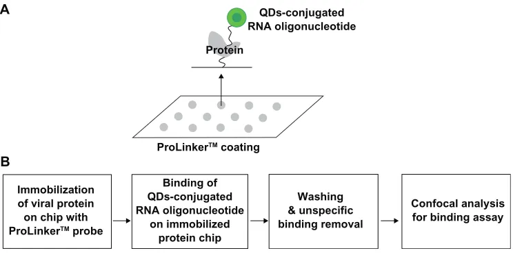
Immobilization of viral protein on chip with
ProLinkerTM probe
B A
Binding of QDs-conjugated RNA oligonucleotide
on immobilized protein chip
Washing & unspecific binding removal
Confocal analysis for binding assay
ProLinkerTM coating
Protein
QDs-conjugated RNA oligonucleotide
Figure 1 Representative scheme for the detection of viral protein on chip. A) Scheme for viral protein assay on chip. B) Procedure for viral protein assay.
International Journal of Nanomedicine downloaded from https://www.dovepress.com/ by 118.70.13.36 on 23-Aug-2020
Roh et al Dovepress
group. Second, the QDs-conjugated RNA oligonucleotide was bound on an immobilized chip. Third, the bound chip was washed to remove nonspecific binding. Fourth, it was analyzed to identify the specific detection of viral protein on the biochip. The targeting process for effective monitoring of the viral protein on the biochip is illustrated in Figure 1. Feasibility of targeting and imaging was achieved by using QDs525 conjugates containing RNA oligonucleotide for specific NS5B viral protein.
QDs-conjugated RNA oligonucleotide
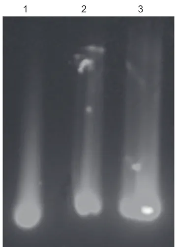
The QDs-supported RNA oligonucleotide was conju-gated via amide formation from coupling of the 5'-end-amine-modified RNA oligonucleotide at the surface of QDs displaying carboxyl groups via standard EDC coupling. Formation of the QDs-conjugated RNA oligonucleotide was confirmed on 2% agarose gel at 100 V in Tris-acetate-EDTA buffer. Agarose gel electrophoresis showed a distinct mobile band pattern between the free QDs and the QDs-conjugated RNA oligonucleotide, confirming the formation of QDs-conjugated RNA oligonucleotide. The mobility shifts are compared in Figure 2. On agarose gel, both the QDs-conjugated NS5Bnor RNA oligonucleotide and the NS5Bmut oligonucleotide showed less mobility shiftthan free QDs, thereby demonstrating conjugation by amide formation between the QDs and RNA oligonucleotide.
By TEM, both the QDs and the QDs-conjugated RNA oligonucleotide appeared spherical and fairly monodispersed (Figure 3). In Figure 3A, only the free QDs nanoparticles are visible. In addition, the nanoparticles appeared to be evenly distributed on the surface. Both the QDs and the RNA oli-gonucleotide were conjugated in the EDC coupling reaction. The QDs (dark spot) was surrounded by a white disk of conjugated RNA oligonucleotide (Figure 3B).
Detection of viral protein on biochip
The recombinant HCV NS5B viral protein in the E. coli expression system was expressed and purified by Ni-NTA affinity chromatography. In the purification step, the protein was eluted in 1 mL fractions with 250 mM imidazole buffer. To verify the purity and homogeneity of the eluted NS5B, aliquots of eluted fractions were analyzed by Coomassie blue staining on SDS-PAGE. Eluates with a purity of 95% were pooled, dialyzed and stored with 50% glycerol in aliquots at-80°C. The HCV NS5B viral protein was purified by a single chromatography step on a Ni2+ affinity column. The
C-termi-nally his-tagged HCV NS5B was visualized with a molecular mass of approximately 66 kDa on SDS-PAGE (Figure 4).
We demonstrated the specific interaction between QDs-conjugated RNA oligonucleotide and immobilized HCV NS5B viral protein on a chip. As illustrated in Figure 5A, the QDs-NS5Bnor RNA oligonucleotide-treated conjugates showed high fluorescent signals on the chip. In Figure 5B, no fluorescence signal was detected with a NS5Bmut RNA oligonucleotide, because of its lack of affinity. The signal of QDs-conjugated NS5Bmut RNA oligonucleotide for protein-binding affinity was very similar to the background signal. Figure 5C shows that QDs-conjugated RNA oligonucleotide is selective for HCV NS5B viral protein at varying protein concentrations. The binding affinity by confocal assay with varying concentrations of the NS5B viral protein was
exam-1 2 3
Figure 2 The conjugation pattern on 2% agarose gel electrophoresis was examined at UV excitation wavelength (345 nm), free QDs (lane 1), QDs-NS5Bnor RNA oligonucle-otide (lane 2), and QDs-NS5Bmut RNA oligonucleoligonucle-otide (lane 3). Carboxyl-terminated QDs are conjugated with RNA oligonucleotide by an amine-carboxyl reaction.
Abbreviations: NS5B, nonstructural protein 5B; QDs, quantum dots;
UV, ultraviolet.
A B
Figure 3 Characterization of QDs by TEM analysis. A) Free QDs. B) QDs-conjugated RNA oligonucleotide.
Abbreviations: QDs, quantum dots; TEM, transmission electron microscopy.
International Journal of Nanomedicine downloaded from https://www.dovepress.com/ by 118.70.13.36 on 23-Aug-2020
Detecting viral protein using RNA oligonucleotide
Dovepress
ined, and it was observed that the signal intensity of the QDs-conjugated RNA oligonucleotide could be detected even at the lowest concentration (1 ng mL-1). The signal intensity
was found to increase gradually by up to 1ng mL-1 of protein
concentration. The insert figure in Figure 5D shows that the detection limit of this method was at the 1 ng mL-1 level.
These results suggest the NS5B RNA oligonucleotide could specifically target the HCV NS5B viral protein on an immobilized chip. These results show that the QDs-RNA oligonucleotide could be effective for the quantification of HCV NS5B viral protein. To confirm a competition reaction of specific binding, BSA was used as a negative control protein. No fluorescence signal was detected with BSA (data not shown). Using the QDs-RNA oligonucleotide, we developed a HCV biosensor prototype biochip that could detect and quantify the HCV NS5B viral protein. Here, the specific RNA oligonucleotide was examined for binding to the HCV NS5B viral protein by using a QDs-conjugated imaging system. HCV-specific targeting and imaging using a QDs-conjugated RNA oligonucleotide agent was dem-onstrated. Preferential binding of QDs-conjugated RNA oligonucleotide for detection of HCV NS5B viral protein was determined by analyzing fluorescence intensity measured by confocal microscopy. The immobilized protein chip showed no binding to NS5Bmut RNA oligonucleotide, thus demon-strating selectivity for HCV NS5B viral protein. Imaging by semiconductor nanocrystal QDs allows for monitoring with intense light emission and accuracy.
70
M 1 2 3 M 4
50
Figure 4 Purification of HCV-NS5B viral protein. The SDS-PAGE 10% gel show-ing NS5B protein with his-tag. M, protein marker; lane 1, before induction form of NS5B; lane 2, total form of NS5B; lane 3, soluble form of NS5B; lane 4, his-Tag form of NS5B.
Abbreviations: HCV,hepatitis C virus; NS5B, non-structural protein 5B; SDS-PAGE, sodium dodecyl sulfate polyacrylamide gel electrophoresis.
A
D
NS5Bmut RNA oligonucleotide
NS5Bmut RNA oligonucleotide 1800
1600 1400 1200 1000 800
400 200 0 600
0.0 0.2 0.4
NS5B protein (µg mL−1)
Relative signal intensit
y 40
35 30 25 20 15 10 5 0
0.00 0.01 0.02 0.03 0.04 0.05 0.06
NS5B protein (µg mL−1)
Relative signal intensity
0.6 0.8 1.0
C (µg mL−1)0.0010.01 0.05 0.1 0.5 1.0
NS5Bnor RNA oligonucleotide
NS5Bnor RNA oligonucleotide
B
Figure 5 A, B) Selective specificity of HCV-NS5B viral protein by QDs-conjugated RNA oligonucleotide. The NS5Bnor and NS5Bmut RNA oligonucleotides were bound on
an immobilized viral protein chip, respectively. C, D) Effect of HCV-NS5B viral protein concentrations on binding assay.
Abbreviations: HCV,hepatitis C virus; NS5B, non-structural protein 5B; NS5Bnor, normal NS5B; NS5Bmut, mutant NS5B; QDS, quantum dots.
International Journal of Nanomedicine downloaded from https://www.dovepress.com/ by 118.70.13.36 on 23-Aug-2020
Roh et al Dovepress
During the last decade, there has been an increasing inter-est in using nanoparticles in biologic applications. The avail-ability of colloidal semiconductor QDs with highly controlled optical properties in the nanometer size range has created widespread interest in the use of biotechnologic systems as a diagnostic platform and for biologic imaging. In this study, we report a QDs-conjugated RNA oligonucleotide probe for screening of HCV with high sensitivity and selectivity.
A RNA oligonucleotide sensor system that could quantify HCV NS5B viral protein using conjugated QDs-RNA oligo-nucleotide could provide an efficient strategy and a promising new platform for monitoring applications.
In summary, we have shown that QDs-RNA oligonucle-otide can specifically recognize the HCV viral protein, and have demonstrated that this conjugated QDs-RNA oligo-nucleotide can interact on a designed biochip specifically and sensitively. This device could generate a QDs-conjugated biosensor prototype biochip for HCV diagnosis. The present visual HCV detection technique may avoid the limitations with its reported methods, namely, its high sensitivity, good specificity, simplicity, speed, and cost-effectiveness. This technique has potential applications in many fields, especially in multiple virus detection chip coupled with nanoparticle will find applications in clinic.
Conclusions
The results of this study indicate that the QDs-conjugated RNA oligonucleotide can recognize the HCV viral protein with a detection limit of 1 ng mL-1 specifically and
sensi-tively on a designed biochip. This device could generate a nanoparticle-conjugated prototype biochip for HCV diagnosis.
Acknowledgments
We thank Professor Dr Myung Heejoon and Profes-sor Dr Seong-Wook Lee for their very kind donation of plasmid pCite. This work was supported by SNU-KAERI Degree and Research Center for Radiation Convergence Sciences from the Korea Research Council of Foundation (KRCF) Science and Technology, South Korea.
Disclosures
The authors report no conflicts of interest in this work.
References
1. Weiss U. Hepatitis. Nature. 2005;436:929.
2. Jameel S, Siddiqui A, Hu KQ. The molecular biology of hepatitis virus.
In: Gitnick G, editor. Principles and Practice of Gastroenterology and
Hepatology. New York, NY: Elsevier; 1994. p. 743–757.
3. Pawlotsky JM. Hepatitis C virus population dynamics during infection.
Curr Top Microbiol Immunol. 2006;299:261–284.
4. Reed KE, Rice CM. Overview of hepatitis C virus genome structure, polyprotein processing, and protein properties. Curr Top Microbiol Immunol. 2000;242:55–84.
5. De Francesco R, Tomei L, Altamura S, et al. Approaching a new era for hepatitis C virus therapy: Inhibitors of the NS3-4A serine
prote-ase and the NS5B RNA-dependent RNA polymerprote-ase. Antiviral Res.
2003;58:1–16.
6. Behrens SE, Tomei L, De Francesco R. Identification and properties of
the RNA-dependent RNA polymerase of hepatitis C virus. EMBO J.
1996;15:12–22.
7. Adachi T, Ago H, Habuka N, et al. The essential role of C-terminal residues in regulating the activity of hepatitis C virus RNA-dependent
RNA polymerase. Biochim Biophys Acta. 2002;1601:38–48.
8. Ferrari E, Wright-Minogue J, Fang JW, et al. Characterization of soluble hepatitis C virus RNA-dependent RNA polymerase expressed
in Escherichia coli. J Virol. 1999;73:1649–1654.
9. Yamashita T, Kaneko S, Shirota Y, et al. RNA-dependent RNA polymerase activity of the soluble recombinant hepatitis C virus
NS5B protein truncated at the C-terminal region. J Biol Chem.
1998;273:15479–15486.
10. Ishii K, Tanaka Y, Yap CC, et al. Expression of hepatitis C virus NS5B protein: Characterization of its RNA polymerase activity and RNA
binding. Hepatology. 1999;29:1227–1235.
11. Ago H, Adachi T, Yoshida A, et al. Crystal structure of the
dependent RNA polymerase of hepatitis C virus. Structure. 1999;7:
1417–14126.
12. Bressanelli S, Tomei L, Roussel A, et al. Crystal structure of the
RNA-dependent RNA polymerase of hepatitis C virus. Proc Natl Acad Sci
U S A. 1999;96:13034–13039.
13. Lohmann V, Overton H, Bartenschlager R. Selective stimulation of hepatitis C virus and pestivirus NS5B RNA polymerase activity by
GTP. J Biol Chem. 1999;274:10807–10815.
14. Dhanak D, Duffy KJ, Johnston VK, et al. Identification and biological characterization of heterocyclic inhibitors of the hepatitis C virus
RNA-dependent RNA polymerase. J Biol Chem. 2002;277:38322–38327.
15. Neerja KB, Alain BW, Tanaji TT, et al. Identification and
characteriza-tion of coumestans as novel HCV NS5B polymerase inhibitors. Nucl
Acids Res. 2008;36:1482–1496.
16. Cullen BR. Immunology. Outwitted by viral RNAs. Science. 2007;
317:329–330.
17. Griffin J, Singh AK, Senapati D, et al. Size- and distance-dependent nanoparticle surface-energy transfer (NSET) method for selective
sensing of hepatitis C virus RNA. Chemistry. 2009;15:342–351.
18. Gaudy C, Thevenas C, Tichet J, et al. Usefulness of the hepatitis C virus core antigen assay for screening of a population undergoing routine
medical checkup. J Clin Microbiol. 2005;43:1722–1726.
19. Alivisatos P. The use of nanocrystals in biological detection. Nat Biotechnol. 2004;22:47–52.
20. Fortina P, Kricka LJ, Surrey S, et al. Nanobiotechnology: The prom-ise and reality of new approaches to molecular recognition. Trends Biotechnol. 2005;23:168–173.
21. Pinaud F, King D, Moore HP, et al. Bioactivation and cell targeting of semiconductor CdSe/ZnS nanocrystals with phytochelatin-related
peptides. J Am Chem Soc. 2004;126:6115–6123.
22. Biju V, Itoh T, Anas A, et al. Semiconductor quantum dots and metal nanoparticles: Syntheses, optical properties, and biological applications.
Anal Bioanal Chem. 2008;391:2469–2495.
23. Ghasemi Y, Peymani P, Afifi S. Quantum dot: Magic nanoparticle for
imaging, detection and targeting. Acta Biomed. 2009;80:156–165.
24. Jun T, Bochu W, Liancai Z. Quantum Dots: A novel tool to discovery.
Pak J Biol Sci. 2006;9:917–922.
25. William WY, Chang E, Drezek R, et al. Water-soluble quantum dots for
bio-medical applications. Biochem Biophys Res Comm. 2006;348:781–786.
26. Medintz IL, Mattoussi H, Clapp AR. Potential clinical applications of
quantum dots. Int J Nanomedicine. 2008;3:151–167.
International Journal of Nanomedicine downloaded from https://www.dovepress.com/ by 118.70.13.36 on 23-Aug-2020
International Journal of Nanomedicine
Publish your work in this journal
Submit your manuscript here: http://www.dovepress.com/international-journal-of-nanomedicine-journal
The International Journal of Nanomedicine is an international, peer-reviewed journal focusing on the application of nanotechnology in diagnostics, therapeutics, and drug delivery systems throughout the biomedical field. This journal is indexed on PubMed Central, MedLine, CAS, SciSearch®, Current Contents®/Clinical Medicine,
Journal Citation Reports/Science Edition, EMBase, Scopus and the Elsevier Bibliographic databases. The manuscript management system is completely online and includes a very quick and fair peer-review system, which is all easy to use. Visit http://www.dovepress.com/ testimonials.php to read real quotes from published authors.
Detecting viral protein using RNA oligonucleotide
Dovepress
Dovepress
27. Schröter S, Feucht M, Zöllner B, et al. Quantitative detection of hepatitis C virus RNA by light cycler PCR and comparison with two different
PCR assays. J Clin Microbiol. 2001;39:3056–3059.
28. Bartolomé J, López-Alcorocho JM, Castillo I, et al. Ultracentrifugation of serum samples allows detection of hepatitis C virus RNA in patients
with occult hepatitis C. J Virol. 2007;81:7710–7715.
29. Riccardi CS, Dahmouche K, Santilli CV, et al. Immobilization of streptavidin in sol-gel films: Application on the diagnosis of hepatitis C
virus. Talanta. 2006;70:637–643.
30. Riccardi CS, Kranz C, Kowalik J, et al. Label-free DNA detection of hepatitis C virus based on modified conducting polypyrrole films at microelectrodes and atomic force microscopy Tip-integrated electrodes.
Anal Chem. 2008;80:237–245.
31. Laemmli UK. Cleavage of structural proteins during the assembly of
the head of bacteriophage T4. Nature. 1970;227:680–685.
32. Bradford M. A rapid and sensitive method for the quantitation of microgram quantities of protein utilizing the principle of protein-dye
binding. Anal Biochem. 1976;72:248–254.
33. Lee Y, Lee EK, Cho YW, et al. ProteoChip: A highly sensitive protein microarray prepared by a novel method of protein immobilization for application of protein-protein interaction studies. Proteomics. 2003;3:2289–2304.
International Journal of Nanomedicine downloaded from https://www.dovepress.com/ by 118.70.13.36 on 23-Aug-2020