Image Denoising using Neural Network with
SVM (Support Vector Machine) and
LDA (Linear Discriminant Analysis)
Ahmad Ali Saihood #
Lecturer in Computer Centre, Thi-Qar University Nassirya,Thi-Qar,Iraq
Abstract
—
the medical figure technology is become a much powerful component for number of applications such as diagnosis; research; and treatment. Medical figure like X-Ray; CT; MRI; PET and SPECT have small information about heart brain and nerves. All of These figures need to be exact and free from noise. To minimize noise plays an important role in medical imaging. The numbers of methods of noise removal such as filter and thresholding based on wavelet. Although this method does not produce good results but also have some limitations. Taking and analyzing the limitations of the previous method; our research presents neural networks as an efficient and robust tool for noise reduction [1,7]. In research; use Neural Network; SVM and LDA as the learning algorithm which follows the supervised learning. The proposed research use both mean and median statistical functions for calculating the output pixels results in terms of PSNR and MSE.Keywords:- Noising, De-noising, Medical images, Neural Network, SVM and LDA
I. INTRODUCTION
Image processing is a form of signal processing for which the input is an image such as a photograph or video frame and the output of image processing may be either an image or the image parameters. Image is a two dimensional function of two real variables. Image= f(x, y) where, x and y are the spatial coordinates known as pixels and f is the amplitude. Before, processing an image is converted into the digital form. The digitization includes; sampling of images and quantization of the sampled values. Therefore after converting the image into bit information the processing is performed. Then processing technique may be image enhancement; image reconstruction and image compression. Image is processed in two ways:
1. Spatial domain: Spatial domain, refers to the image plane itself; it is based on the direct manipulations of the pixels in the image.
2. Frequency domain: In frequency domain, image is processed in form of sub bands. All types of transformations are applied in frequency domain. e.g DWT, DFT etc.
The aim of image processing is divided into five groups: 1. Visualization: Observe the objects that are not visible. 2. Image Sharpening and Restoration: To create a better image.
3. Image Retrieval: Seek for the image of interest.
4. Measurement of the Pattern: Measure various objects in an image.
perceived to designate the set of techniques that non-invasively produce images of the internal aspect of the body. [8].
The paper describe as in section II shows medical image denoising. Section III tell about NN and section IV gives idea about LDA and section V discuss SVM. At last in section VI detailed about result and section VII about conclusion.
II. MEDICAL IMAGE DE-NOISING
The arrival of digital medical imaging technologies such as positron emission tomography (PET), magnetic resonance imaging (MRI), computerized tomography (CT) and ultrasound Imaging has revolutionized modern medicine. Today, many patients no longer need to go through invasive and often dangerous procedures to diagnose a wide variety of illnesses. Therefore widespread use of digital imaging in medicine today; the quality of digital medical images becomes an important issue. To achieve the best possible diagnosis it is important that medical images be sharp; clear; and free of noise and artifacts. The technologies for acquiring digital medical images continue to improve; resulting in images of higher and higher resolution and quality, removing noise in these digital images remains one of the major challenges in the study of medical imaging, because they could mask and blur important subtle features in the images; many proposed de-noising techniques have their own problems. Image de-noising still remains a challenge for researchers because noise removal introduces artifacts and causes blurring of the images. Noise modelling in medical images is greatly affected by capturing instruments; data transmission media; image quantization and discrete sources of radiation. Therefore different algorithms are used depending on the noise model. Then most of images are assumed to have additive random noise which is modelled as a white Gaussian noise. Medical images such as magnetic resonance imaging (MRI) and ultrasound images have been widely exploited for more truthful pathological changes as well as diagnosis. They suffer from a number of shortcomings and these includes: acquisition noise from the equipment; ambient noise from the environment; the presence of background tissue; other organs and anatomical influences such as body fat; and breathing motion.
III.NEURAL NETWORKS
Artificial neural networks are composed of interconnecting artificial neurons (programming constructs that mimic the properties of biological neurons). Therefore Artificial neural networks may either be used to gain an understanding of biological neural networks; or for solving artificial intelligence problems without necessarily creating a model of a real biological system. Therefore real; biological nervous system is highly complex: artificial neural network algorithms attempt to abstract this complexity and focus on what may hypothetically matter most from an information processing point of view. Good performance (e.g. as measured by good predictive ability; low generalization error); or performance mimicking
source of evidence towards supporting the hypothesis that the abstraction really captured something important from the point of view of information processing in the brain. Other incentive for these abstractions is to reduce the amount of computation required to simulate artificial neural networks; so as to allow one to experiment with larger networks and train them on larger data sets. [4,6].
IV.LINEAR DISCRIMINANT ANALYSIS:
Linear Discriminant Analysis (LDA) is a techniques used for data classification and dimensionality reduction. In PCA; then the shape and the location of the original data sets changes when transformed to a different spaces whereas LDA doesn’t change the location but only tries to provide more class separability and draw decision between the given classes.
The discriminant analysis; two scatter matrices; called within-class (Sw) and between-class (Sb) matrices, are defined to quantify the quality[12].
The concept of Fuzzy Logic (FL) was conceived by LotfiZadeh; a professor at the University of California at Berkley; and presented not as a control methodology; but as a way of processing data by allowing partial set membership rather than crisp set membership or non-membership. And this approach to set theory was not applied to control systems until the 70's due to insufficient small-computer capability prior to that time. Therefore Professor Zadeh reasoned that people do not require precise; numerical information input; and yet they are capable of highly adaptive control. And if feedback controllers could be programmed to accept noisy; imprecise input; they would be much more effective and perhaps easier to implement. The U.S. manufacturers have not been so quick to embrace this technology while the Europeans and Japanese have been aggressively building real products around it. In this context, FL is a problem-solving control system methodology that lends itself to implementation in systems ranging from simple; small; embedded micro-controllers to large; networked; multi-channel PC or workstation-based data acquisition and control systems. This can be implemented in hardware; software; or a combination of both technique. [13].
V.SVM
algorithm that depends on the data only through dot-products. This is the case; the dot product can be replaced by a kernel function which computes a dot product in some possibly high dimensional feature space. It has two advantages: First; the ability to generate non-linear decision boundaries using methods designed for linear classifiers. Then second; the use of kernel functions allows the user to apply a classifier to data that have no obvious fixed-dimensional vector space representation. Therefore prime example of such data in bioinformatics are sequence; either DNA or protein; and protein structure. Using SVMs effectively requires an understanding of how they work. When training an SVM the practitioner needs to make a number of decisions: how to preprocess the data, what kernel to use; and finally; setting the parameters of the SVM and the kernel [1]. Uninformed choices may result in severely reduced performance. We purpose to provide the user with an intuitive understanding of these choices and provide general usage guidelines.
A. Properties of SVM:
1. Flexibility in choosing a similarity function
2. Sparseness of solution when dealing with large data sets
3. Ability to handle large feature spaces
4. Over fitting can be controlled by soft margin approach
VI RESULT DISCUSSION
Figure 1: GUI front layout
Figure 2: GUI 2nd layout to load CT image
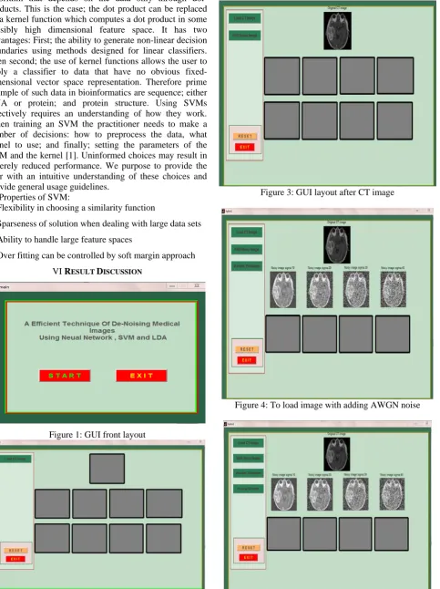
Figure 3: GUI layout after CT image
Figure 4: To load image with adding AWGN noise
Figure 6: To perform NN, SVM and LDA
Figure 7: Neural Network view
Figure 8: Performance analyze by using NN
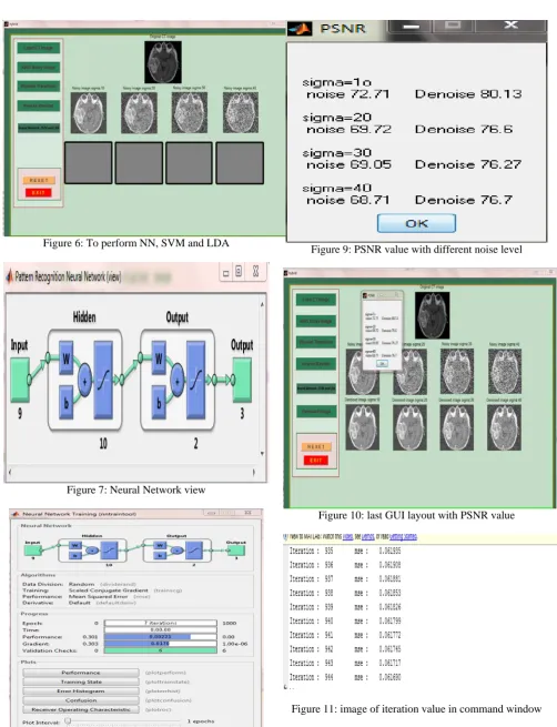
Figure 9: PSNR value with different noise level
Figure 10: last GUI layout with PSNR value
Figure 11: image of iteration value in command window
VII.CONCLUSION
Medical figure technology is becoming an important component of large number of applications such as the diagnosis research and treatment. Noise reduction therefore becomes very important. The main techniques of image de-noising are filters wavelets and neural networks. The LDA based approach is a powerful and effective method for image de-noising. Considering and analyzing the drawbacks of the previous methods we propose a new improved LDA approach to de-noise the medical images. This approach includes using PSNR value to show that denoising is good by using NN, SVM and LDA technique.
ACKNOWLEDGMENT
I would like to thank my guide Mr.Rakesh Kumar in MMEC,Mullana for his supporting and lovely help he offered , I also many thanks to Mr. Hazeem Baqer in Thi-Qar university ,Iraq .
REFERENCES
[1] Abuzoum Mohamed Saleh “Efficient analysis of medical image de-noising for MRI andUltrasound Images”, (2012).
[2] Akutagawa Mastake, ChanYongjia, Katayama Masato, Yohsuke Kinouchi, Qinyu Zhang,“Additive and multiplicative noise reduction by back propagation neural network”, Proceedings of the 29th Annual International Conference of the IEEE EMBS Internationale, Lyon, France August 23-26, 2007 IEEE(2007).
[3] “Image de-noising using neural network based non-linear filter in wavelet domain”, 2012, 0-7803-8874-7/05/IEEE(2012),S.Zhang, E.Salari.
[4] Dr. T.Santhanam, S.Radhika, “Applications of neural networks for noise and filterclassification to enhance the image quality”, IJCSI International Journal of Computer Science Issues, Vol. 8, Issue 5, No 2, September 2011 (IJCAI 2011).
[5] E.Salari, S. Zhang ,“Image de-noising using neural network based non-linear filter inwavelet domain”, 0-7803-8874-7/05/IEEE(2005) [6] F.Marvasti, N.sadati, S.M.E Sahraeia, “ Wavelet image De-noising
based on neural network and cycle spinning”
1424407281/07/IEEE(2007).
[7] Gupta Manoj, KumarPapendra, KumarSuresh (IJCA-2010)
“Performance comparison of median and the weiner filter in image de-noising.”
[8] KaurJappreet, KaurManpreet, KaurManpreet, KaurPoonamdeep
“Comparative analysis of image de-noising techniques.”
(IJETAE2012)
[9] Leavline E.Jebamalar Sutha S, Singh D.Asir Anton Gnana (IJCA-2011) “Wavelet domain shrinkage methods for noise removal in mages.”
[10] Mr. S. Hyder Ali, Dr.(Mrs.) R. Sukanesh, Ms. K. Padma Priya “ Medical image de-noising using neural networks”.
[11] Rehman Amjad, Sulong Ghazali, Saba Tanzila “An intelligent approach to imagedenoising”, (JATIT 2005-2010).
[12] Sontakke Trimbak R, Rai RajeshKumar, “Implementation of image de-noising using thresholding techniques”, IJCTEE.