Growth and patterning from the engrailed interface
ALICIA HIDALGO*
Department of Genetics, University of Cambridge, Cambridge, United Kingdom
ABSTRACT The Drosophila wing is divided into anterior and posterior compartments, the latter characterized by the expression of the engrailed gene. A comparative analysis is presented here, and suggests that a primary conserved role of engrailed is to drive growth of limbs along the proximo-distal axis. The Apical Ectodermal Ridge in vertebrate limbs resembles the Antero/ Posterior compartment boundary in fly wings, particularly in molecular aspects. Multiple evidence suggests that the fly wing Antero/Posterior boundary is not the result of differential cell affinities between all anterior and posterior cells, but responds to the area of cell communication between anterior and posterior compartments. Arguments are presented here to support the notion that the compartment boundary is a consequence of decapentaplegic function in the control of growth. Patterning, on the other hand, requires the participation of several genes, among which are engrailed, invected and hedgehog. Finally, regulatory interactions between en/En-1 and hh/Shh may be significant in the context of morphogenetic regulation during normal development.
KEY WORDS:
engrailed, compartments, selector genes, AER, decapentaplegic
0214-6282/97/$10.00 © UBC Press
Printed in Spain
*Address for reprints: Department of Genetics, University of Cambridge, Downing Street, Cambridge CB2 3EH, United Kingdom. FAX: 44-(0) 1223 333992. e-mail:
Introduction
When Velázquez painted Las Meninas (Fig. 1) he taught us that the content of an image can flip depending on how we perceive it. So, we can either see a portrait of the princesses, or a portrait of the King and Queen (who are reflected on the mirror) or a portrait of the artist himself. Images are present in living organisms, as whole or part of structures (e.g., eyes) and patterns (e.g., of hairs, of gene expression) which can be seen by naked eye or with sophisticated methods. Developmental geneticists look at these images to try to understand how a fly is made, as an architect from the future would look at our buildings to work out how we made houses from bricks, windows, roofs and doors. Sometimes these images are so aston-ishing that they make us want to stay within the most immediate plane. The story of the engrailed (en) gene and its conceptual trail is a fascinating one in developmental biology, and to Antonio we owe vision in creating a whole area of research.
At a time when no developmental gene had yet been cloned, García-Bellido observed that mutations in the en gene produce a transformation of posterior pattern of the wing into anterior (García-Bellido and Santamaría, 1972). Furthermore, he observed that the domain of pattern transformation coincides with a partition along the wing which could only be visualized by clonal analysis, as it restricts the proliferation of cells to either side of it (Fig. 2) (García-Bellido et al., 1973). García-(García-Bellido named compartment each of the wing halves separated by this line. He predicted that compart-ments would be under the genetic control of selector genes (García-Bellido et al., 1973). A binary switch would determine that a given group of cells would either express one of these genes or
not, and this decision would be maintained by all the progeny of these founder cells (García-Bellido, 1975). The beauty of this proposal lied on the fact that we could understand morphology as an addition of lineages, and morphogenesis as the piece-meal process in which these lineages become different from each other in terms of shape, size and pattern by expressing a given combi-nation of selector genes. This proposal was visionary, because it was later shown that only cells of the posterior compartment of the wing express the en gene, which like other genes whose mutations cause homeotic transformations, contains a homeobox . Further-more, it has also been shown that, as predicted, interfaces be-tween compartments are instructive in the control of growth. In fact, work from many labs has described a network of genes that function to establish this interface. Now, following Antonio’s fre-quent exhortation to people working with him, we must look at cell behavior and also beyond Drosophila to try to understand how a fly is made.
Conservation of engrailed function: enabling
proximo-distal growth
engrailed has been studied in leech (Wedeen and Weisblat, 1991; Lans et al., 1993), insects and crustaceans (Patel et al.,
Abbreviations used in this paper: A, anterior; P, posterior; D/V, Dorso/Ventral;
1989a,b; Scholtz et al., 1993,1994; Scholtz and Dohle, 1996), amphioxus (Holland et al., 1997) and vertebrates (Joyner et al., 1991; Wurst et al., 1994; Loomis et al., 1996; Logan et al., 1997). In segmented organisms, en behaves in ways surprisingly similar to how it does in Drosophila. Within the trunk, it is invariably expressed in stripes around where each segment boundary will form. These stripes are defined by positional cues, not by lineage (Scholtz et al., 1994; Scholtz and Dohle, 1996). In their onset, the anterior end of the en segmental stripe is frequently straight, whereas the posterior end is wiggly as some cells switch off en expression (Fig. 2) (Patel et al., 1989a; Scholtz et al., 1993). In Drosophila too, en expression is not clonal in the embryo, but depends on the distance from wingless (wg) signaling cells (Vin-cent and O’Farrell, 1992). After the establishment of the anterior non-en / en interface, more posterior cells switch on en de novo within each segment, to define the segment boundary (Patel et al., 1989a; Scholtz et al., 1993) (Fig. 2). These later en-expressing cells are not necessarily related by lineage to the more anterior en-cells (Scholtz and Dohle, 1996). A dramatic example of the temporal regulation of en expression is found in the leech, where en-expressing cells demarcate segment boundaries, despite switch-ing en on and off at different times along each stripe (Lans et al.,
1993). The fact that en is switched off in cells that are too distant from an inductive signal and switched on de novo in cells not related by lineage is relevant, because it was thought that a key feature of the role of en in morphogenesis was that it was switched on once in the embryo and maintained clonally thereafter. That is, the state of expression of en was thought to be a mechanism of cell memory essential for the building process. Comparative analysis tells us that en functions in the trunk in two conserved processes: the establishment of the antero-posterior interface and the defini-tion of the segment boundary.
In all arthropods examined to date, the anterior straight interface of non-en (wg) and en cells demarcates the point from which limbs will develop (Fig. 2). This, in Drosophila, corresponds to the A/P compartment boundary. In some crustaceans, trunk cells along this anterior-edge of the en domain can also cease to express en (Scholtz and Dohle, 1996), although it is not known whether this also happens to en-expressing cells within the limb. It would be interesting to see if there is a straight compartment boundary in all arthropod limbs. This would tell us whether proximo-distal growth necessarily correlates with the presence of an A/P boundary. Furthermore, we still don’t know whether proximo-distal growth in other arthropods is linked to clonal subdivisions. And similarly, whereas en expression is maintained in insect and crustacean limbs, there is no knowledge as to whether it is through lineage (Patel et al., 1989a,b). Answers to these comparative questions would be very revealing to our understanding of the control of growth. Unfortunately, we cannot extrapolate our understanding of en regulation during segmentation to limb formation, because the latter requires extensive cell division along the proximo-distal axis. Consequently, the contribution of compartments, hence lineage, to morphogenesis has to be evaluated independently in these two different cellular contexts. So far, we rely on Drosophila data to unravel the cellular aspects of arthropod limb formation.
In vertebrates, where the trunk is not segmented, en is still required for limb formation. However, its expression pattern is somewhat different, and might reveal the general role of compart-ments in morphogenesis. In vertebrates, En-1 is expressed along a longitudinal stripe that runs along the length of the embryo and divides the limb into dorsal (Wint7a-expressing) and ventral (En-1 expressing) domains (Fig. 2) (Logan et al., 1997). This means that expression of En-1 in vertebrates is shifted by 90° compared to Drosophila. The interface between these domains gives rise to the Apical Ectodermal Ridge. The AER is essential for proximo-distal limb growth and induces proliferation in the underlying mesenchy-mal cells (the progress zone). Recent molecular data have shown that the AER is remarkably similar to the fly A/P compartment boundary. Firstly, they are both established by the interface be-tween homologous wingless/Wint and en/En-1- expressing cells (Loomis et al., 1996; Logan et al., 1997). Moreover, both the A/P boundary and the AER express decapentaplegic (dpp)/BMP2 homologous proteins of the TGF-β family, which are otherwise repressed by en/En-1 in adjacent cells (Loomis et al., 1996). Furthermore, in both cases the expression of dpp/BMP2 is induced by hedgehog (hh)/Sonic hedgehog (Shh) from neighboring cells (Marigo et al., 1996). In Drosophila these are posterior cells, where hh expression depends directly on en. In the vertebrate limb, Shh is expressed in a posterior domain adjacent to and maintained by the AER, still depending indirectly on En-1 function (Hammerschmidt et al., 1997; Logan et al., 1997). A significant difference between
hh in Drosophila and Shh in vertebrates is that the domain of Shh expression overlaps but does not coincide with the domain of En-1, and is in fact shifted by 90°. It would be interesting to know if there are other arthropods with a similar organization of en and hh patterns as in the vertebrate limb. This could tell us whether en is required in non-vertebrates for the establishment of posterior compartments, or more generally for proximo-distal growth. The observation that in vertebrates both normal digits and ectopic limbs form at the interface between Wint7a and En-1 cells (Logan et al., 1997), just as both branches of crustacean biramous limbs form along the non-en/en interface (Panganiban et al., 1995), strongly suggests that a conserved function of en is to enable growth.
Consistent with this idea, in Drosophila, complete homeotic transformations of posterior (P) into anterior (A) (without an in-crease in growth) following complete removal of both en and inv functions have not yet been found. This is because if en function is
never present in disc development, dpp expression will not be spatially restricted and the disc will not grow normally. This implies that a distinct role of en is to establish the domain of dpp, to allow growth and field regulation (reviewed in Hidalgo, 1996). This is reminiscent of the role of En-1 in the vertebrate limb. Loss of function mutations in En-1 in mouse cause ventral expansion of the AER, with the consequent formation of distal structures in proximal locations of limbs (Loomis et al., 1996). Also, the AER can be damaged and disrupt proliferation of mesenchymal cells leading to truncation of digits (Wurst et al., 1994). Conversely, ectopic ex-pression of En-1 in chick affects the AER causing a decrease in proximo-distal outgrowth (Logan et al., 1997). Hence, a primary role of en is to enable proximo-distal limb growth.
The pattern of growth
The cellular mechanism by which the wing grows remains a great mystery to Drosophila developmental geneticists. It has long been believed that all wing disc cells proliferate equally, because clones of cells can be induced anywhere in the disc at any time (García-Bellido and Merriam, 1971). However, there may be proliferation centres which change with time, because the distribu-tion of clones is not identical at all times (García-Bellido and Merriam, 1971; González-Gaitán et al., 1994). Furthermore, clus-ters of synchronous cells have been observed at different times during wing development (Milan et al., 1996a, b). Intriguingly, cells in these clusters are not clonally related and in the early phase of wing growth these clusters cannot clearly be correlated to particu-lar wing regions. In vertebrates, the AER is the leading edge of the growing bud, and induces proliferation in the underlying progress zone (Fig. 3). This in turn maintains the neighboring AER, thus establishing a positive feedback loop that controls growth. Cells leaving the progress zone earlier will form proximal structures, whereas cells that divide for a longer time within this zone will make distal structures. Hence, growth is directional.
We are used to looking at Drosophila wing discs from a frontal view: the flat aspect of the disc towards us, the A/P boundary running within the flat disc. However, if we flip the disc so that we now have the folds of the wing pouch in a frontal view, and the compartment boundary on the edge, the fly disc looks like a vertebrate limb bud (Fig. 3). Now en/En-1 expression looks the same in both limbs, the AER ridge corresponds to the A/P boundary and hh/Shh expression is oriented with regards to veins and digits. In flies, dpp is expressed along the A/P boundary, just as BMP2 is along the AER. There is abundant evidence ascribing dpp a role in the control of wing disc growth from this expression domain. Perhaps the most elegant demonstration is the induction of clones expressing dpp ectopically in wings that practically lack dpp function (Fig. 4) (Zecca et al., 1995). dpp expressed in these clones alone is able to induce proximo-distal growth and wings of remarkably normal appearance in terms of growth. Similar effects have also been observed when clones lacking smoothened function redirect the distribution of dpp expression more anteriorly than its normal domain (Chen and Struhl, 1996). Conversely, lack of dpp function in mutant flies and in clones along the A/P boundary prevents wing growth (Posakony et al., 1991). And ectopic expression of dpp induces cell proliferation (Capdevila and Guerrero, 1994; Burke and Basler, 1996). These experiments demonstrate that dpp expressed along the A/P boundary is
necessary to direct cell proliferation in the wing disc along the proximo-distal axis.
An important consequence of directional growth is that cells that fall beyond the reach of dpp should make proximal structures, whereas cells that continue to divide closer to the source should make distal structures. A glimpse of this situation is revealed by the features of clones of en mutant cells. A very intriguing aspect of the homeotic transformations caused by loss of en function in P compartments of the wing is that they are complete only if clones are extremely large, reach the wing margin and hinge region and duplicate a compartment boundary (see below) (Fig. 5) (Tabata et al., 1995). That is, a new morphogenetic field (generated from ectopic both anterior and posterior compartments) must be estab-lished in order for controlled growth and patterning to take place. The limb field in vertebrates is defined by the homogeneous expression of Wint7a (Parr and McMahon, 1995) (Fig. 2), and similarly in the fly wing the homolog wg, is expressed throughout the wing field in the early stages of the wing disc development (Couso et al., 1993). Later on, wg expression is restricted to several domains, which include a ring of cells at the base of the wing (Couso et al., 1993), perhaps defining the edge of the wing field. en/ En-1 expression bisects the field in vertebrates and flies, defining the domain of expression of BMP2/dpp (Sanicola et al., 1995; Loomis et al., 1996) (Fig. 3). In order to induce an ectopic anterior compartment within the normal posterior, clones of en mutant cells must bisect again the field in two points across its border and consequently induce a new stripe of dpp expression (Fig. 5). These two boundaries function as centers for the organization of normal and duplicated wing growth. This situation has never been reported with X-rays induced clones, presumably because the frequency of
mitotic recombination induced by X-rays is too low to affect more than one cell within the initial polyclone. However, with the flip-FRT system, the frequency of recombination is increased considerably, and can affect a small group of adjacent cells.
These clones have two important implications: 1) complete wing duplications are only caused by those en mutant clones that were able to reproduce an ectopic A/P boundary. 2) That because in order for this to occur the field must be bisected through its border, early (hence older and larger) clones are more likely to span proximal areas than later clones. This situation is surprisingly similar to the one found in vertebrate limbs, where older cells are located in proximal regions. Hence, it would imply that in Dro-sophila growth may also be directional, being equally driven by dpp from the A/P boundary. Very remarkably, despite the spatially restricted expression of dpp along the A/P boundary, dpp receptors are required throughout the wing blade to fulfill the role of dpp in the control of cell proliferation (Burke and Basler, 1996). Moreover, this long range requirement for dpp changes as the disc grows. Removal of the dpp receptor Tkv causes more dramatic effects earlier on in disc development and closer to the source of Dpp (Burke and Basler, 1996). This also suggests that growth may be directional in Drosophila as it is in vertebrates. In this context, it is important to bear in mind that the A/P boundary spans the whole disc along its major axis. It is very revealing that ectopic expression of dpp can induce growth only in the wing blade (Burke and Basler, 1996). Perhaps dpp regulates growth preferentially as a conse-quence of the intersection of the A/P axis with the D/Vaxis at the
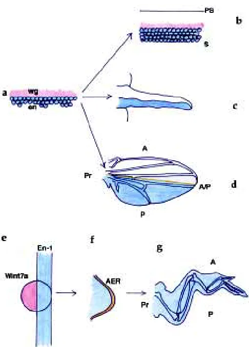
Fig. 3. Growth from the AER and A/P boundary. (a) Wing disc, classical frontal view. (b) Wing disc, flipped, lateral view. (c) Vertebrate limb bud. (d) Establishment of the wing disc field in the Drosophila embryo by the wingless/engrailed interface. (e) Wing disc, diagrammatic representation. en and hh are expressed in posterior cells, patched anteriorly, dpp along the A/P boundary. (f) Vertebrate limb, diagrammatic representation. Shh, ptc and BMP2 expressed posteriorly, En-1 ventrally, BMP2 along the AER. Blue, en and En-1; Red, wingless; Orange, dpp, A/P boundary, AER, BMP2; Yellow, hedgehog, Shh; Green, hh + en, Shh + En-1.
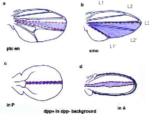
Fig. 4. dpp directs growth and establishes the A/P boundary. (a) Clone of ptc-en double mutant cells causing an anterior ectopic compartment boundary. This boundary is not flanked by en expressing cells on either side. (b) Clone of smo mutant cells between the normal compartment boundary and vein 3. This clone duplicates the area between L3 and L1 and establishes an ectopic compartment boundary just posteriorly to L3. The normal boundary lies at the posterior edge of the clone, posterior to ectopic L1'. (c) Clone of cells expressing dpp ectopically within the posterior compartment in a background mutant for dpp. Notice the remarkable straightness of the clone. The normal A/P boundary (both in c and d) lies proximally, although it is practically imperceptible. (d) Clone of cells expressing dpp ectopically in A, in a dpp mutant background. Clones in purple.
L1
L2
L3
presumptive wing margin (Meinhardt, 1986; Diaz-Benjumea and Cohen, 1993; Williams et al., 1994; Neumann and Cohen, 1997) or the P/D axis in the hinge region (Neumann and Cohen, 1996). This situation is similar to the vertebrate limb, in which the AER is restricted distally. It is extremely intriguing that both in flies and vertebrates proximo-distal limb growth requires the intersection of A/P and D/V axes, having conserved the molecules, even if their distribution has changed. In flies, and subsequently to the estab-lishment of the limb primordia, en and hh define the P ment, whereas wg and fringe are restricted to the ventral compart-ment, opposing the expression of apterous in dorsal cells. In vertebrates, En-1 is expressed in ventral cells, opposing dorsal cells expressing Wint7a and the fringe (r-Fng) and apterous (Lmx-1) homologs, whereas Shh is expressed in posterior cells (Johnson and Tabin, 1997). These comparable orthogonal distributions reveal constraints for proximo-distal growth.
The compartment boundary as a consequence of dpp
function and growth
Because posterior clones of cells mutant for en1 can cross the A/P boundary into anterior territory, it was proposed that en regulates cell-autonomously the expression of cell adhesion mol-ecules which prevent anterior cells from mixing with posterior cells (García-Bellido, 1975). Hence, cells lacking en function acquire anterior features, and mingle with cells of the anterior compart-ment. Consequently, the compartment boundary was considered
a line which results from differential cell affinities expressed by all anterior or posterior cells (García-Bellido, 1975; Morata and Law-rence, 1975).
Over recent years we have seen that the A/P boundary does not simply behave as an adhesion barrier to proliferating cells. In fact, clones of cells mutant for en can cross the boundary from the posterior side, but they can also cross it from the anterior side, and they can equally respect it and not cross it at all (Hidalgo, 1994; Blair and Ralston, 1997). Furthermore, clones of cells lacking the function of either smoothened or cubitus-interruptus, two genes which are expressed in the anterior compartment, can cross into posterior territory, also indicating that the A/P boundary is not established exclusively by genes downstream of en (Domínguez et al., 1996; Blair and Ralston, 1997; Rodriguez and Basler, 1997). Moreover, when clones of cells lacking en function are found in their entirety within A, they do not mingle with anterior cells (Blair and Ralston, 1997). Instead, they have round edges, as if avoiding contact with surrounding cells. The same clone shapes are found when cells lacking smo or ci functions are located within P (Domínguez et al., 1996; Blair and Ralston, 1997). Finally, to date no cell adhesion molecules have been found to be expressed differentially in A or P compartments. These results suggest that, whereas it cannot be ruled out that en may regulate expression of adhesion molecules, differential cell affinities expressed by A and P posterior cells are not clearly part of the compartmentalization process. This provokes the question of why then should normal clones never cross the compartment boundary.
There is genetic evidence indicating that the compartment bound-ary depends on dpp function (Hidalgo, 1994) (Fig. 4). The most dramatic is the fact that, when clones of dpp expressing cells in a dpp mutant background induce the formation of wings, these clones are unusually long, thin and straight (Fig. 4) (Zecca et al., 1995). The straightness of these clones resembles the normal A/P boundary. Furthermore, ectopic expression of dpp in patched-engrailed double mutant clones can induce an ectopic boundary within A (Hidalgo, 1994) (Fig. 4). And similarly, smo clones within A can displace the expression of dpp more anteriorly, also generating an ectopic compartment boundary within A (Chen and Struhl, 1996) (Fig. 4). And finally ectopic expression of the dpp activated receptor tkv generates clones of straight boundaries that avoid contact with neighboring cells (Burke and Basler, 1996). In all these cases, the ectopic boundaries are flanked on both sides by cells that do not express en. Conversely, loss of dpp function alone damages the integrity of the A/P boundary (Hidalgo, 1994). Remarkably, ectopic boundaries are present, and the endogenous boundary can be damaged, despite the fact that en expression remains unaltered. This means that, rather than en, dpp function is necessary and sufficient to establish the A/P compartment boundary.
This view implies that clones of mutant cells that cross the A/P boundary must in some ways affect dpp expression. This is consistent with the observations that en1 mutations cause the ectopic expression of dpp throughout P (Raftery et al., 1991). Ectopic expression of dpp is also caused by complete loss of en/ inv functions and by ptc en double mutant clones (Hidalgo, 1994; Sanicola et al., 1995; Tabata et al., 1995). Similarly clones of smo and ci also interfere with the regulation of dpp, either by reducing or displacing it from its normal position (Chen and Struhl, 1996; Domínguez et al., 1996). Clones from all these mutant genotypes can cross the A/P boundary.
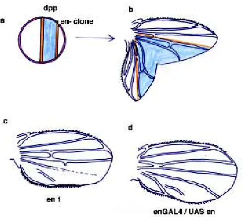
Fig. 5. Pattern transformations caused by alterations in engrailed expression. (a) Hypothetical transection of the wing field early in develop-ment by a clone of en/inv mutant cells that will later on give rise to organized growth and pattern transformation. (b)Observed FRT-hsFLIP induced clone of en/inv mutant cells causing the formation of an ectopic A/P boundary, a duplicated wing with A (mutant) and P (wild-type) compart-ments. (c)Homeotic transformation observed with en1, a neomorphic
Blair has shown that the A/P boundary can also be visualized in terms of cell morphology, as these cells are more elongated than their neighbors (Blair, 1992).
This suggests that dpp or hedgehog (acting directly or via dpp), rather than directly en, may regulate the expression of A/P bound-ary cell adhesion molecules (Blair and Ralston, 1997; Rodriguez and Basler, 1997). The existence of enhancer trap lines expressed along this boundary suggest that this is not unlikely. The compart-ment boundary could thus be stabilized by cell adhesion between cells at either side of the boundary, but not necessarily beyond.
A puzzling aspect of cell behavior is revealed by twin spot analysis. Mutant en clones have been found in their entirety within A, whereas their twin lied in P. According to the compartment hypothesis, en mutant cells crossed completely into A territory (Blair and Ralston, 1997). Although migration of wing clones has not been observed in vivo, if the A/P boundary were to behave like the AER, it would be difficult to understand how twin clones could be found in different compartments other than through cell migra-tion. The AER is a specialized group of cells that can be easily distinguished from surrounding cells and that does not exchange cells with its surroundings. Consequently, twin clones should not be found in different compartments as a consequence of cell proliferation. On the other hand, if differential cell affinities between all A and P do not play a role in compartmentalization, it is unclear why should mutant clones migrate at all.
Because dpp function has been shown to induce cell prolifera-tion (Posakony et al., 1991; Zecca et al., 1995), this implies that the straight A/P compartment boundary is a consequence of the control of growth. That is, the A/P boundary of the wing is telling us how the wing disc grew. In dpp mutants in which the wing disc does not grow to its normal size, the A/P boundary is not defined (Hidalgo, 1994). Furthermore, very suggestive is the observation that a proximal nubbin clone causing a dramatic reduction in wing
size does not respect the A/P boundary (Ng et al., 1995). It would be interesting to know if further mutations that reduce the normal growth of the wing also show abnormal cell behavior at the compartment boundary. To conclude, the A/P boundary is a foot-print of the pattern of growth. In the near future, it will be very exciting to learn more about this mysterious line, its relationship with growth and about how far we can generalize our understand-ing of the control of growth to different morphologies.
Patterning: shifting hh and en domains according to
morphology
en also plays an important role in patterning posterior cells (see Hidalgo, 1996). Complete loss of en and inv functions in small clones reproduces anterior pattern elements (Hidalgo, 1994; Tabata et al., 1995). Also, homeotic transformations not linked to altera-tions in growth have been observed when en is expressed ectopically in P, indicating that en can repress its own expression to mimic partially the loss of function phenotype (Fig. 5) (Guillén et al., 1995). Finally, duplicated wings produced by the ectopic expression of dpp within A grow well but do not reproduce posterior patterning (Fig. 4) (Zecca et al., 1995). However, the patterning role of en is not restricted to the posterior compartment. In fact, en is expressed in anterior cells lying between L3 and L4 (Blair, 1992), where it affects the size of this region and represses enervation at the wing margin (Hidalgo, 1994). This implies that during normal develop-ment patterning is not absolutely restricted by compartdevelop-mental subdivisions. Furthermore, A vs P identity is not a clear cut cell-autonomous consequence of en/inv functions. In fact hh, ex-pressed in P and direct target of en, is necessary to define vein 3, which lies within A (Mullor et al., 1997). This role cannot be accomplished by dpp, a frequent target of hh (Mullor et al., 1997).
Fig. 6. Patterning of digits and veins from the source of Shh/hh. (a) Vertebrate limb field, frontal view. (b) Vertebrate limb bud, lateral view. Digits are specified perpendicularly to the AER. Shh is expressed in a posterior domain, En-1 ventrally. (c) Adult vertebrate limb. Final skeletal morphology of digits. (d) Fly wing disc field. en and hh are coexpressed in P. (e)Fly wing disc. Veins appear in the wing disc parallel to the A/P boundary. (f)The adult fly wing, vein pattern. Blue, engrailed and En-1; Yellow, Shh and hh; Green, Shh and En-1, hh and en; Orange, AER and A/ P boundary.
In vertebrates, loss of function and ectopic expression of En-1 cause partial transformations of dorso-ventral patterning, suggest-ing that although En-1 is involved in D/V patternsuggest-ing, it is not sufficient (Wurst et al., 1994; Loomis et al., 1996; Logan et al., 1997). Furthermore, En-1 also regulates expression of Shh in neighboring cells, where it plays an important role patterning the digits (see (Hammerschmidt et al., 1997). Consequently, specifica-tion of digits is linked to D/V patterning and assigning patterning roles exclusively to En-1 is not straight forward.
Reminiscent of the situation in flies, Shh regulates BMP2 expression in posterior cells, but digit patterning cannot be achieved by BMP2 .Shh is expressed in a posterior domain, only partly overlapping the En-1 domain, and perpendicular to the AER (Hammerschmidt et al., 1997) (Fig. 6). Digits are also perpendicu-lar to the AER. In the fly wing, veins are instead parallel to the A/ P boundary and they are at least partly specified by hh (Mullor et al., 1997) (Fig. 6). Intriguingly, this might at least partly explain why hh expression coincides with the domain of en. It is fascinating that these patterning roles of Shh/hh have been conserved, correlating with a shift in their expression domains to allow patterning of digits (perpendicular to the AER) or veins (parallel to the A/P boundary) (Fig. 6). Further comparative studies will tell us how far the patterning roles of hh can be separated from its role in growth control.
The intimate relation between en, inv and hh is not only interest-ing to our notion of identity, but also to our understandinterest-ing of how compartments are established, and, in essence, of what they really are. Compartments are considered units of lineage. Consequently, en expression is thought to be maintained cell autonomously (García-Bellido, 1975). However, it has been shown that en ex-pression between veins L3 and L4 within A is induced by hh from posterior cells (de Celis and Ruiz-Gómez, 1995; Guillén et al., 1995) (Fig. 7). Furthermore, ectopic expression of hh in all en cells causes the anterior domain of en to expand dramatically (Mullor et al., 1997) (Fig. 7). And finally, anterior smo clones in this region, which cannot receive hh signaling, produce neurons typical of loss of en expression (Blair and Ralston, 1997). These data show that en expression in A is induced non-autonomously by hh. So far, although it is thought that en expression in posterior cells is maintained cell autonomously through lineage, this has not been shown.
It is not known how En-1 expression is maintained in ventral cells of the vertebrate limb either, nor for how long. However, it is known that En-1 protein is not required for En-1 expression, suggesting that cell-autonomous autoregulation is not employed (Logan et al., 1997). Remarkably, despite the regulation of Shh expression by a homeobox gene (Johnson and Tabin, 1997), no clonal restrictions have been found in vertebrates within the dorsal (non-en) or ventral (en) compartments. That is, expression of Shh does not seem to be restricted by a clonal mechanism either. It would be interesting to know how the expression of en and En1 are maintained in fly and vertebrate limbs.
The plasticity of morphogenetic fields
Growth and patterning in the wing disc may take place in a manner more similar to vertebrates than had been anticipated. Rather than lineage and irreversible determination of cell fate, cell communication seems to be the driving force of morphogenesis.
Not surprisingly, cell communication provides cell populations with the plasticity required to adjust to change (regulation within a morphogenetic field). Upon interference with normal development, a field can regulate cell number and cell size to ensure that global normal proportions, size and shape are reached. The wing disc behaves as a field, because it can regulate autonomously when grown in the larval abdomen. Compartments can also regulate, but only as long as there is an A/P interface (Wilcox and Smith, 1980; Weigmann et al., 1997) - that is, their regulation is not autonomous. Regulation is not restricted by compartmental subdivisions either, since following removal or transplantations of fractions of wing disc, the disc regulates by inducing cell proliferation and patterning irrespectively of where compartment boundaries lie (Bryant, 1975). Nevertheless, there is abundant evidence showing that the A/P and D/V boundaries and the proximal edge of the wing field are important in the control of growth during normal development (see Edgar and Lehner, 1996). Cells must sense the dimensions of the field from these three regions.
Whereas a non-autonomous view of compartment regulation would make the notion of lineage dispensable to our understanding of morphogenesis, it would not turn compartments into less stable domains. On the contrary, regulation insures morphological stabil-ity. In the future, it will be very enlightening to learn more about the regulation of cell proliferation and size. As Antonio has also foreseen, perhaps we may soon be able to look at a wing and see beyond the image of the geometric design the now mysterious plane of controlled growth.
Acknowledgments
I would like to thank D.Weisblat and G.Scholtz for sharing with me their interest on engrailed; M.Averof and J.Zrzavy for discussions on crusta-ceans; A.Martinez-Arias for comments on the manuscript. A.H.’s work is funded by the Wellcome Trust.
References
BLAIR, S. (1992). engrailed expression in the anterior lineage compartment of the developing wing blade of Drosophila. Development 115: 21-33.
BLAIR, S.S. and RALSTON, A. (1997). Smoothened-mediated Hedgehog signalling is required for the maintenance of the antero-posterior lineage restriction in the developing wing of Drosophila. Development 124: 4053-4063.
BRYANT, P.J. (1975). Regeneration and duplication in imaginal discs. Ciba Found. Symp. 71-93.
BURKE, R. and BASLER, K. (1996). Dpp receptors are autonomously required for cell proliferation in the entire developing Drosophila wing. Development 122: 2261-2269.
CAPDEVILA, J. and GUERRERO, I. (1994). Targeted expression of the signalling molecule decapentaplegic induces pattern duplications and growth alterations in Drosophila wings. EMBO J. 13: 4459-4468.
CHEN, Y. and STRUHL, G. (1996). Dual roles for Patched in sequestering and trasnducing Hedgehog. Cell 87: 553-563.
COUSO, J.P., BATE, M. and MARTINEZ-ARIAS, A. (1993). A wingless-dependent polar coordinate system in Drosophila imaginal discs. Science 259: 484-489.
DE CELIS, J.F. and RUIZ-GOMEZ, M. (1995). groucho and hedgehog regulate engrailed expression in the anterior compartment of the Drosophila wing. Devel-opment 121: 3467-3476.
DIAZ-BENJUMEA, F.J. and COHEN, S.M. (1993). INteraction between dorsal and ventral cells in the imaginal disc directs wing development in Drosophila. Cell 75: 741-752.
EDGAR, B.A. and LEHNER C.P. (1996). Developmental control of cell cycle regulatiors: a fly's perspertive. Science 274: 1646-1652.
GARCIA-BELLIDO, A. (1975). Genetic control of wing disc development in Dro-sophila. Cell patterning, Ciba Found. Symp. 161-182.
GARCIA-BELLIDO, A. and MERRIAM, J.R. (1971). Parameters of the wing imaginal disc development of Drosophila melanogaster. Dev.Biol. 24: 61-87.
GARCIA-BELLIDO, A. and SANTAMARIA, P. (1972). Developmental analysis of the wing disc in the mutant engrailed of Drosophila melanogaster. Genetics 72: 87-104.
GARCIA-BELLIDO, A., RIPOLL, P. and MORATA, G. (1973). Developmental compartmentalisation of the wing disc of Drosophila. Nature New Biol. 245: 251-253.
GONZALEZ-GAITAN, M.A.F., CAPDEVILA, M.P. and GARCIA-BELLIDO, A. (1994). Cell proliferation patterns in the wing imaginal disc of Drosophila. Mech. Dev. 46: 183-200.
GUILLEN, I., MULLOR, J.L., CAPDEVILA, J., SANCHEZ-HERRERO, E., MORATA, G. and GUERRERO, I. (1995). The function of the engrailed gene and the specification of the Drosophila wing pattern. Development 121: 3447-3456.
HAMMERSCHMIDT, M., BROOK, A. and MCMAHON, A. (1997). The world accord-ing to hedgehog. Trends Genet. 13: 14-21.
HIDALGO, A. (1994). Three distinct roles for the engrailed gene in Drosophila wing development. Curr. Biol. 4: 1087-1098.
HIDALGO, A. (1996). The roles of engrailed. Trends Genet. 12: 1-3.
HOLLAND, L.Z., KENE, M., WILLIAMS, N.A. and HOLLAND, N.D. (1997). Sequence and embryonic expression of the amphioxus engrailed gene (AmphiEn): the metameric pattern of transcription resembles that of its segment-polarity homolog in Drosophila. Development 124: 1723-1732.
JOHNSON, R.L. and TABIN, C.J. (1997). Molecular models for vertebrate limb development. Cell 90: 979-990.
JOYNER, A., HERRUP, K., AUERBACH, B.A., DAVIS, C.A. and ROSSANT, J. (1991). Subtle cerebellar phenotype in mice homozygous for a targeted deletion of the En-2 homeobox. Science 251: 1239-1243.
LANS, D., WEDEEN, C.J. and WESIBLAT, D.A. (1993). Cell lineage analysis of the expression of an engrailed homolog in leech embryos. Development 117: 857-871.
LOGAN, C., HORNBRUCH, A., CAMPBELL, I. and LUMSDEN, A. (1997). The role of Engrailed in establishing the dorsoventral axis of the chick limb. Development 124: 2317-2324.
LOOMIS, C.A., HARRIS, E., MICHAUD, J., WURST, W., HANKS, M. and JOYNER, A.L. (1996). The mouse Engrailed-1 gene and ventral limb patterning. Nature 382: 360-363.
MARIGO, V., SCOTT, M.P., JOHNSON, R.L., GOODRICH, L.V. and TABIN, C.J. (1996). Conservation in hedgehog signalling: induction of a chicken patched homolog by Sonic hedgehog in the developing limb. Development 122: 1225-1233.
MEINHARDT, H. (1986). The threefold subdivision of segments and the initiation of legs and wings in insects. Trends Genet 2: 36-41.
MILAN, M., CAMPUZANO, S. and GARCIA-BELLIDO, A. (1996a). All cycling and patternod cell proliferation in the wing primordium of Drosophila. Proc. Natl. Acad. Sci. USA 93: 640-645.
MILAN, M., CAMPUZANO, S. and GARCIA-BELLIDO, A. (1996b). Cell cycling and patterned cell proliferation in the Drosophila wing during metamorphosis. Proc. Natl. Acad. Sci. USA. 93: 11687-11692.
MORATA, G. and LAWRENCE, P.A. (1975). Control of compartment development by the engrailed gene in Drosophila. Nature 255: 614-617.
MULLOR, J.L., CALLEJA, M., CAPDEVILA, J. and GUERRERO, I. (1997). Hedgehog activity, independent of decapentaplegic, participates in wing disc patterning. Development 124: 1227-1237.
NEUMANN, J.C. and COHEN, S.M. (1996). Distinct mitogenic and cell fate specifica-tion funcspecifica-tions of wingless in different regions of the wing. Development 122: 1781-1789
NEUMANN,C.J. and COHEN, S.M. (1997). Long-range action of Wingless organises the dorso-ventral axis of the Drosophila wing. Development 124: 871-880.
NG, M., DIAZ-BENJUMEA, F.J. and COHEN, S.M. (1995). nubbin encodes a POU-domain protein required for proximo-distal patterning in the Drosophila wing. Development 121: 589-599.
PANGANIBAN, G., SEBRING, A., NAGY, L. and CARROLL, S. (1995). The develop-ment of crustacean limbs and the evolution of arthropods. Science 270: 1363-1366.
PARR, B.A. and MCMAHON, A.P. (1995). Dorsalizing signal Wnt7a required for nomral polarity of D-V and A-P axes of mouse limb. Nature 374: 350-353.
PATEL, N.H., KORNBERG, T.B. and GOODMAN, C.S. (1989a). Expression of engrailed during segmentation in grasshopper and crayfish. Development 107: 201-212.
PATEL, N.H., MARTIN-BLANCO, E., COLEMAN, K.G., POOLE, S.J., ELLIS, M.C., KORNBERG, T.B. and GOODMAN, C.S. (1989b). Expression of engrailed proteins in arthropods, annelids and chordates. Cell 58: 955-968.
POSAKONY, L.G., RAFTERY, L.A. and GELBART, W.M. (1991). Wing formation in Drosophila melanogaster requires decapentaplegic gene function along the antero-p[osterior compartment boundary. Mech. Dev. 33: 69-82.
RAFTERY, L.A., SANICOLA, M., BLACKMAN, R.K. and GELBART, W.M. (1991). The relationship of decapentaplegic and engrailed expression in Drosophila imaginal disks: do these genes mark the antero-posterior compartment bound-ary? Development 113: 27-33.
RODRIGUEZ, I. and BASLER, K. (1997). Control of compartment affinity boundaries by Hedgehog. Nature 389: 614-617.
SANICOLA, M., SEKELSKY, J., ELSON, S. and GELBART, W.M. (1995). Drawing a stripe in Drosophila imaginal disks - negative regulation of decapentaplegic and patched expression by engrailed. Genetics 139: 745-756.
SCHOLTZ, G. and DOHLE, W. (1996). Cell lineage and cell fate in crustacean embryos - a comparative approach. Int. J. Dev. Biol. 40: 211-220.
SCHOLTZ, G., DOHLE, W., SANDEMAN, R. and RICHTER, S. (1993). Expression of engrailed can be lost and regained in cells of one clone in crustacean embryos. Int. J. Dev. Biol. 37: 299-304.
SCHOLTZ, G., PATEL, N.H., and DOHLE, W. (1994). Serially homologous engrailed stripes are generated via different cell lineages in the germ band of amphiopod crustaceans (Malacostraca, Peracarida). Int. J. Dev. Biol. 38: 471-478.
TABATA, T., SCHWARTZ, C., GUSTAVSON, E., ALI, Z. and KORNBERG, T.B. (1995). Creating a Drosophila wing de novo, the role of the engrailed gene, and the compartment border hypothesis. Development 121: 3359-3369.
VINCENT, J.P. and O’FARRELL, P.H. (1992). The state of engrailed expression is not clonally transmitted during early Drosophila development. Cell 68: 923-931.
WEDEEN, C.J. and WEISBLAT, D.A. (1991). Segmental expression of an engrailed-class gene during early development and neurogenesis in an annelid. Develop-ment 113: 805-814.
WEIGMANN, K., COHEN, S.M. and LEHNER, C.F. (1997). Cell cycle progression, growth and patterning in imaginal discs despite inhibition of cell division after inactivation of Drosophila Cdc2 kinase. Development 124: 3555-3563.
WILCOX, M. and SMITH, R.J. (1980). Compartments and distal outgrowth in the Drosophila wing imaginal disc. Roux Arch. Dev. Biol. 188: 157-161.
WILLIAMS, J.A., PADDOCK, S.W., VORWECK, K. and CARROLL, S.B. (1994). Organization of wing formation and induction of a wing-patterning gene at the dorso-ventral compartment boundary. Nature 368: 299-305.
WURST, W., AERBACH, A.B. and JOYNER, A.L. (1994). Multiple developmental defects in Engrailed-1 mutant mice: an early mid-hindbrain deletion and patterning defects in forelimbs and sternum. Development 120: 2065-2075.