Western University Western University
Scholarship@Western
Scholarship@Western
Electronic Thesis and Dissertation Repository
4-13-2015 12:00 AM
Exploring the Structural and Functional Organization of the Dorsal
Exploring the Structural and Functional Organization of the Dorsal
Zone of Auditory Cortex in Hearing and Deafness
Zone of Auditory Cortex in Hearing and Deafness
Melanie A. Kok
The University of Western Ontario
Supervisor
Dr. Stephen Lomber
The University of Western Ontario Graduate Program in Neuroscience
A thesis submitted in partial fulfillment of the requirements for the degree in Doctor of Philosophy
© Melanie A. Kok 2015
Follow this and additional works at: https://ir.lib.uwo.ca/etd
Part of the Neurosciences Commons
Recommended Citation Recommended Citation
Kok, Melanie A., "Exploring the Structural and Functional Organization of the Dorsal Zone of Auditory Cortex in Hearing and Deafness" (2015). Electronic Thesis and Dissertation Repository. 2735.
https://ir.lib.uwo.ca/etd/2735
This Dissertation/Thesis is brought to you for free and open access by Scholarship@Western. It has been accepted for inclusion in Electronic Thesis and Dissertation Repository by an authorized administrator of
EXPLORING THE STRUCTURAL AND FUNCTIONAL
ORGANIZATION OF THE DORSAL ZONE OF AUDITORY CORTEX
IN HEARING AND DEAFNESS
(Thesis format: Integrated Article)
by
Melanie A. Kok
Graduate Program in Neuroscience
A thesis submitted in partial fulfillment of the requirements for the degree of
Doctor of Philosophy
The School of Graduate and Postdoctoral Studies The University of Western Ontario
London, Ontario, Canada
ii
Abstract
Recent neuroscientific research has focused on cortical plasticity, which
refers to the ability of the cerebral cortex to adapt as a consequence of
experience. Over the past decade, an increasing number of studies have
convincingly shown that the brain can adapt to the loss or impairment of a
sensory system, resulting in the expansion or heightened ability of the remaining
senses. A particular region in cat auditory cortex, the dorsal zone (DZ), has been
shown to mediate enhanced visual motion detection in deaf animals. The
purpose of this thesis is to further our understanding of the structure and function
of DZ in both hearing and deaf animals, in order to better understand how the
brain compensates following insult or injury to a sensory system, with the
ultimate goal of improving the utility of sensory prostheses.
First, I demonstrate that the brain connectivity profile of animals with early- and
late-onset deafness is similar to that of hearing animals, but the projection
strength to visual brain regions involved in motion processing increases as a
consequence of deafness. Second, I specifically evaluate the functional impact of
the strongest auditory connections to area DZ using reversible deactivation and
electrophysiological recordings. I show that projections that ultimately originate in
primary auditory cortex (A1) form much of the basis of the response of DZ
neurons to auditory stimulation. Third, I show that almost half of the neurons in
DZ are influenced by visual or somatosensory information. I further demonstrate
that this modulation by other sensory systems can have effects that are opposite
in direction during different portions of the auditory response. I also show that
techniques that incorporate the responses of multiple neurons, such as multi-unit
and local field potential recordings, may vastly overestimate the degree to which
multisensory processing occurs in a given brain region. Finally, I confirm that
individual neurons in DZ become responsive mainly to visual stimulation
iii
Together, these results shed light on the function and structural
organization of area DZ in both hearing and deaf animals, and will contribute to
the development of a comprehensive model of cross-modal plasticity.
Keywords: Hearing, deafness, multisensory, neuroplasticity, auditory cortex,
iv
Statement of Co-Authorship
All data collection, analysis and writing of the manuscripts that comprise this
doctoral thesis dissertation were primarily conducted by the author of this thesis,
Melanie Kok. Dr. Nicole Chabot assisted with experimental design and data
analysis, as well as editing of the manuscript for Chapter 2. Dr. Daniel Stolzberg
provided assistance with the experimental design for Chapter 3, and in addition,
assisted with data collection and edited the manuscript for this chapter, along
with Dr. Trecia Brown. Drs. M. Alex Meredith and Andres Carrasco assisted with
the experimental design and edited the manuscript for Chapters 4 and 5. Dr.
Stephen Lomber provided expert advice and supervision during all phases of this
v
Acknowledgements
First and foremost, I would like to thank my supervisor, Dr. Stephen Lomber, for quite simply, everything. I feel incredibly fortunate to have been able to mentor under someone who is not only a class scientist, but a world-class person. I truly couldn’t have ended up in a better lab.
I would also like to thank the members of my advisory committee, Dr. Scott MacDougall-Shackleton and Dr. David Sherry for always providing thoughtful insight and constructive criticism throughout my graduate career.
Words can’t adequately express the gratitude I feel towards all of the members of the Lomber lab, past and present (Dr. Trecia Brown, Dr. Blake Butler, Dr. Andres Carrasco, Dr. Nicole Chabot, Amee Hall, Pam Nixon, Dr. Daniel Stolzberg, and Carmen Wong). Whether providing serious scientific feedback during all aspects of every project I undertook, or sharing a laugh (by which I mean tolerantly listening to me as I spout off about something), I quite literally could not have done this without you. Special thanks to Pam Nixon for making sure that I always had healthy and happy participants for my experiments.
vi
Table of contents
Abstract ... ii
Statement of Co-Authorship ... iv
Acknowledgements ... v
Table of contents. ... vi
List of Figures ... xi
List of Abbreviations and Symbols ... xiv
Chapter 1: General Introduction ... 1
1.1 Overview ... 1
1.2 The auditory system ... 2
1.2.1 Acquisition of auditory information by the nervous system ... 2
1.2.2 Interpretation of auditory information by the nervous system ... 3
1.3 Sensory loss and the cerebral cortex ... 5
1.3.1 Short-term removal of sensory input ... 6
1.3.2 Blindness and visual deprivation ... 7
1.3.3 Deafness and auditory deprivation ... 10
1.3.4 General principles of cross-modal plasticity ... 13
1.3.5 Developmental considerations ... 14
1.4 Thesis overview... 15
1.5 References ... 17
Chapter 2: Cross-modal reorganization of cortical afferents to dorsal auditory cortex following early- and late-onset deafness ... 27
2.1 Abstract. ... 27
2.2 Introduction ... 28
2.3 Materials and Methods ... 30
2.3.1 Deafening Procedures ... 33
vii
2.3.3 Histological processing ... 36
2.3.4 Areal border delimitation ... 37
2.3.5 Data analysis. ... 39
2.4 Results ... 40
2.4.1 Tracer deposits ... 40
2.4.2 Labeling of cortical afferents ... 42
2.4.3 Comparisons between modalities ... 49
2.4.4 Auditory cortical projections ... 51
2.4.5 Visual cortical projections ... 52
2.5 Discussion ... 53
2.5.1 Spatial processing in DZ ... 53
2.5.2 Localization of injection sites ... 53
2.5.3 Auditory cortical projections to DZ in hearing animals... 54
2.5.4 Comparison to late- and early-deafened animals ... 55
2.5.5 Visual cortical projections to DZ in hearing animals ... 56
2.5.6 Comparison to late- and early-deafened animals ... 58
2.5.7 Other considerations ... 62
2.5.8 Summary and conclusions ... 64
2.6 References ... 67
Chapter 3: Dissociable influences of primary auditory cortex and the posterior auditory field on neuronal responses in the dorsal zone of auditory cortex ... 75
3.1 Abstract. ... 75
3.2 Introduction ... 76
viii
3.3.1 Overview. ... 78
3.3.2 Surgical procedures ... 79
3.3.3 Stimulus generation and presentation. ... 80
3.3.4 Data acquisition. ... 81
3.3.5 Histological procedures ... 84
3.3.6 Data analysis. ... 85
3.4 Results ... 87
3.4.1 Comparison of DZ responses to A1 and AAF responses ... 87
3.4.2 Noise burst responses during cooling deactivation ... 88
3.4.3 Noise RIF responses during cortical cooling ... 91
3.4.4 Responses to tones during reversible deactivation ... 91
3.4.5 Results summary ... 101
3.5 Discussion ... 101
3.5.1 Comparison of DZ responses to previously published findings ... 101
3.5.2 Effects of reversible deactivation in DZ ... 103
3.6 References ... 107
Chapter 4: Diametric modulation of early and late components of acoustically-evoked activity in the dorsal zone of auditory cortex by visual and tactile stimulation ... 110
4.1 Abstract ... 110
4.2 Introduction ... 111
4.3 Materials and Methods ... 112
4.3.1 Overview ... 112
4.3.2 Surgical Preparation ... 113
ix
4.3.4 Preparation for recording ... 114
4.3.5 Stimulus generation and presentation ... 116
4.3.6 Data acquisition ... 118
4.3.7 Histological Procedures ... 118
4.3.8 Data Analysis ... 119
4.4 Results ... 123
4.4.1 Overview ... 123
4.4.2 Multisensory integration in DZ neurons ... 124
4.4.3 Response characteristics of single units in DZ ... 130
4.4.4 Timing of the visual stimulus ... 135
4.4.5 Comparison of SU data with MU and LFP activity ... 138
4.4.6 Summary of findings ... 141
4.5 Discussion ... 141
4.6 References ... 146
Chapter 5: Visual and somatosensory cross-modal reorganization in the dorsal zone of auditory cortex following perinatal deafness ... 152
5.1 Abstract ... 152
5.2 Introduction ... 153
5.3 Materials and methods ... 154
5.3.1 Deafening procedures ... 154
5.3.2 Electrophysiological recordings ... 156
5.3.3 Data acquisition and stimulus presentation ... 158
5.3.4 Histological procedures ... 159
5.3.5 Data analysis ... 159
x
5.4.1 Area DZ identification. ... 161
5.4.2 Single unit responses ... 162
5.4.3 Comparison to multiunit responses and LFP activity ... 165
5.5 Discussion ... 167
5.6 References ... 170
Chapter 6: General Discussion ... 174
6.1 Main findings and conclusions ... 174
6.1.1 DZ receives strong projections from visual cortex in hearing animals ... 174
6.1.2 The strength of visual cortical projections to DZ is increased in deaf animals... 174
6.1.3 DZ neurons rely on input from A1, whereas PAF may modulate DZ responses ... 175
6.1.4 Almost half of DZ neurons are multisensory ... 175
6.1.5 Population-based measures of neural activity may overestimate the degree of multisensory processing in a cortical area ... 176
6.1.6 DZ neurons in deaf animals respond mainly to visual stimulation ... 176
6.2 Conclusions ... 177
6.2.1 DZ may be homologous to caudal auditory fields in the primate ... 177
6.2.2 DZ is the most extensively-documented model of cross-modal plasticity in mammalian cortex to date ... 180
6.3 Future directions... 181
6.4 References ... 182
Appendix A………...……..……….187
xi
List of Figures
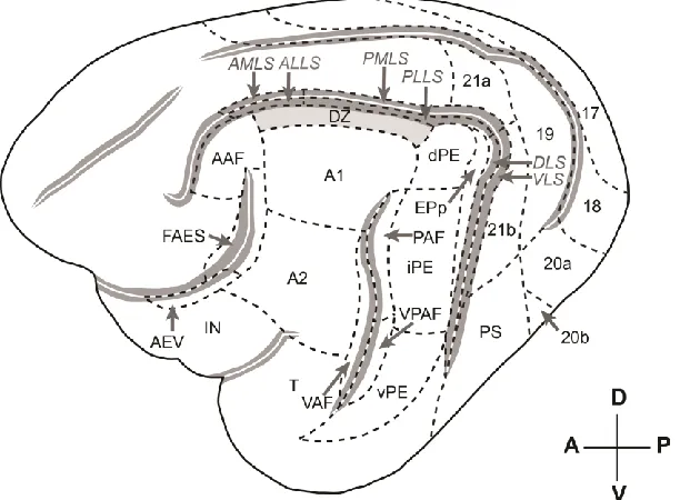
Figure 2.1 The auditory and visual cortices and sub-fields of the cat. ... 31
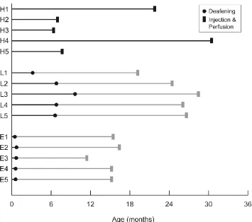
Figure 2.2 Timeline of deafening and other procedures performed on each group. ... 32
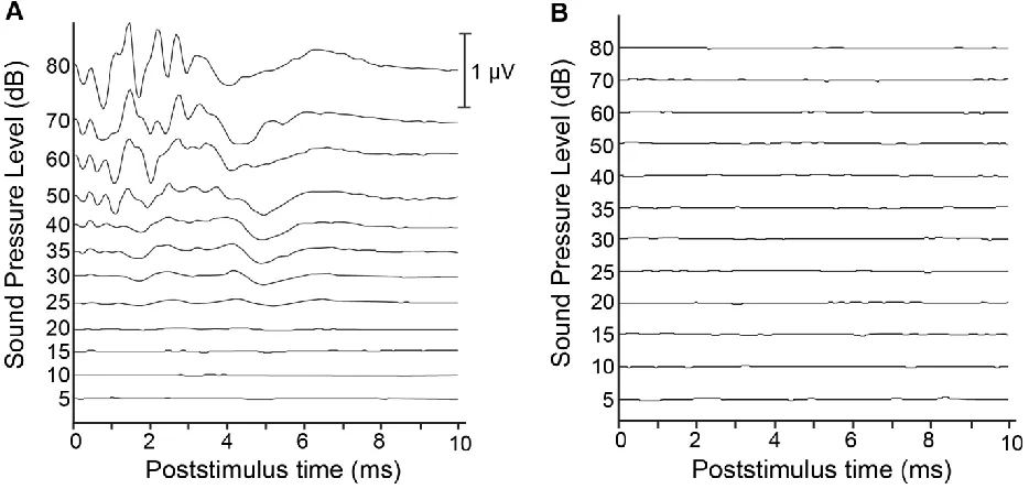
Figure 2.3 Pre- and post-deafening auditory brainstem responses. ... 34
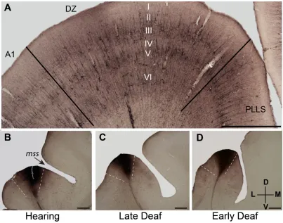
Figure 2.2.4 Cortical border delimitation. ... 38
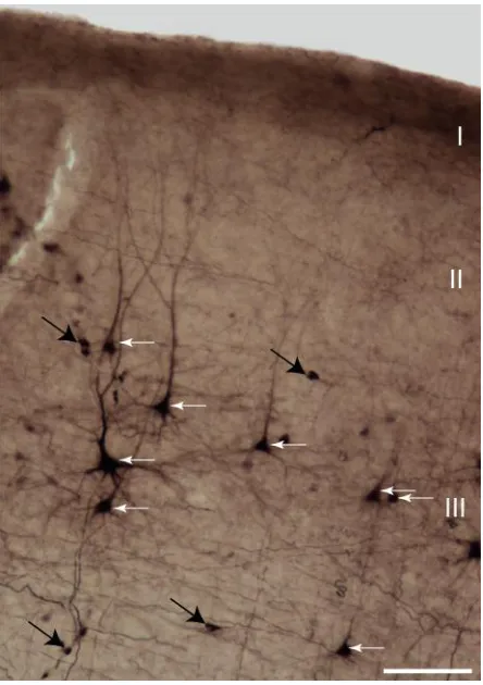
Figure 2.5 BDA-labeled neurons in DZ. ... 41
Figure 2.6 Injection sites and labeling in a hearing animal. ... 42
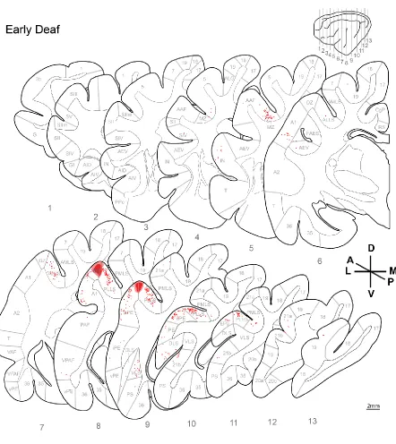
Figure 2.7 Labeled neurons and injection sites from a late-deafened animal (L4). ... 44
Figure 2.8 Labeled neurons and injection sites from an early-deaf animal (E2). 45 Figure 2.9 Injection sites and neuronal labeling for a hearing animal. ... 46
Figure 2.10 Standardized injection sites and neuronal labeling for a late-deafened animal. ... 47
Figure 2.11 Injection sites and labeled neurons on std. sections... 48
Figure 2.12 Proportion of projections by cortical area. ... 50
Figure 2.13 Pattern of labeling within the middle suprasylvian sulcus. ... 59
Figure 2.14 Projections present in hearing animals and change in projection strength following late- and early-deafness. ... 65
Figure 3.1 Organization and hierarchical connections of cat auditory cortex. .... 77
Figure 3.2 Position of cryoloops and extent of cortical deactivation. ... 82
Figure 3.3 Population level effects of reversible deactivation on DZ responses to 65 dB noise bursts. ... 89
Figure 3.4 Effects of reversible deactivation during noise burst presentation on individual sites. ... 90
Figure 3.5 Noise Rate-intensity functions and monotonicity ratios. ... 92
Figure 3.6 Population level effects of reversible deactivation on DZ responses to tones. ... 93
xii
Figure 3.8 Representative example of tuning curves recorded in fields A1, AAF and DZ. ... 97
Figure 3.9 Summary of changes in DZ receptive field properties as a function of reversible deactivation. ... 98
Figure 3.10 Summary of changes in threshold and CF at individual sites in DZ. ... 100
Figure 3.11 Representative example of a recording site in DZ in response to various stimuli. ... 102
Figure 4.1 Location of recording sites within DZ. ... 115
Figure 4.2 Waveforms and typical profile of single unit and multiunit activity at a representative site following auditory noise burst stimulation ... 120
Figure 4.3 Representative examples of rasters, PSTHs and bar graphs of single unit responses for the four classes of neurons recorded in DZ... 125
Figure 4.4 Summary of multisensory integration for individual single units. ... 127
Figure 4.5 Summary of multisensory integration across the population of single units. ... 129
Figure 4.6 Location of bimodal and integrative neurons in DZ... 131
Figure 4.7 Summary of differences in response characteristics among classes of neurons in DZ. ... 132
Figure 4.8 Distributions showing the enhancement and additivity indices for responses to combined-modality stimulation for each class of neuron
encountered... 134
Figure 4.9 Analysis of visual onset asynchronies. ... 136
Figure 4.10 Representative site showing neuronal responses at different scales of activity ... 137
Figure 4.11 Summary of results for single unit (SU), multiunit (MU) and local field potential (LFP) responses... 139
Figure 5.1 Photomicrographs of the craniotomy, electrode penetrations and SMI-32 stained sections in hearing and deafened animals ... 155
Figure 5.2 Auditory brainstem responses (ABRs) for a hearing and a deaf animal. ... 157
xiii
Figure 5.4 Representative examples of sensory neurons recorded in DZ of hearing and deaf animals. ... 163
Figure 5.5 Organization of bimodal versus unimodal neurons in DZ of deaf
animals ... 164
Figure 5.6 Comparison of single unit, multiunit and LFP responses in DZ of
xiv
List of Abbreviations and Symbols
A1 primary auditory cortex A2 second auditory cortex AAF anterior auditory field
ABR auditory brainstem response AES anterior ectosylvian sulcus AEV anterior ectosylvian visual area
ALLS anterolateral lateral suprasylvian area AMLS anteromedial lateral suprasylvian area ANOVA analysis of variance
BDA biotinylated dextran amine
CF characteristic frequency
dB SPL decibels sound pressure level
df degrees of freedom
DLS dorsal lateral suprasylvian area
dPE dorsal posterior ectosylvian area of auditory cortex DZ dorsal zone of auditory cortex
EEG electroencephalogram
EPp posterior part of the posterior ectosylvian gyrus
fAES field of the anterior ectosylvian sulcus fMRI functional magnetic resonance imaging
GABA gamma-aminobutyric acid
Hz hertz
i.m. intramuscular i.v. intravenous IN insular cortex
iPE intermediate posterior ectosylvian area of auditory cortex
kg kilograms
kHz kilohertz
L litres
LFP local field potential
MEG magnetoencephelogram
mg milligrams
xv
mm millimeters
ms milliseconds
MT middle temporal area
MU multiunit
MW molecular weight
PAF posterior auditory field
PLLS posterolateral lateral suprasylvian area PMLS posteromedial lateral suprasylvian area PS posterior suprasylvian area
PSTH peri-stimulus time histogram
R rostral field of auditory cortex RIF rate-intensity function
s.c. subcutaneous
SU single unit
T temporal cortex
V1 primary visual cortex (area 17) VAF ventral auditory field
VLS ventral lateral suprasylvian area VPAF ventral posterior auditory field
vPE ventral posterior ectosylvian area of auditory cortex
ºC degrees Celsius
% percent
1
Chapter 1: General Introduction
1.1 Overview
Until relatively recently, it was thought that the structure of the brain was
largely immutable following the closure of developmental critical periods (Gross,
2001). This was based largely on the knowledge that outside of specialized
regions of the hippocampus and olfactory system, neurons do not regenerate
once lost, and damage to the brain, whether degenerative or traumatic, is for the
most part, irreversible. However, multiple avenues of research over the last
quarter-century have demonstrated that the brain adapts to environmental input
throughout life, and that its connectivity can be both structurally and functionally
altered as a consequence of experience. This phenomenon is referred to as
plasticity.
While much research on cortical plasticity has focused on normal
adaptation to environmental input (e.g. development and maturation, learning), a
growing body of research is focused on understanding the conditions under
which the cerebrum is capable of rewiring itself following the loss or impairment
of a sensory system. This rewiring is a compensatory mechanism that has been
shown to take place across sensory modalities, and is therefore referred to as
cross-modal plasticity. It is generally understood that this process not only helps
to compensate for the lost sensory modality, but additionally results in enhanced
behavioral performance in the remaining sensory modalities. This plasticity has
important consequences for the use of sensory prostheses (i.e. cochlear
implants), as it is thought that cross-modal reorganization may limit the
reintroduction of missing sensory information by colonizing the deprived region of
cortex for the processing of other sensory functions (Lee et al., 2001).
As such, a complete framework for understanding how and why the brain
is able to reorganize following sensory loss must include both an understanding
2
characterization of the changes that occur under conditions of sensory loss or
impairment. Because the body of work that comprises this thesis was conducted
in auditory cortex, I first review what is known about the sense of hearing and
how sound is processed by the auditory system. I then go on to discuss the
consequences of the removal of sensory input on sensory systems in the brain.
1.2 The auditory system
1.2.1 Acquisition of auditory information by the nervous system
The basic mechanisms underlying how the nervous system acquires
auditory information from the environment are relatively well known at this point.
Sound waves in the environment are detected as mechanical pressure by the
tympanic membrane or eardrum, and these vibrations are passed along by
middle ear structures to the cochlea. The cochlea is a specialized structure within
the inner ear containing the basilar membrane, whose properties change as a
function of length. Because of this, the basilar membrane responds differently
based on the spectral information of the incoming sound wave. Sounds of high
frequency do not propagate far along the basilar membrane, reaching a peak
displacement at the base, while sounds of low frequency travel further along the
membrane and reach a peak displacement at the apex. Sounds of intermediate
frequency are represented orderly and continuously along the length of the
basilar membrane on a logarithmic scale. The local movement of the basilar
membrane is converted to electrical impulses by specialized sensory hair cells,
which synapse with auditory nerve fibers. In this way, hair cells and auditory
nerve fibers from a particular location along the basilar membrane fire in
response to sound of a particular frequency. This position-based spectral
organization is referred to as a tonotopic map, and the place theory states that it
is this tonotopic organization of the basilar membrane that gives rise to pitch
perception. Sound intensity is also coded by the firing rate of auditory nerve
fibers, i.e. maximal displacement of the basilar membrane is represented by a
saturated neuronal response. Thus, the frequency of the incoming sound is
3
represented by the firing rate of those fibers. This information is then propagated
along the auditory pathway to the brainstem (cochlear nuclei and superior olivary
nuclei), then to the midbrain (nuclei of the lateral lemniscus and inferior
colliculus), to the thalamus (medial geniculate nucleus; MGN) and finally, to
auditory cortex. It should be noted that the tonotopic map previously discussed is
preserved throughout these subcortical stations and in some regions of auditory
cortex.
1.2.2 Interpretation of auditory information by the nervous system
Unlike the visual system, acoustical information is processed in parallel at
the subcortical level, and significant auditory processing occurs before
information reaches primary auditory cortex (A1). Specifically, while the spectral
content of incoming sound is represented in the auditory system, the location of it
is not, and must be reconstructed in order to decipher the source of the sound in
space. This reconstruction is done in the superior olivary nuclei by comparing the
input arriving from each ear. Neurons in the medial superior olive code for the
difference in sound arrival time at each ear, referred to as the interaural time
difference. Neurons in the lateral superior olive code for the difference in sound
intensity arriving at each ear, or the interaural level difference.
Beyond the level of the superior olivary nuclei, specific functional
designations for structures in the auditory pathway are less clear-cut. For
example, a portion of the inferior colliculus receives both auditory and
somatosensory inputs, and a putative role in sound localization has also been
suggested based on the high numbers of neurons sensitive to interaural timing
and level differences. It should be noted that there is no homolog of the inferior
colliculus in any of the other sensory systems (Winer et al., 2005). At the level of
the thalamus, clear structural and functional differences exist between the
ventral, dorsal and medial subregions of MGN (Banks and Smith, 2011) and the
current opinion is that auditory thalamus is not a simple relay station, but rather
provides important modification of incoming information based on the state of the
4
The ascending auditory pathway terminates in auditory cortex, which
consists of central core areas, surrounded by belt and para-belt regions in
mammals. The core includes A1, which is a cytoarchitectonically distinct region
that is tonotopically organized and has been described in detail in many species.
Core auditory cortical fields share a number of characteristics. Core fields receive
strong projections from ventral MGN (Kaas et al., 1999), are densely
interconnected with one another, are characterized by robust, short latency
responses to pure tones with sharp frequency tuning curves, and function in
parallel with one another (i.e. lesions of one core region do not abolish responses
to pure tones in the remaining core regions). Core regions outside of A1 include
the rostral (R) and rostrotemporal fields in the primate and the anterior auditory
field (AAF) in the cat, ferret, gerbil, and rat (Hackett, 2011).
The core is surrounded by several other regions, which vary in number
based on the species under study, as well as in terms of the response properties
of the neurons located there. Some of these fields maintain tonotopic
organization while others do not. These fields may also show response specificity
for more complex sounds compared to pure tones (such as conspecific
vocalizations or the rate or direction of frequency-modulated sweeps), or may
display more complex receptive field tuning. While significant subcortical
processing of interaural timing and level differences is known to occur, sound
localization behavior is dependent on an intact auditory cortex, as ablation and
reversible deactivation studies have conclusively demonstrated. From these
studies, a number of non-primary fields have been identified as playing a role in
the spatial processing of sound in the cat, namely, the dorsal zone (DZ), the
auditory field of the anterior ectosylvian sulcus (fAES), the posterior auditory field
(PAF). Similarly, caudal fields in the monkey also show more spatial sensitivity
than do rostral fields.
In conjuction with these findings, as well as emerging structural and
functional investigations from multiple species, a dual-stream model of auditory
5
auditory objects and the guidance of movement in space, namely the ‘what’ and ‘where’ pathways, similar to those that exist in the visual system (Romanski et
al., 1999; Rauschecker and Tian, 2000; Lomber and Malhotra, 2008).
Hierarchical models of auditory cortical processing incorporating these features,
in conjuction with known connectivity have been introduced for both the primate
and the cat.
Overall, the neuroanatomy and connectivity of auditory cortical and
subcortical structures have been fairly well-documented in a number of species
(de la Mothe et al., 2006 a, b; Lee and Winer, 2008 a, b). However, our
understanding of the organization of function in auditory cortex seems
comparatively lacking, especially when compared to the serial, hierarchical
organization of the visual system, in which visual features of increasing
complexity are processed in an orderly fashion. While the basic perceptual
features of sound (i.e. loudness, pitch, duration, timbre) have been investigated
in auditory cortex, no one region has been identified as being specialized for the
processing of that particular function to the exclusivity of other regions. Rather,
representations of auditory features appear to be distributed across auditory
cortex, and many of these features are present in subcortical regions as well.
From these observations, it seems clear that although many parallels can be
drawn between the processing of auditory and visual information, important
differences also exist.
1.3 Sensory loss and the cerebral cortex
Neuroscientists have long used loss-of-function techniques in order to
evaluate which structures in the brain are responsible for the mediation of
particular behaviors or functions. For more than a century, researchers have
evaluated case studies of individuals who had either had naturally occurring
lesions of the brain (e.g. due to stroke) or experimentally induced permanent
damage to the brain via the ablation or aspiration of tissue in animal models.
short-6
term, reversible removal of input using pharmacological or cryogenic reversible
deactivation. With respect to sensory loss, both short-term deactivation, as well
as investigations of long-term removal of sensory input (i.e. blindness, deafness),
have been used to probe sensory function in the brain. Because investigations of
sensory removal in the somatosensory system have largely resulted in changes
in local cortical maps, but not cross-modal plasticity (Merzenich et al., 1984;
Chen et al., 2002), I focus my review on the effects of sensory loss on the
auditory and visual systems.
1.3.1 Short-term removal of sensory input
Short-term sensory deprivation has been a method of choice for
investigations of local plasticity and assessment of function within sensory
systems for decades. In fact, investigations of the effects of early visual
deprivation on cat visual cortex (Wiesel and Hubel 1963, 1965 a, b) were critical
in establishing a role for experience in the development of sensory systems and
directly shaped our understanding of critical periods for sensory input (Hubel and
Wiesel 1970). There are a number of advantages associated with reversible
deactivation of sensory areas, including the use of within-subject comparisons
and the ability to experimentally control regions of deactivation with a high
degree of precision (Lomber, 1999). Reversible deactivation techniques have
directly led to the localization of functions to regions of visual cortex (e.g. Girard
et al., 2002), assessments of the role played by feedback connections to visual
cortical regions (e.g. Bullier et al, 2001), and functional evaluation of visual
cortical hierarchical organization (e.g. Girard et al., 1991). Similarly,
pharmacological (e.g. Nodal et al., 2012) and cryogenic (e.g. Lomber and
Malhotra, 2008) deactivation of specific regions in auditory cortex have been
shown to impair localization behavior in ferrets and cats, and have allowed for
direct assessment of the dependence of higher-order fields of auditory cortex on
core fields (e.g. Carrasco and Lomber, 2009). Hierarchical assessments of
somatosensory regions of the brain have also been evaluated using reversible
7
1.3.2 Blindness and visual deprivation
The first neuronal evidence of cross-modal compensation following visual
deprivation was shown in the superior colliculus, a multimodal midbrain structure
that contains spatial maps of auditory, tactile, and visual space in register with
one another. These studies showed a decrease in the number of visually
responsive neurons, with a corresponding increase in auditory- and
somatosensory-responsive neurons in dark-reared rats (Vidyasagar 1978) and
binocularly deprived cats (Rauschecker and Harris, 1983). Similar reorganization
was shown in area 7 of parietal cortex in binocularly deprived monkeys
(Hyvarinen 1981). More recently, a series of behavioral and electrophysiological
investigations showed auditory and somatosensory reorganization of a normally
visually-responsive region in the multimodal anterior ectosylvian area (AES) of
binocularly deprived cats (Rauschecker and Korte, 1993). These animals also
showed concomitant improvements in auditory localization behavior
(Rauschecker and Kniepert, 1994) and auditory spatial tuning of neurons in that
area (Korte and Rauschecker, 1993). Together, these observations provide
cellular evidence of cross-modal reorganization in polymodal areas that are part
of the same cerebral network responsible for multimodal processing in
non-deprived animals.
But what happens to the brain regions that are primarily involved in
processing the missing sense? Do these regions of the brain effectively lie
dormant or are they reorganized for some other purpose? While anecdotal
reports of enhanced sensory abilities in blind individuals have been circulating for
more than a century, only recently has concrete behavioral evidence attesting to
this arisen. Blind subjects have been shown to outperform sighted individuals on
selected tactile discrimination tasks (Stevens et al. 1996; Van Boven et al. 2000;
Goldreich and Kanics 2003; Alary et al. 2008, 2009; Legge et al. 2008; Wong et
al. 2011), as well as auditory spatial (Lessard 1998; Röder et al. 1999; Voss et al.
2004) and pitch discrimination tasks (Gougoux et al. 2004; Wan et al., 2010).
8
2009), suggesting that these compensatory behaviors are not restricted to
auditory and tactile functions.
However, this raises the question of whether these enhanced abilities are
mediated by supra-normal processing within the auditory and somatosensory
cortices themselves, or potentially in polymodal or other cortical regions. Early
investigations showed corresponding changes in somatosensory (Pascual-Leone
and Torres 1993, Sterr et al., 1998 a,b) and auditory (Elbert et al., 2002, Stevens
and Weaver, 2009) cortices in the blind, but also showed evidence of posterior
activation in blind subjects performing sound localization (Kujala et al., 1992) and
discrimination (Alho et al, 1993) tasks, suggesting that regions of the brain
involved in visual processing in sighted individuals may be recruited for the
processing of stimuli from other sensory modalities. Since then, a host of
functional imaging studies has confirmed the latter (Sadato et al., 1996, 1998;
Büchel et al., 1998; Weeks et al., 2000; Burton et al, 2002, 2004; Gougoux et al.,
2005; Ptito et al., 2005; Poirier et al., 2006; Voss et al., 2008), and further
evidence has shown that transcranial magnetic stimulation-induced disruptions to
occipital cortex interfere with Braille reading in blind individuals (Cohen et al.,
1997; Hamilton and Pascual-Leone, 1998; Kupers et al., 2007), directly
demonstrating a functional role for occipital cortex in the performance of
compensatory behaviors in the blind. While a range of visual cortical areas were
activated in these studies, it is important to note that many of these investigations
demonstrated V1 activation in congenitally or early-blind individuals.
Electrophysiological investigations in animal models largely corroborate
these findings. Auditory evoked potentials have been found in visual cortex of
mice lacking photoreceptors (Bonaventure and Karli, 1968), bilaterally
enucleated hamsters (Izraeli et al., 2002), and dark-reared cats (Sanchez-Vives
et al., 2006). Multiunit responses during active tactile object manipulation have
been observed in area 19 of monkeys following one year of binocular deprivation
(Hyvarinen et al., 1981). Auditory, but not somatosensory, single unit responses
9
(Izraeli et al., 2002). An increase in the number of neurons responding to auditory
stimulation was found in the anterior lateral suprasylvian areas of both
binocularly deprived and enucleated cats compared to hearing controls (Yaka et
al., 1999). Primary visual cortex (V1) itself has been shown to respond to
auditory and somatosensory stimuli in bilaterally enucleated mice and opossums
(Kahn and Krubitzer, 2002; Karlen et al., 2006), and to auditory stimulation in
binocularly enucleated (Yaka et al., 2000) and dark-reared cats (Sanchez-Vivez
et al., 2006). Finally, auditory responses in visual cortex (Heil et al., 1991),
including V1 (Bronchti et al, 1992), have been observed in the congenitally blind
mole rat, although it should be noted that drawing meaningful conclusions from
these two studies is constrained by the lack of an appropriate sighted control, as
was present in all of the previously cited studies.
Interestingly, despite the plethora of electrophysiological, behavioral and
functional imaging evidence of cross-modal plasticity following blindness or visual
deprivation, the anatomical substrates of these plastic changes have remained
largely uninvestigated. While a number of conflicting volumetric, metabolic, and
morphological changes in visual cortex of blind humans have been reported
(reviewed in Noppeney, 2007), only one study has evaluated changes in
connectivity of the blind human brain using dynamic causal modeling, which
suggested that cortico-cortical multimodal feedback projections may constitute
the main input to blind V1 (Fujii et al., 2009). In animal models of blindness,
projections from auditory, somatosensory, and multimodal regions of thalamus
and cortex are present in visual cortex of the bilaterally enucleated opossum, but
not in sighted controls (Karlen et al., 2006). Two separate studies in the
congenitally blind mole rat (Doron and Wollberg, 1994) and the binocularly
enucleated hamster (Izraeli et al., 2002) have found no evidence of
cortico-cortical connectional changes, but have documented novel inferior colliculus
projections to visual thalamus.
It has been suggested that regions of the brain that are known to receive
10
cross-modal reorganization following the loss of one sense (Rauschecker and
Korte, 1993). For this reason, anatomical and electrophysiological studies of
multisensory processing in non-deprived animals also provide information
pertinent to the investigation of the mechanisms underlying cross-modal
plasticity. Consistent with this hypothesis, a growing number of studies have
shown that even primary sensory areas receive multimodal projections – V1
receives direct projections from auditory cortex in the primate (e.g. Falchier et al.,
2002), cat (e.g. Hall and Lomber, 2008), prairie vole (e.g. Campi et al., 2010), rat
(e.g. Miller and Vogt, 1984) and mouse (e.g. Charbonneau et al., 2012). Although
electrophysiological investigations of multisensory processing in visual cortex are
generally lacking, modulation of visually responsive neurons by auditory
stimulation has been demonstrated in cat extrastriate cortex (Allman and
Meredith, 2007).
While important progress has been made by studying visual deprivation,
some important caveats should be noted. For example, in many of the imaging
studies cited above, a range of visual cortical areas are activated in blind
compared to sighted individuals. Additionally, there are two problems with the
two most commonly studied animal models of visual deprivation. First, binocular
deprivation is accomplished by suturing the eyelids shut, which still allows for
some light penetration through the eyelids, resulting in an incomplete impairment.
Second, while binocular enucleation ensures that the animal receives no light
exposure, the enucleation itself is traumatic, resulting in widespread atrophy of
the retinocortical pathway, including complete degeneration of the optic nerve
and optic chiasm (e.g. Yaka et al., 1999). This trauma could have unintended
consequences for spontaneous activity in visual cortex, which may affect
subsequent reorganization, and which may not be generalizable to congenital
blindness in humans.
1.3.3 Deafness and auditory deprivation
In comparison to the fairly extensive documentation of enhanced auditory
11
documented. Like blind individuals, the early deaf have shown enhanced tactile
sensitivity (Levanen and Hamdorf, 2001), and deaf individuals have been shown
to respond faster and more accurately to visual motion than hearing controls
(Hauthal et al., 2013). Converging evidence from a number of studies has also
suggested enhanced peripheral visual processing in deaf individuals (reviewed in
Bavelier et al., 2006).
As with cross-modal visual cortical activation in the blind, the activation of
hearing-related areas of the brain by other sensory modalities has been
documented in the deaf. Activation of deaf auditory cortex has been shown in
response to vibrotactile (Levanen et al., 1998; Auer et al., 2007), and visual
(Finney et al., 2001, 2003) stimulation. Sign language has also been shown to
activate auditory cortex (Nishimura et al., 1999; Lambertz et al., 2005), as well as
speech-related areas (Petitto et al., 2000) of deaf individuals. Visual motion
stimuli also evoke responses in the auditory cortex of deaf signers, whereas
hearing signers or non-signers do not show auditory cortical activation (Fine et
al., 2005), suggesting that this cross-modal activation is not the consequence of
sign language use.
Electrophysiological evidence of cross-modal reorganization following
deafness has also been documented in the animal literature. With respect to A1
itself, there are conflicting reports. An early study by Rebillard and colleagues
(1977) showed visually-evoked activity in A1 of congenitally deaf and
cochleotomized cats. However, more recent studies have found no
visually-evoked potentials or spiking activity in A1 of congenitally deaf cats (Kral et al.,
2003), while core auditory areas A1 and AAF in the congenitally deaf mouse
showed both visual and tactile responses (Hunt et al., 2006). This finding has
been confirmed for AAF of early-deafened cats (Meredith et al., 2011), while
late-deafened ferrets or animals with early hearing impairment only show tactile
reorganization of A1 and AAF (Allman and Meredith, 2009; Meredith and Allman,
2012). Beyond core auditory cortex, only one non-primary region of auditory
12
reorganization: fAES becomes responsive mainly to visual stimulation in early
deaf animals, but also responds extensively to tactile and bimodal visual-tactile
stimulation (Meredith et al., 2011).
Importantly, the neural loci of enhanced visual motion detection and
peripheral localization abilities in deaf cats have recently been determined.
Reversible deactivation of auditory cortical area DZ abolishes enhanced visual
motion detection behavior, whereas deactivation of PAF abolishes enhanced
peripheral localization behavior (Lomber et al., 2010). Similarly, deficits in
contralateral visual orienting behavior were shown when fAES was reversibly
deactivated, confirming a functional role for the visual reorganization previously
mentioned (Meredith et al., 2011). As all three of these areas are involved in
auditory spatial localization, these findings suggest that original function of these
reorganized cortical areas may be maintained following sensory deprivation,
even though the sensory modality that mediates the function has changed. As
such, the spatially-related functionality of these areas appears to be supramodal – although the sensory modality of the input changes, these areas remain
dedicated to the spatial processing of environmental stimuli.
As with visual deprivation, structural investigations of the changes in
auditory cortical connectivity that subserve these plastic changes are lacking.
Ferrets deafened late in life showed no evidence of structural changes in
connectivity that could account for the tactile cross-modal changes that were
electrophysiologically observed (Allman and Meredith, 2009). Similarly, evidence
of weak novel projections to DZ in deaf animals from visual areas 19 and 20, as
well as from somatosensory area IV (Barone et al., 2013) are unlikely to account
for the enhanced visual motion detection mentioned above (Lomber et al., 2010).
Novel projections from the retina to regions of auditory thalamus and the superior
colliculus have been documented in congenitally deaf mice (Hunt et al., 2005),
however, whether these findings are generalizable to other phylogenetically
13
Multisensory processing in auditory cortex of animal models has been
better documented than it has in visual cortex. Evidence of
auditory-somatosensory and auditory-visual processing have been demonstrated in the
primate using functional imaging, multiunit and field potential activity (Schroeder
et al., 2001; Schroeder and Foxe, 2002; Kayser et al., 2007; Lakatos et al.,
2007). Single unit studies have shown auditory-visual integration in both primary
and higher-order regions of ferret (Bizley et al., 2007; Bizley and King, 2008,
2009) and macaque (Kayser et al., 2008) auditory cortex, as well as overt
somatosensory responses in non-primary regions of macaque auditory cortex
(Fu et al., 2003).
1.3.4 General principles of cross-modal plasticity
As with comparisons between the processing of visual and auditory
information in the brain, similarities and differences between cross-modal
reorganization in visually- and auditory-deprived cortices exist. Taken together,
similarities between these studies can hint at generalized principles of
cross-modal plasticity in cerebral cortex. For example, both deaf and blind individuals
show enhanced abilities for the performance of specific sensory tasks,
suggesting that the brain develops compensatory mechanisms following the loss
of a sense. These enhanced abilities do not appear to be related to superior
perception, since sensory thresholds are not altered in deaf individuals (reviewed
in Bavelier et al., 2006). In both the blind and deaf, as well as in humans and
animals, the deprived sensory cortices are recruited by the remaining senses,
and furthermore, the deprived region may maintain its characteristic functional
specialization following deprivation (reviewed in Dormal and Collignon, 2011).
Evidence of multisensory processing in higher-order regions of cortex that
have traditionally been considered unimodal is mounting for auditory and visual
cortices alike (e.g. see review of Macaluso, 2006). Furthermore, evidence of
extra-modal responses is being documented at increasingly earlier stages of
sensory processing in unimodal areas, even as early as primary fields (e.g. see
14
for cross-modal influences to build on in the absence of a sense, and represent
an important gap in understanding how unimodal areas reorganize following
sensory deprivation (reviewed in Bavelier and Neville, 2002).
Despite these similarities, some notable differences between auditory and
visual cross-modal reorganization exist. For example, in the blind, converging
evidence from functional imaging, electrophysiological, and anatomical evidence
suggests that V1 itself becomes reorganized. However, conflicting reports exist
for A1, which has led to the proposal of a model in which auditory deprivation
leads to deficits in the interaction of primary with higher-order cortical areas (Kral,
2007). If A1 truly does not reorganize following deafness, this would represent a
fundamental difference between reorganization in the visual and auditory
systems. Some of the differences in cross-modal plasticity among and between
the studies of blind and deaf individuals above could be due to heterogeneity in
the etiology of the deficit (Bavelier et al., 2006), and furthermore, some of the
enhanced abilities documented may not be generalizable to blindness or
deafness of differing etiology (Merabet and Pascual-Leone, 2010).
1.3.5 Developmental considerations
It is well-known that the visual, auditory, and somatosensory systems of
mammals undergo a critical period during which typical development and
maturation of the system is dependent on specific inputs received during that
time window (Hensch, 2004). It is also known that functional recovery following
cochlear implant in humans is affected by the length of time that lapses between
the onset of hearing loss and implantation (Lee et al., 2003; Doucet et al., 2006),
as well as the age of the individual at implantation (Lee et al., 2001; Harrison et
al., 2005; Sharma et al., 2005). It has been suggested that the degree to which
cross-modal reorganization has occurred may account for these effects, as the
cortical real estate devoted to processing information from the remaining sensory
modalities may limit the adaptation needed to occur following the reintroduction
15
While it is generally accepted that sensory deprivation that occurs early in
life leads to task-specific cross-modal compensations (Kujala et al., 2000),
whether such changes occur following damage to mature sensory systems
remains under debate. To this end, a number of studies in humans have sought
to evaluate the differences following sensory deprivation early versus later in life
with ambiguous results. Some studies have reported no evidence of
cross-modal plasticity in cases of late sensory deprivation (Cohen et al., 1999), while
others have reported similar changes after late sensory deprivation to that
observed following early (Kujala et al., 1997), and still others have reported
cases of late sensory deprivation that differ from both early-deprived and control
participants (Büchel et al., 1998). A number of studies in animal models support
these findings, and have shown that reorganization following damage to mature
sensory systems can occur (Rebillard et al., 1977; Shepherd et al., 1999; Allman
et al., 2009; Park et al., 2010).
1.4 Thesis overview
This thesis aims to shed light on the structural and functional properties of DZ
of cat auditory cortex in hearing and deafness. Chapters 2 through 5
chronologically outline the research questions and experiments undertaken in
order to accomplish this goal.
First, in Chapter 2, I evaluate the structural changes that occur following
long-term removal of auditory input at two different stages of development.
In order to achieve this, a neuronal retrograde tracer was injected into DZ
of hearing, early-, and late-deafened animals, and the cortical connectivity
patterns within and outside of auditory cortex were examined. Specifically,
because a role in visual motion processing has been identified for DZ in
congenitally deaf animals (Lomber et al., 2010), I hypothesized that
changes in connectivity with cortical visual areas might be found in
early-deaf animals.
Second, I evaluate the role of DZ within the auditory cortical hierarchy in
16
combination, using reversible deactivation in combination with multiunit
electrophysiology. The consequences of the short term removal of input
from these areas on neuronal firing patterns in DZ are reported. As
mentioned above, a major distinguishing feature of the ascending auditory
pathway is the amount of parallel versus serial processing that occurs,
especially in comparison to the visual pathway. Because of this, I did not
expect abolition of auditory responses in DZ following cooling of either A1
or PAF; however, because it is thought that information in auditory cortex
arrives first to auditory cortical areas, and is subsequently disseminated to
surrounding belt and para-belt regions, and because of the strong
tonotopic organization in A1, I expected reduced responses to pure tonal
stimuli in DZ as a consequence of A1 deactivation.
Third, I assess multisensory processing in DZ of hearing animals in Chapter 4, using electrophysiological recording techniques. I describe how
auditory responses are modulated by the presence of visual and
somatosensory stimulation, and I compare single unit, multiunit and local
field potential measures of multisensory processing. Because Chapter 2
showed strong visual projections from adjacent extrastriate visual areas to
DZ in hearing animals, I expected that visual stimulation might influence
auditory cortical responses.
Finally, in Chapter 5, I investigate cross-modal reorganization at the neuronal level in DZ of early-deafened animals. I compare these findings
at multiple scales of activity to sensory responses in hearing animals.
Again, since DZ of deaf animals has been shown to mediate enhanced
visual motion processing abilities (Lomber et al., 2010), I expected that this would be reflected at the neuronal level in deaf animals – DZ neurons
would have to respond to visual stimuli in order to mediate this behavioral
advantage. However, because DZ is not known to receive strong
projections from any region involved in tactile processing, I did not expect
that DZ neurons would respond to somatosensory stimulation in deaf
17
Together, these findings are expected to significantly advance our
understanding of the structural and functional organization in area DZ of the cat.
In particular, these advances may lead to the establishment of a homology
between DZ and auditory cortical regions in other species, and will further
provide a comprehensive description of cross-modal plasticity in a higher-order
region of mammalian cortex.
1.5 References
Alary F, Duquette M, Goldstein R, Chapman CE, Voss P, La Buissonnière-Ariza V, Lepore F (2009) Tactile acuity in the blind: a closer look reveals superiority over the sighted in some but not all cutaneous tasks. Neuropsychologia 47:2037-2043.
Alary F, Goldstein R, Duquette M, Chapman CE, Voss P, Lepore F (2008) Tactile acuity in the blind: a psychophysical study using a two-dimensional angle discrimination task. Exp Brain Res 187:587-594.
Alho K, Kujala T, Paavilainen P, Summala H, Näätänen R (1993) Auditory processing in visual brain areas of the early blind: evidence from event-related potentials. Electroen Clin Neuro 86:418-427.
Allman BL, Meredith MA (2007) Multisensory processing in “unimodal” neurons: cross-modal subthreshold auditory effects in cat extrastriate visual cortex. J Neurophysiol 98:545-549.
Auer Jr. ET, Bernstein LE, Sungkarat W, Singh M (2007) Vibrotactile activation of the auditory cortices in deaf versus hearing adults. Neuroreport 18:645-648.
Banks MI, Smith PH (2011) Thalamocortical relations. In JA Winer & CE Schreiner (eds.), The Auditory Cortex (pp. 75-97). New York, NY: Springer.
Bavelier D, Dye MW, Hauser PC (2006) Do deaf individuals see better? Trends Cogn Sci 10:512-518.
Bavelier D, Neville HJ (2002) Cross-modal plasticity: where and how? Nat Rev Neurosci 3:443-452.
Bizley JK, King AJ (2008) Visual–auditory spatial processing in auditory cortical neurons. Brain Res 1242:24-36.
18
Bizley, JK, Nodal FR, Bajo VM, Nelken I, King AJ (2007) Physiological and anatomical evidence for multisensory interactions in auditory cortex. Cereb cortex 17:2172-2189.
Büchel C, Price C, Frackowiak RS, Friston K (1998) Different activation patterns in the visual cortex of late and congenitally blind subjects. Brain 121:409-419.
Bullier J, Hupé JM, James AC, Girard P (2001) The role of feedback connections in shaping the responses of visual cortical neurons. Prog Brain Res 134:193-204.
Burton H, Snyder AZ, Conturo TE, Akbudak E, Ollinger JM, Raichle ME (2002) Adaptive changes in early and late blind: a fMRI study of Braille reading. J Neurophysiol 87:589-607.
Burton H, Sinclair RJ, McLaren DG (2004) Cortical activity to vibrotactile stimulation: an fMRI study in blind and sighted individuals. Hum Brain Mapp 23:210-228.
Campi KL, Bales KL, Grunewald R, Krubitzer L (2010) Connections of auditory and visual cortex in the prairie vole (Microtus ochrogaster): evidence for multisensory processing in primary sensory areas. Cereb Cortex 20:89-108.
Carrasco A, Lomber SG (2009) Evidence for hierarchical processing in cat auditory cortex: nonreciprocal influence of primary auditory cortex on the posterior auditory field. J Neurosci 29:14323-14333.
Charbonneau V, Laramée ME, Boucher V, Bronchti G, Boire D (2012) Cortical and subcortical projections to primary visual cortex in anophthalmic, enucleated and sighted mice. Eur J Neurosci 36:2949-2963.
Chen R, Cohen LG, Hallett M (2002) Nervous system reorganization following injury. Neuroscience 111:761-773.
Cohen LG, Celnik P, Pascual-Leone A, Corwell B, Faiz L, Dambrosia J, Honda M, Sadato N, Gerloff C, Catala MD, Hallett M (1997) Functional relevance of cross-modal plasticity in blind humans. Nature 389:180-183.
Cuevas I, Plaza P, Rombaux P, De Volder AG, Renier L (2009) Odour discrimination and identification are improved in early blindness. Neuropsychologia 47:3079-3083.
19
de la Mothe LA, Blumell S, Kajikawa Y, Hackett TA (2006) Thalamic connections of the auditory cortex in marmoset monkeys: core and medial belt regions. J Comp Neurol 496:72-96.
Dormal G, Collignon O (2011) Functional selectivity in sensory-deprived cortices. J Neurophysiol 105:2627-2630.
Doron N, Wollberg Z (1994) Cross-modal neuroplasticity in the blind mole rat Spalax ehrenbergi: a WGA-HRP tracing study. Neuroreport 5:2697-2702.
Doucet ME, Bergeron F, Lassonde M, Ferron P, Lepore F (2006) Cross-modal reorganization and speech perception in cochlear implant users. Brain 129:3376-3383.
Elbert T, Sterr A, Rockstroh B, Pantev C, Müller MM, Taub E (2002) Expansion of the tonotopic area in the auditory cortex of the blind. J Neurosci 22:9941-9944.
Falchier A, Clavagnier S, Barone P, Kennedy H (2002) Anatomical evidence of multimodal integration in primate striate cortex. J Neurosci 22:5749-5759.
Fine I, Finney EM, Boynton GM, Dobkins KR (2005) Comparing the effects of auditory deprivation and sign language within the auditory and visual cortex. J Cogn Neurosci 17:1621-1637.
Finney EM, Clementz BA, Hickok G, Dobkins KR (2003) Visual stimuli activate auditory cortex in deaf subjects: evidence from MEG. Neuroreport 14:1425-1427.
Finney E M, Fine I, Dobkins K R (2001) Visual stimuli activate auditory cortex in the deaf. Nat Neurosci 4:1171-1173.
Fu K, MG, Johnston TA, Shah, AS, Arnold L, Smiley J, Hackett TA, Garraghty PE, Schroeder CE (2003) Auditory cortical neurons respond to somatosensory stimulation. J Neurosci 23:7510-7515.
Fujii T, Tanabe HC, Kochiyama T, Sadato N (2009) An investigation of cross-modal plasticity of effective connectivity in the blind by dynamic causal modeling of functional MRI data. Neurosci Res 65:175-186.
Ghazanfar AA, Schroeder CE (2006) Is neocortex essentially multisensory? Trends Cogn Sci 10:278-285.
Girard P, Lomber SG, Bullier J (2002) Shape discrimination deficits during reversible deactivation of area V4 in the macaque monkey. Cereb Cortex 12:1146-1156.shep
20
Goldreich D, Kanics I M (2003) Tactile acuity is enhanced in blindness. J Neurosci 23:3439-3445.
Gougoux F, Lepore F, Lassonde M, Voss P, Zatorre R J, Belin P (2004) Neuropsychology: pitch discrimination in the early blind. Nature 430:309-309.
Gougoux F, Zatorre RJ, Lassonde M, Voss P, Lepore F (2005) A functional neuroimaging study of sound localization: visual cortex activity predicts performance in early-blind individuals. PLOS Biol 3:e27.
Gross C (2000) Neurogenesis in the adult brain: death of a dogma. Nat Rev Neurosci 1:67-73.
Hackett T (2011) Information flow in the auditory cortical network. Hear Res 271:133-146.
Hall AJ, Lomber SG (2008) Auditory cortex projections target the peripheral field representation of primary visual cortex. Exp Brain Res 190:413-430.
Hamilton RH, Pascual-Leone A (1998) Cortical plasticity associated with Braille learning. Trends Cogn Sci 2:168-174.
Harrison RV, Gordon KA, Mount RJ (2005) Is there a critical period for cochlear implantation in congenitally deaf children? Analyses of hearing and speech perception performance after implantation. Dev Psychobiol 46:252-261.
Hauthal N, Sandmann P, Debener S, Thorne J D (2013) Visual movement perception in deaf and hearing individuals. Adv Cogn Psychol 9:53-61.
Hensch TK (2004) Critical period regulation. Annu Rev Neurosci 27:549-579.
Hubel DH, Wiesel TN (1970) The period of susceptibility to the physiological effects of unilateral eye closure in kittens. J Physiol 206:419-436.
Hunt DL, King B, Kahn DM, Yamoah EN, Shull GE, Krubitzer L (2005) Aberrant retinal projections in congenitally deaf mice: how are phenotypic characteristics specified in development and evolution? Anat Rec Part A 287:1051-1066.
Hunt D L, Yamoah E N, Krubitzer L (2006) Multisensory plasticity in congenitally deaf mice: how are cortical areas functionally specified? Neurosci 139:1507-1524.
21
Hyvärinen L, Linnankoski I (1981) Modification of parietal association cortex and functional blindness after binocular deprivation in young monkeys. Exp Brain Res 42:1-8.
Izraeli R, Koay G, Lamish M, Heicklen‐Klein AJ, Heffner HE, Heffner RS, Wollberg Z (2002) Cross‐modal neuroplasticity in neonatally enucleated hamsters: structure, electrophysiology and behaviour. Eur J Neurosci 15:693-712.
Kaas JH, Hackett TA, Tramo MJ (1999) Auditory processing in primate cerebral cortex. Curr Opin Neurobiol 9:164-170.
Kahn D M, Krubitzer L (2002) Massive cross-modal cortical plasticity and the emergence of a new cortical area in developmentally blind mammals. PNAS 99:11429-11434.
Karlen S J, Kahn D M, Krubitzer L (2006) Early blindness results in abnormal corticocortical and thalamocortical connections. Neurosci 142:843-858.
Kayser C, Petkov CI, Augath M, Logothetis NK (2007) Functional imaging reveals visual modulation of specific fields in auditory cortex. J Neurosci 27:1824-1835.
Kayser C, Petkov CI, Logothetis NK (2008) Visual modulation of neurons in auditory cortex. Cereb Cortex 18:1560-1574.
Korte M, Rauschecker JP (1993) Auditory spatial tuning of cortical neurons is sharpened in cats with early blindness. J Neurophysiol 70:1717-1721.
Kral A (2007) Unimodal and cross-modal plasticity in the ‘deaf’ auditory cortex. Int J Audiol 46:479-493.
Kral A, Schröder JH, Klinke R, Engel AK (2003) Absence of cross-modal reorganization in the primary auditory cortex of congenitally deaf cats. Exp Brain Res 153:605-613.
Kupers R, Pappens M, de Noordhout AM, Schoenen J, Ptito M, Fumal A (2007) rTMS of the occipital cortex abolishes Braille reading and repetition priming in blind subjects. Neurology 68:691-693.
Kujala T, Alho K, Paavilainen P, Summala H, Näätänen R (1992) Neural plasticity in processing of sound location by the early blind: an event-related potential study. Electroen Clin Neuro 84:469-472.
22
Lambertz N, Gizewski ER, de Grieff A, Forsting M (2005) Cross-modal plasticity in deaf subjects dependent on the extent of hearing loss. Cog Br Res 25:884-890.
Lee DS, Lee JS, Oh SH, Kim SK, Kim JW, Chung JK, Lee MC, Kim CS (2001) Deafness: cross-modal plasticity and cochlear implants. Nature 409:149-150.
Lee JS, Lee DS, Oh SH, Kim CS, Kim JW, Hwang CH, Koo J, Kang E, Chung JK, Lee MC (2003) PET evidence of neuroplasticity in adult auditory cortex of postlingual deafness. J Nucl Med 44:1435-1439.
Lee CC, Winer JA (2008) Connections of cat auditory cortex: I. Thalamocortical system. J Comp Neurol 507:1879-1900.
Lee CC, Winer JA (2008) Connections of cat auditory cortex: III. Corticocortical system. J Comp Neurol 507:1920-1943.
Legge GE, Madison C, Vaughn BN, Cheong AM, Miller JC (2008) Retention of high tactile acuity throughout the life span in blindness. Percept Psychophys 70: 1471-1488.
Lessard N, Pare M, Lepore F, Lassonde M (1998) Early-blind human subjects localize sound sources better than sighted subjects. Nature 395:278-280.
Levänen S, Hamdorf D (2001) Feeling vibrations: enhanced tactile sensitivity in congenitally deaf humans. Neurosci Lett 301:75-77.
Levänen S, Jousmäki V, Hari R (1998) Vibration-induced auditory-cortex activation in a congenitally deaf adult. Curr Biol 8:869-872.
Lomber SG (1999) The advantages and limitations of permanent of reversible deactivation techniques in the assessment of neural function. J Neurosci Meth 86:109-117.
Lomber SG, Malhotra S (2008) Double dissociation of 'what' and 'where' processing in auditory cortex. Nat Neurosci 11:609-616.
Lomber SG, Meredith MA, Kral A (2010) Cross-modal plasticity in specific auditory cortices underlies visual compensations in the deaf. Nat Neurosci 13:1421-1427.
Macaluso E (2006) Multisensory processing in sensory-specific cortical areas. Neuroscientist 12:327-338.