DOI: 10.1534/genetics.109.113746
Mitotic Expression of Spo13 Alters M-Phase Progression and Nucleolar
Localization of Cdc14 in Budding Yeast
Elisa Varela,*
,1Ulrich Schlecht,
†,2Anca Moina,
†James D. Fackenthal,
‡,3Brian K. Washburn,
‡,4Christa Niederhauser-Wiederkehr,
†Monika Tsai-Pflugfelder,* Michael Primig,
†,5,6Susan M. Gasser*
,7and Rochelle E. Esposito
‡,6*Friedrich Miescher Institute for Biomedical Research, Maulbeerstrasse 66, CH-4058 Basel, Switzerland,†Biozentrum and Swiss Institute of Bioinformatics, University of Basel, Klingelbergstrasse 50-70, CH-4056 Basel, Switzerland, and
‡Department of Molecular Genetics and Cell Biology, University of Chicago, Chicago, Illinois 60637
Manuscript received December 31, 2009 Accepted for publication April 7, 2010
ABSTRACT
Spo13 is a key meiosis-specific regulator required for centromere cohesion and coorientation, and for progression through two nuclear divisions. We previously reported that it causes a G2/M arrest and may delay the transition from late anaphase to G1, when overexpressed in mitosis. Yet its mechanism of action has remained elusive. Here we show that Spo13, which is phosphorylated and stabilized at G2/M in a Cdk/Clb-dependent manner, acts at two stages during mitotic cell division. Spo13 provokes a G2/M arrest that is reversible and largely independent of the Mad2 spindle checkpoint. Since mRNAs whose induction requires Cdc14 activation are reduced, we propose that its anaphase delay results from inhibition of Cdc14 function. Indeed, the Spo13-induced anaphase delay correlates with Cdc14 phosphatase retention in the nucleolus and with cyclin B accumulation, which both impede anaphase exit. At the onset of arrest, Spo13 is primarily associated with the nucleolus, where Cdc14 accumulates. Significantly, overexpression of separase (Esp1), which promotes G2/M and anaphase progression, suppresses Spo13 effects in mitosis, arguing that Spo13 acts upstream or parallel to Esp1. Given that Spo13 overexpression reduces Pds1 and cyclin B degradation, our findings are consistent with a role for Spo13 in regulating APC, which controls both G2/M and anaphase. Similar effects of Spo13 during meiotic MI may prevent cell cycle exit and initiation of DNA replication prior to MII, thereby ensuring two successive chromosome segregation events without an intervening S phase.
S
PO13, a gene expressed uniquely in meiosis (Buckingham et al. 1990), was identified in abudding yeast strain found in nature that generated two-spored asci (Klapholzand Esposito1980a).
Sub-sequent analysis showed that it is required for faithful centromere segregation at MI (Klapholzand Esposito
1980b; Rutkowskiand Esposito2000), protection of
Rec8 cleavage by separase during MI (Kleinet al. 1999;
Shonn et al. 2002), the recruitment of monopolin to
centromeres (Tothet al. 2000; Rabitschet al. 2003; Katis et al. 2004; Leeet al. 2004), and the establishment of two
successive chromosome segregation events in wild-type cells (Klapholzand Esposito 1980b; Wagstaff et al.
1982; Rutkowskiand Esposito2000; Shonnet al. 2000).
Although the action of Spo13 in centromere cohesion and coorientation has been studied extensively, the mechanism by which it regulates the timing and pro-gression of meiotic cell divisions is unclear.
In earlier overexpression studies, it was proposed that Spo13 acts as a negative regulator of cell division, based on the finding that it caused a G2/M arrest in mitosis, when overexpressed from a heterologous promoter. In meiosis, Spo13 was thought to act transiently at two stages: first at G2/M to delay meiotic segregation until the machinery for reductional segregation is assembled, and second, at anaphase/mitotic exit (A/exit) to allow a second round of segregation without an intervening S phase (McCarrolland Esposito1994). This
hypoth-esis was based on the observation that SPO13 over-expression during sporulation delayed completion of MI and that the capacity for two consecutive divisions in a spo13deletion mutant could be partially restored by slowing down meiosis. Moreover,SPO13overexpression
Supporting information is available online athttp://www.genetics.org/ cgi/content/full/genetics.109.113746/DC1.
1Present address:Spanish National Cancer Research Centre (CNIO), Melchor Ferna´ndez Almagro 3, E-28029 Madrid, Spain.
2Present address:Stanford Genome Technology Center, 855 California Ave., Palo Alto, CA 94304.
3Present address: Department of Medicine and Center for Clinical Cancer Genetics, University of Chicago, Box MC2115, Chicago, IL 60637. 4Present address: Department of Biological Science, Florida State University, 89 Chieftain Way, Tallahassee, FL 32306.
5Present address:Inserm, U625, Rennes, France; Universite´ de Rennes 1, IFR140, GERHM, Rennes, F-35042, France.
6These authors contributed equally to this work.
7Corresponding author: Friedrich Miescher Institute for Biomedical Research, Maulbeerstrasse 66, CH-4058 Basel, Switzerland.
E-mail: [email protected]
during meiosis suppressed the failure ofcdc28-1cells to execute MII at semi-permissive temperature, a defect that generates dyads with two diploid MI spores (Shuster
and Byers 1989). Instead, mostly four-spored tetrads
appeared, suggesting that Spo13 compensates for re-duced CDK activity sufficiently to promote entry into a second M phase (MII) after MI, without passage through G1/S (McCarrolland Esposito1994).
Progression through mitotic M phase and through the two meiotic divisions requires the activation of Cdc14 phosphatase. This coincides with the release of Cdc14 from its nucleolar inhibitor Net1 (Shou et al. 1999;
Visintinet al. 1999) upon Net1 phosphorylation (Azzam et al. 2004; reviewed in Stegmeierand Amon2004; Toth et al. 2007). Two different pathways positively regulate this process: the FEAR pathway (Cdc fourteen early anaphase release), which is active during the mitotic M phase, as well as the meiotic MI phase (Stegmeier et al. 2002;
Marstonet al. 2003), and the MEN (mitotic exit network
(Geymonat et al. 2002; Stegmeier and Amon 2004)).
Separase, a protease encoded byESP1, is part of the FEAR pathway (Stegmeieret al. 2002), which couples cohesin
cleavage with the early release of Cdc14 from the nucleolus (Queralt et al. 2006), thus promoting the
G2/M transition. Separase is also implicated in MEN pathway activation and the promotion of the A/exit transition (Queraltet al. 2006).
Here we investigate how Spo13 overexpression pro-vokes mitotic arrest. We exploited Spo13’s behavior dur-ing mitosis to study its mechanism of action, since the ability to synchronize mitotic division, coupled with ex-tensive knowledge of metaphase, anaphase, and mitotic exit events, allows questions to be addressed that are difficult to examine in meiosis. While overexpression studies can result in phenotypes that do not reflect
normal activity, they have also provided important in-sights into gene function. Finally, we believe that the findings described for Spo13-dependent cell cycle arrest in mitotic cells may shed light on its normal meiotic func-tion, since several hundred PCR-generated mutants in the
SPO13open reading frame selected for being deficient in mitotic arrest were also deficient for meiotic division (J. D. Fackenthal and R. E. Esposito, unpublished
observation). This suggests that the meiotic and mitotic functions of Spo13 are closely related, if not inseparable.
MATERIALS AND METHODS
Strains and growth conditions:Table 1 lists the yeast strains used in this work. Most studies used isogenic homozygous diploids containing SPO13 or spo13fs fused to the GAL1 promoter (McCarrolland Esposito1994) and integrated into the genome of W303 strains at theURA3locus (this study). The nativeSPO13 gene is not expressed in mitosis and was therefore not deleted in these strains. Other tagged and deletion strains were constructed using standard transforma-tion and PCR-based techniques (Longtine et al. 1998). Plasmids and overexpression constructs used are described in previous studies (Tinker-Kulberg and Morgan 1999; Bystricky et al. 2005). Standard growth and sporulation media were used (Williams et al. 2002). To induce Spo13 overexpression, cells were pregrown to mid/late log phase in YP with 3% raffinose. Galactose (2%) was then added and cells were sampled at various times after induction. Where in-dicated, nocodazole was added to cultures adjusted to 1% DMSO at a final concentration of 8mg/ml.In vivo phosphor-ylation was carried out with a 15-min pulse of phosphate32Pi (0.25 mm; 108cpm/mmol phosphate). Spo13 was immunopre-cipitated and analyzed by SDS-PAGE. Calf intestinal phospha-tase (CIP), protein phosphaphospha-tase 2A (PP2A), a Ser/Thr phosphatase, and protein tyrosine phosphatase (PTP), with or without the phosphatase inhibitors (0.1mNa2HPO4, 20mm okadaic acid, or 200 mm Na-vanadate) were used to assess phosphorylation.
TABLE 1
Yeast strains
Strain name Relevant genotype (W303 background)
GA-583 MATa/MATaGAL-spo13fs-URA3/GAL-spo13fs-URA3 MAD2Tmad2-KanMX/MAD2Tmad2-KanMX GA-662 MATa/MATaGAL-SPO13-URA3/GAL-SPO13-URA3 MAD2Tmad2-KanMX/MAD2Tmad2-KanMX GA-4318 MATa/MATaGAL-spo13fs-URA3/GAL-spo13fs-URA3
GA-3419 MATa/MATaGAL-SPO13-URA3/GAL-SPO13-URA3
GA-4175 MATa/MATaGAL-SPO13-GFP-KanMX/GAL-SPO13-KanMX pNOP1-CFP-HIS3 GA-4355 MATacdc28Tcdc28-as1-URA3GAL-SPO13-13Myc-URA3
GA-4388 MATa/MATaGAL-spo13fs-URA3/GAL-spo13fs-URA3cdc14TCDC14-5GFP-TRP1/cdc14TCDC14-5GFP-TRP1 pNOP1-CFP-HIS3
GA-4390 MATa/MATaGAL-SPO13-URA3/GAL-SPO13-URA3 cdc14TCDC14-5GFP-TRP1/cdc14TCDC14-5GFP-TRP1 pNOP1-CFP-HIS3
GA-4393 MATa/MATaGAL-spo13fs-URA3/GAL-spo13fs-URA3 cdc14TCDC14-6HA-TRP1/cdc14TCDC14-6HA-TRP1 GA-4394 MATa/MATaGAL-SPO13-URA3/GAL-SPO13-URA3 cdc14TCDC14-6HA-TRP1/cdc14TCDC14-6HA-TRP1 GA-5230 MATaGAL-SPO13 GAL-ESP1 cdc14TCDC14-6HA-TRP1
GA-5406 MATaGAL-ESP1 cdc14TCDC14-6HA-TRP1 GA-5441 MATaGAL-SPO13 cdc14TCDC14-6HA-TRP1 GA-5826 MATaGAL-spo13fs-URA3 pds1TPDS1-18MYC-TRP1 GA-5829 MATaGAL-SPO13-URA3 pds1TPDS1-18MYC-TRP1
Western blot analysis:Western blot analysis was performed with HRP-conjugated secondary antibodies. The signal was acquired using Quantity One software (Bio-Rad). Antibody dilutions were as follows: anti-Spo13 1/5000, anti-Clb2 1/2000 (Santa Cruz, SC 9071), anti-Sic1 1/100 (Santa Cruz SC 6712), and anti-tubulin (TAT-1) 1/2000. Antibodies against TrpE-Spo13 and GST-TrpE-Spo13 fusions were used for Western blot analyses of the immunoprecipitated protein.
Immunofluorescence and live imaging:Cells were grown as described above and fixed with a final concentration of 4% paraformaldehyde (PFA) for 20 min at room temperature. PFA was removed by washing with YPD at room temperature and cells were spheroplasted with lyticase and zymolyase. Cells were spotted onto a microscope slide, permeabilized, and processed for immunodetection as previously described (Gottaet al. 1996). Live imaging was performed using exponentially growing cultures at a density of 5–83106cells/ml. Cells were bound to concanavalinA-coated coverslips in a Ludin chamber (Life Imaging Services) and imaged in synthetic complete (SC) media at 30°using a Zeiss LSM510 confocal microscope. Step size for focal stacks was 200 nm. Imaging was performed as previously described (Varelaet al. 2009).
Total RNA isolation, cRNA target synthesis, and GeneChip hybridization: Culture conditions and array hybridization protocols were carried out as published, with a few modifica-tions (Hochwagenet al. 2005). cRNA target molecules were prepared from 50-ml cultures at 3–53107cells/ml. Samples were hybridized to yeast S98 GeneChips (Affymetrix). Quality of total RNA and cRNA was monitored using RNA Nano 6000 chips processed using the 2100 BioAnalyzer (Agilent). A 220-ml hybridization cocktail containing heat-fragmented and biotin-labeled cRNA at a concentration of 0.05 mg/ml was injected into GeneChips and incubated at 45°on a rotator in a Hybridization Oven 640 (Affymetrix) overnight at 60 rpm. The arrays were washed and stained with a streptavidin-phycoerythrin conjugate (SAPE; Molecular Probes). The Gene Chips were processed in a GeneArray Scanner (Agilent) using the default settings.
Microarray data processing and statistical analysis: CEL files containing the raw data were computed from DAT array image files using the statistical algorithm implemented in MAS 5.0 (Affymetrix). Data processing and quality control were done as previously described (Schlecht et al. 2008). First, probe sets for which we observed a signal $100 (empirical conservative background threshold) in at least one condition were identified. From these probe sets, the genes differentially expressed were identified by ANOVA. Genes were filtered using a P-value ,0.005 and a standard deviation .0.3. Differentially expressed genes were then clustered using the k-means algorithm. Gene ontology was established by search-ing expression clusters for enrichment of functions ussearch-ing annotation, mapping, expression, and network (AMEN) analysis tool; see SourceForge at http://sourceforge.net/ projects/amen), a microarray data analysis and visualization software suite (Chalmeland Primig2008). For MIAME com-pliance, CEL feature-level data files are available via the EBI ArrayExpress public data repository athttp://www.ebi.ac.uk/ arrayexpress under accession no. E-MEXP-1459. The data are also accessible via the GermOnline database at http:// www.germonline.org(Gattikeret al. 2007).
RESULTS
Inhibition of mitotic nuclear division by SPO13 is reversible and largely independent of Mad2:Spo13 has been proposed to act as a cell-cycle regulator dur-ing meiosis (McCarroll and Esposito 1994).
Loss-of-function mutants undergo a single division generat-ing dyads with mostly equational sister–centromere segregation. Tetrad formation can be restored by slow-ing down meiosis with low temperatures or addslow-ing hydroxyurea (HU) to the medium (McCarroll and
Esposito1994). One model to explain this phenotype
proposes that wild-type Spo13 acts as a transient negative regulator that slows down entry into and/or exit from M phase, until centromere cohesion, coor-ientation, and segregation occur, allowing the MI and MII divisions to proceed properly. One might expect, therefore, that overexpression of Spo13 from a heterol-ogous promoter would provoke a gain-of-function phenotype in mitosis, such as a G2/M cell-cycle arrest. Indeed, this was found to be the case (McCarrolland
Esposito1994; Leeet al. 2002).
To examine more closely how overexpressed Spo13 affects the mitotic cell cycle, we created isogenic homozygous diploid cells bearing two integrated copies of wild-type SPO13or a frame-shift mutation (spo13fs) under control of the inducibleGAL1-10promoter (here-after called GAL-SPO13 or GAL-spo13fs). Log-phase cultures growing in raffinose were induced for 2 or 4 hr with galactose and stained with 49 ,6-diamidino-29-phenylindol-dihydrochloride (DAPI). Using fluores-cence imaging, we observed four distinct classes of arrest after 2 hr on galactose, in cells expressing the wild-type but not the mutant gene (see supporting information, Figure S1). In class 1, the nucleus was present as a single body in the mother cell (G2/M phenotype); in class 2, the nucleus extended between mother and daughter cells (‘‘transition’’ state); in class 3, a single nucleus migrated into the daughter cell (‘‘migration’’); in class 4, an elongated nucleus extended from mother to daughter cell, typical of anaphase (Figure 1A). It should be emphasized that the transition nuclei represent an ambiguous class, which could either reflect G2/M arrested cells or G2/M delayed cells moving toward nuclear migration. It is unlikely to reflect nuclei caught in the act of division since cell number remained constant during Spo13 overexpression. The appearance of long spindles in class 4 indeed indicates that these cells are arrested in anaphase (see below).
toward migration (class 3), as considered earlier, then class 2 might be expected to decrease with time as migration increases, which does not occur.
Significantly, cell growth continued in all classes even though cytokinesis was inhibited, resulting in a pseudo-hyphal or hyperpolarized growth morphology (Figure 1A). Given that the arrest was not observed in cells overexpressing thespo13fsgene, we conclude that these phenotypes result from full-length Spo13 expression and not from growth on galactose (McCarroll and
Esposito1994). Subsequent immunofluorescence
anal-ysis for tubulin confirmed the predicted cell-cycle arrest points, by revealing a vast predominance of short, misoriented spindles (2.5 mm) with elongated cyto-plasmic microtubules, even by 2 hr on galactose (Figure 1D). Only a small number of anaphase spindles were detected (Figures 4C and 7A).
To investigate whether the G2/M arrest is mediated by and dependent on the spindle checkpoint, we
generated mad2 deletion strains in the GAL-SPO13
background. Again,SPO13was overexpressed on galac-tose and the phenotypes were scored after 2 hr and 4 hr of induction. No substantial differences between the
mad2andMAD2strains were found by 2 hr, suggesting that the initial arrest phenotype is largely independent of the spindle checkpoint (Figure 1B), and therefore are not due to impaired kinetochore function. By 4 hr, we note a threefold increase in anaphase cells in the
mad2 mutant, possibly indicating a role for Mad2 in blocking progression from G2/M into a second arrest point in anaphase.
We further showed that the arrest due to SPO13
overexpression is reversible and that arrested cells can efficiently resume growth upon repression of theGAL1
promoter. Specifically, after 4 hr on galactose,SPO13 -expressing cells were washed twice in glucose and then cultured in raffinose to repressGAL-SPO13expression. After just 2 hr on raffinose, cells started to rebud. By
24 hr, cell morphology was identical to that observed beforeSPO13induction (Figure 1C). Coincident with the reversal of arrest, Spo13 protein levels decreased significantly (see below and Figure 5A). These results are consistent with earlier observations of rapidSPO13
transcript turnover during transcriptional repression (Wanget al. 1987; Suroskyand Esposito1992), and
the instability of Spo13 protein in meiosis once tran-scription ceases after MI (Katis et al. 2004; Sullivan
and Morganet al. 2007; J. D. Fackenthal and R. E.
Esposito, unpublished results).
On the basis of these initial results, we conclude that Spo13-dependent mitotic arrest prevents cell-cycle pro-gression but not growth, stalling cells both at G2/M as well as in late mitosis in a reversible manner. Nuclei are none-theless able to migrate into resulting pseudohyphae. The frameshift and reversibility controls argue that cell-cycle arrest stems directly from the high levels of Spo13 protein.
Spo13 is phosphorylated in vivo and depends on Cdc28/Clb for stability: Immunoprecipitation of Spo13 from cells arrested in mitosis yielded multiple forms, which migrated more slowly than the major Spo13 band (Figure 2A, anti-Spo13). This suggested that Spo13 might be post-translationally modified. Be-cause cdc28 mutant cells overexpressing SPO13 break through the mitotic arrest (McCarrolland Esposito
1994), we reasoned that Spo13 might be a target of a Cdk kinase and/or that the Cdc28/Clb complex might control Spo13 stability. Indeed, Spo13 contains 10 Cdk target motifs (SP/TP), as well as the conserved cyclin recognition site RLLEL, which enhances phosphoryla-tion by cyclin/Cdk complexes when present on sub-strates (Brownet al. 1999).
To test this hypothesis, Myc-tagged SPO13 was ex-pressed under control of the GAL1 promoter in the presence of 32P-labeled phosphate. Spo13-Myc was
se-lectively immunoprecipitated and a32P-labeled protein
comigrating with Spo13 was recovered in the precipitate (Figure 2A). The incorporated label in the immuno-precipitated complex was sensitive to CIP and PP2A, but not PTP, suggesting that the protein does indeed carry Ser/Thr phosphorylation (Figure 2A).
To test whether Cdc28 was required for Spo13 stability, we generated a strain carryingGAL-SPO13-Myc andcdc28-as1, an allele of the unique Cdk in yeast that allows selective inhibition of Cdc28 by the addition of a small molecule inhibitor, 1NM-PP1 (Bishopet al. 2000).
After induction ofSPO13, the cells were switched to YPD media to repressSPO13expression, and half the culture was incubated for 3.5 hr in the presence of 1NM-PP1 (10
mmfinal concentration), to inhibit Cdc28. Compared to
the half that was exposed to solvent only, the culture incubated in 1NM-PP1 showed a strongly reduced Spo13-Myc signal, suggesting that Spo13 stability re-quires Cdc28 activity (Figure 2B). Consistent with this observation, we noted that Spo13-GFP levels were extremely low in G1-phase cells monitored by quantita-tive live microscopy (data not shown), and that in GAL-SPO13-Myc cells arrested in G1 bya-factor growth on galactose did not lead to Spo13-Myc accumulation (Figure 2C). Since Cdc28/Clb is absent in G1, we conclude that Cdc28/Clb kinase activity either directly or indirectly promotes Spo13 stability. These results are compatible with an independent study proposing that Spo13 is a target of the anaphase promoting complex (APC)-mediated degradation pathway in G1-phase cells (Sullivanand Morgan2007).
Spo13 overexpression leads to reduced transcript levels of Ace2- and Swi5-dependent cell cycle genes:
While there is no evidence that Spo13 is or interacts with a transcription factor, we reasoned that genome-wide mRNA profiling analysis during SPO13 overexpression could reveal indirect effects that might shed light on why Spo13 provokes a mitotic arrest. To investigate this, we
used high-density oligonucleotide microarrays (S98 Gen-eChips; Affymetrix bearing probes for 6430 annotated, protein-coding genes) and mRNA isolated from cells expressing either the GAL-SPO13 or GAL-spo13fs allele (Figure S1).
Independent cultures grown in duplicate on YP1 raffinose were subjected to galactose induction of Spo13. Total RNA was isolated from samples at 0, 2, and 4 hr after the addition of galactose. RNA Nano6000 Chips and the 2100 BioAnalyzer (Agilent) were used to control the quality and homogeneity of total RNA and cRNA samples (Figure S2, A and B). Log2-transformed signals compared across the sample set using density and box plots were very similar and within the expected range, confirming reproducibility. Because global ex-pression signals were similar at 0 and 2 hr, but distinct after 4 hr of Spo13 induction (in contrast to the frameshift control), at least some of the transcriptional changes appeared to be a consequence of the Spo13-induced cell-cycle arrest (Figure S3).
Using a statistical test (ANOVA) with stringent param-eters, we identified 58 genes that displayed signal changes during the time course (seematerials and methods). K-means clustering revealed 37 genes with reduced mRNA concentration and 21 displaying increased transcript concentration inGAL-SPO13 vs. GAL-spo13fscells (Figure 3, A and B). The cluster of genes for which reduced signals were obtained (for convenience referred to as down-regulated genes, irrespective of whether direct or indirect effects cause their signal reduction) included loci involved in cell-cycle progression (AMN1,DSE3,PCL9, andSIC1), chromatin assembly (HTB2), cytokinesis (BUD9, CTS1, and DSE2), DNA replication (CDC46/MCM5, CDC47/ MCM7, CDC54/MCM4, and MCM3), and formation of the cell wall (SCW11) (Figure 3, A and C). The group of genes for which increased mRNA concentrations were detected (referred to as upregulated, regardless of cause or mechanism) included, as expected, SPO13 itself and galactose inducible loci (GAL2,GAL7, andGAL10) (Figure 3C). In addition, we detected genes encoding metabolic functions (BNA2,CAT2,OPI3,SNO2, andSNZ2), a heat-shock protein (HSP26), and NDT80, which encodes a sporulation-specific transcription factor required for mid-dle-meiotic gene expression and activation of its own expression via a positive feedback loop (Pakand Segall
2002). In this context, it is noteworthy that three of the upregulated genes (NDT80, BNA2, and YJR079W) were previously reported to be repressed by Sum1, a DNA-binding transcription factor that represses Ndt80 and a subset of Ndt80-induced genes during vegetative growth (Hepworthet al. 1998; Xieet al. 1999; McCordet al. 2003;
Jollyet al. 2005). However, theSUM1transcript itself is not
altered under these conditions, suggesting that the protein may be regulated by post-transcriptional mechanisms.
A substantial number of genes whose mRNA con-centration dropped below the threshold of detection during Spo13-induced cell-cycle arrest contain binding
sites for the DNA-binding transcription factors Ace2 combined with Swi5, Fkh1, or Fkh2 (AMN1,DSE2, and
SCW11), or Swi5 alone (PCL9 and SIC1). Since the mRNAs of these genes are not normally downregulated below detection limits of microarrays at the G2/M phase of the mitotic cell cycle, the decreasing signal intensi-tites are likely due to impaired activator function and not simply a consequence of cells accumulating at the arrest stage (Choet al. 1998; Granovskaiaet al. 2010).
We suspected these transcription factors might be post-translationally regulated since neither ACE2nor SWI5
transcript levels were detectably altered (Figure 3D). Indeed, the nuclear localization of Ace2 and Swi5 is known to be promoted by Cdc14 phosphatase and Net1 phosphorylation during the cell cycle (Visintin et al.
1998; O’Conallainet al. 1999). These results, coupled
with the Spo13-induced cell-cycle arrest phenotype shown in Figure 1 (see also McCarrolland Esposito
1994), led us to propose that Spo13 may delay mitotic exit by antagonizing the release of Cdc14 from its nucleolar inhibitor Net1, thereby preventing the nu-clear localization of Ace2 and Swi5. This explanation would not only account for the decreased mRNA concentration of their target genes (Figure 3D), but would provide a mechanism for the Spo13-induced mi-totic arrest, given the important role of Cdc14 phos-phatase in progression through mitosis.
Spo13 overexpression inhibits nucleolar release of Cdc14 phosphatase:To test the status of Cdc14 activa-tion upon Spo13 overexpression, we introduced a
CDC14-GFPgene fusion intoGAL-SPO13orGAL-spo13fs
cells for live analysis. The nucleolus was visualized by expression of a plasmid-borne NOP1-CFP fusion (Bystrickyet al. 2005). After a 2-hr induction of either GAL-SPO13 or GAL-spo13fs, cells were examined by confocal microscopy. In Spo13-arrested cells, Cdc14 colocalized with Nop1 in the mother cell nucleolus (Figure 4A, a; G2/M arrest phenotype), or in the rDNA domain spanning from mother to daughter nuclei in anaphase (Figure 4A, b). There was no detectable re-lease of Cdc14. In contrast, in cells expressingspo13fs, Cdc14 was released from its interphase colocalization with the nucleolus (Nop1) in late mitosis, and cells continued through the cell cycle (Figure 4A, c and d).
nucleolus, unlike late anaphase cells expressingspo13fs
(Figure 4). These data support the conclusion that Spo13 directly or indirectly interferes with Cdc14 release and thereby delays the downregulation of the mitotic Cdk, an event essential for the A/exit transition.
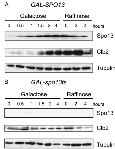
Spo13 overexpression affects the stability of Clb2:
To examine the molecular effects of Spo13 overexpres-sion further, we monitored the reversibility of changes at a biochemical level. After 4 hr on galactose, strains harboring GAL-SPO13 and GAL-spo13fs were shifted
Figure3.—Genome-wide mRNA profiling during Spo13-dependent mitotic arrest. (A and B) Heat maps and graphical displays with signal distributions for each time point, displayed in duplicate. Data are shown for overexpression of wild type (GAL-SPO13, GA-3419) at 0 hr, 2 hr, and 4 hr, and frameshift (GAL-spo13fs, GA-4318) at 4 hr. Left dots indicate genes whose expression depends upon the specific transcription factor given at bottom. Right dots indicate participation in the biological process (given at bot-tom). Log2-transformed signal intensities are color coded as given in the scale. (C) Bar diagrams summarize linear signal inten-sities (y-axis)vs. color-coded samples (x-axis) for typical cases of down- and upregulated genes, as well as control genes. The samples are described below the graphs. (D) Model of the pathways that contribute to the Spo13-dependent sequestration or Net1-dependent release of Cdc14 from the nucleolus. Diagram shows regulation of Ace2 and Swi5 transcription factors by Cdc14, which leads to the transcription of their target genes (top). Bar diagrams below show linear expression signals for four selected genes; samples are color coded as in C.
back to glucose and released into a medium containing raffinose, in which the GAL1 promoter remains re-pressed. Samples were taken at the indicated time points, and the instability of Clb2, which normally increases as cells progress through mitosis, was assayed by Western blot (Figure 5). As expected, the Spo13 protein accumulated duringGAL-SPO13induction and was not present in cells expressing the frameshift allele (spo13fs; Figure 5, A and B). Spo13 persisted, albeit at a reduced level, for 4 hr even after theGAL1promoter was repressed, consistent with it being stabilized by the M-phase Cdk. Clb2 also accumulated to high levels during Spo13-induced arrest (Figure 5A; see Lee et al. 2002)
and remained high during 4 hr on raffinose, suggesting that APC-dependent degradation of Clb2 was impaired. Neither the accumulation of Clb2 on galactose, nor its degradation once Spo13 protein levels decreased, were observed inGAL-spo13fscells (Figure 5B)
Both Cdc14 activation and a functional APC are necessary for Clb2 degradation in late mitosis (Stegmeierand Amon2004; Queraltet al. 2006; Toth et al. 2007). Thus the persistent accumulation of Clb2 in the presence of overexpressed Spo13 is consistent with the proposal that a high level of Spo13 in mitosis inhibits Cdc14 activation and directly or indirectly impairs activation of the APC.
ESP1separase overexpression bypasses the cell-cycle arrest imposed by Spo13: To determine how Spo13 might inhibit Cdc14 release, we investigated the relation-ship between overexpressed Spo13 and the separase Esp1. Besides its role in cleaving cohesin, Esp1 functions in the FEAR (Stegmeieret al. 2002) and MEN (Queralt et al. 2006) pathways, leading to the phosphorylation of Net1 and subsequent release of Cdc14 from the nucle-olus, which in turn promotes exit from mitosis. Esp1 acts in this context by inactivating PP2ACdc55 phosphatase to
allow phosphorylation of both Net1 by Cdc28 kinase (FEAR activation) and the GAP complex (Bub2/Bfa1) promoting MEN activation (Queralt et al. 2006).
In-triguingly, Esp1 overexpression is sufficient to trigger Cdc14 activation, through a mechanism independent
of its proteolytic activity on cohesin (Sullivan and
Uhlmann 2003). To distinguish whether Spo13
inter-feres with Cdc14 release by acting upstream or down-stream of Esp1, we used similar suppression analyses.
Using strains carrying eitherGAL-SPO13,GAL-ESP1, or both, we tested whetherSPO13induction overrides the release of Cdc14 provoked byGAL-ESP1induction (Tinker-Kulberg and Morgan 1999; Sullivan and
Uhlmann2003). To ensure that cells have progressed
properly through S phase without premature sister separation, we blocked cells at the G2/M boundary by exposure to nocodazole under noninducing conditions. Galactose was then added for 1 hr, cells were washed with YP raffinose, and released from nocodazole into galac-tose. Cell-cycle progression was monitored by both fluorescence imaging and Western blot analysis (Figure 6, A–D). As expected, GAL-ESP1cells passed through mitotic exit and began to rebud 40 min after release, coinciding with the degradation of Clb2. In contrast,
GAL-SPO13cells remained arrested, with high Clb2 and Spo13 levels. The persistence of metaphase and ana-phase spindles throughout the experiment in these cells agreed with our earlier results on mitotic arrest.
In contrast to overexpression of Spo13 alone, the coordinated overexpression of both Esp1 and Spo13 allowed metaphase spindles to disappear quickly de-spite the presence of high levels of Spo13. Thus, Esp1 overexpression clearly suppressed the Spo13-induced arrest at G2/M. These cells also accumulated anaphase
TABLE 2
Spo13 inhibits Cdc14 release from the nucleolus
Cdc14-Nop1 colocalization/number examined
Strain Exp G1 S G2/M
Extended nucleolus
GAL-spo13fs Live 184/184 86/86 7/7 0/5 GAL-SPO13 Live — — 66/66 12/12 GAL-spo13fs IF 43/43 45/45 22/22 0/22 GAL-SPO13 IF — — 93/93 17/20
A summary of the frequency of observed phenotypes after 2 hr of induction ofGAL-SPO13on galactose from the experi-ments shown in Figure 4, A and B. Experimental details are described in the legend to Figure 4. IF, immunostaining; live, live GFP imaging.
spindles rapidly after release from nocodazole, illustrat-ing the effect of Spo13 on anaphase/exit, which was less visible in asynchronous cells. By 80 min the anaphase spindles were disassembled, demonstrating more rapid progression in the presence of both Esp1 and Spo13, compared to the presence of Spo13 alone (Figure 6A).
Consistently, Clb2 degradation also occurred faster in strains overexpressing both genes than in cells with
GAL-SPO13 only (Figure 6B). These data indicate a suppression of the Spo13-induced arrest by Esp1, which is known to confer Cdc14 release and activation. In-terestingly, in theESP1 SPO13double-expressing strain
Figure6.—ESP1 overex-pression suppresses Spo13-induced arrest. (A) Graphs show cell counts for bud-ding index, metaphase or anaphase spindles on strains expressing GAL-SPO13, GAL-ESP1, and bothGAL-SPO13and GAL-ESP1 as well as CDC14-6HA (GA-5441, GA-5406, and GA-5230, respectively). Cells were blocked at G2/M with nocodazole and re-leased in the presence of galactose. (B) Western blot analysis of the indicated proteins at the indicated time points from the exper-iment described in A. (C and D) Representative mi-crographs showing immu-nofluorescence analysis of the indicated proteins and strains either during galac-tose induction on nocoda-zole (C) or on cells released from nocodazole arrest for 40, 60, or 80 min on galactose (D). GAL-SPO13 refers to strain GA-5441, GAL-ESP1is GA-5406, and GAL-SPO13 1
low levels of Clb2 were still detectable at 80, 100, and 120 min, consistent with incomplete APC activation.
Immunofluorescence studies performed for Cdc14 on these same cells showed that Cdc14 is released both in nocodazole-arrested and in anaphase cells in the presence of Esp1, either when expressed alone or together with Spo13 (Figure 6, C and D). As expected, cells expressing GAL-SPO13 alone accumulated ana-phase spindles, and Cdc14 release from the nucleolus was less efficient (Figure 6D). On galactose, Cdc14 was released in 97% of cells when Esp1 was overexpressed alone, in 20% of cells when Spo13 was overexpressed alone, and in 56% of cells when both were expressed. Given that the expression of Esp1 overrides the Spo13-induced arrest at both stages of mitosis, and reverses the Spo13-induced sequestration of Cdc14, these data strongly suggest that Spo13 acts upstream of Esp1 (or in parallel to it) to regulate both the FEAR (G2/M) and MEN (A/exit) pathways.
Finally, to test whether Spo13 localization provides any further insight into its mode of action, we monitored the subcellular distribution of overexpressed Spo13 in living cells. At 2 hr of induction ofGAL-SPO13-GFP80% of the Spo13-GFP foci colocalized with the nucleolar marker Nop1 (n ¼ 120; Figure 6E, right), providing novel evidence for its association with the nucleolus. The distribution of the Spo13 signal was nonetheless distinct from the uniform nucleolar staining of Cdc14 (Figure 6E). By 3 hr of induction multiple large nuclear foci of Spo13-GFP were observed, which did not colocalize with either the mitotic spindle or the nucleolus (Figure 6F). The significance of this localization with respect to Esp1 and Cdc14 activation remains unclear.
Spo13 interferes with the APC pathway: The above data suggest that Spo13 may act upstream of separase (Esp1) to impair the release of Cdc14 from its inhibitor Net1, which is required for progression through mitosis. Given that separase is activated by APC cleavage of securin (Pds1) (Cohen-Fix et al. 1996), it seems a
plausible hypothesis that APC function is diminished in the presence of Spo13 (Katiset al. 2004). To test this,
we investigated the stability of securin (Pds1), a key APC substrate, during Spo13 overexpression. Pds1 was tagged with 18-Myc epitopes in strains carrying GAL-SPO13orGAL-spo13fs.After 4 hr of galactose induction, samples were taken and processed for immunofluores-cence (Figure 7A). In thespo13-fsstrain, Pds1 was present in G2/M cells but not detectable in cells at anaphase. In contrast, Pds1 was detectable at all cell-cycle phases in cells overexpressing Spo13. Intriguingly, cells with long spindles usually showed a heterogeneous pattern, with Pds1 signal being stronger in one of the two stretched nuclei. This suggests that Pds1 is partially degraded over time and/or is being resynthesized in a nucleus that has progressed further in the exit pathway.
We examined this further by studying Pds1 stability in synchronized cultures. TheGAL-SPO13andGAL-spo13-fs
strains were grown in YP1raffinose to reach mid-log phase (53106cells/ml), at which point, nocodazole was
added for 1.5 hr to block cells in G2/M. Cells were then washed twice with YP-raffinose, released into drug-free YP-galactose to induce Spo13 expression, and samples analyzed every 30 min and analyzed by Western blot (Figure 7B). In the control strain expressing GAL-spo13fs, Pds1 levels started to drop 60 min after release from nocodazole and were undetectable by 90 min. In
contrast, significant amounts of stabilized Pds1 (30– 50% of the initial level) were seen in cells expressing Spo13. These results confirm that normal degradation of both Pds1 and Clb2, two key substrates for APC, is inhibited by Spo13 in mitotic cells, providing further support for the notion that Spo13 either directly or indirectly inhibits APC function (Katiset al. 2004).
DISCUSSION
It has been proposed that Spo13 acts as a transient negative regulator or timer, slowing meiotic events to (1) allow proper establishment of the reductional machin-ery for centromere cohesion/coorientation prior to the first M phase, allowing homologs to segregate properly, and (2) impede the anaphase to MI exit, thereby ensuring two successive meiotic divisions without an intervening S phase. This permits haploid products to form (McCarroll and Esposito1994). The present
study exploited the mitotic overexpression phenotype of Spo13 to probe Spo13’s mechanism of action in regu-lating cell cycle progression.
When expressed in mitotically dividing cells, the Spo13 protein was stabilized in both S- and M-phase cells, and induced a cell-cycle arrest that is largely independent of the spindle checkpoint gene MAD2. Importantly, Spo13 does not accumulate to detectable levels in G1-arrested cells, and Spo13-induced arrest can be reversed by repressing Spo13 expression or pro-voking Spo13 degradation through inhibition of the Cdc28/Clb kinase complex. The use of an analog-sensitive Cdc28 gene suggests that Spo13 stability is directly dependent on the mitotic activity of the Cdk/ Clb complex. Indeed, Spo13, which has 10 SP/TP consenses, is phosphorylated (Figure 2). This would argue for a reciprocal positive feedback loop between Spo13 and Cdc28. Namely, SPO13 overexpression re-sults in the accumulation of Clb2 and an enhancement of Cdc28/Clb activity, which in turn stabilize Spo13 (McCarrolland Esposito1994). Consistent with this
model, a recent report suggests that Spo13 is a target of APC, which may degrade unphosphorylated Spo13 in G1 (Sullivanand Morgan2007).
Our data provide compelling evidence that Spo13 delays mitotic cell-cycle progression at two different points: at the G2/M transition and late in anaphase. The effect at anaphase is particularly clear if Spo13 is induced in synchronized cells released from a metaphase arrest. We see enrichment of anaphase spindles and poor degradation of Pds1 securin by both immunofluores-cence and Western blot analysis. These findings suggest that the level of Pds1 degradation that occurs in the presence of Spo13 is insufficient to allow the Esp1-mediated cohesin cleavage needed for the G2/M transi-tion, as well as downregulation of PP2ACdc55, which is
necessary for Cdc14 release from the nucleolus and the segregation of rDNA at anaphase (Sullivanet al. 2004).
Genome-wide microarray analysis provided useful clues as to how Spo13 overexpression might arrest cells in late anaphase. We observed progressively decreasing mRNA concentrations for a number of cell cycle-regulated genes that are under the control of Ace2 and Swi5 transcription factors, including some cases where the mRNAs became undetectable in arrested cells. This is not seen in other G2/M-arrested cells. Both Ace2 and Swi5, which are substrates of the Cdc14 phosphatase, require dephosphorylation during late anaphase to activate their function (F. Uhlmann,
personal communication). This led us to examine whether Spo13 might inhibit A/exit by negatively regu-lating Cdc14. Indeed, we find that ectopic expression of Spo13 prevents the release of Cdc14 from the nucleolus, an event that correlates with Cdc14 activation and subsequently with exit from mitosis (Stegmeier et al.
2002; Buonomoet al. 2003; Marstonet al. 2003). Given
that the proper balance of Cdc14 and Cdk/Clb activities is known to be crucial for triggering mitotic exit (Toth et al. 2007), we conclude that high levels of Spo13 must also ultimately interfere with this balance.
Of special relevance for interpreting the effects of Spo13 is the finding that separase (Esp1) plays a key role in coordinating cohesin cleavage at G2/M with subsequent Cdc14 release during anaphase. Esp1 not only cleaves Scc1 releasing cohesion, but also down-regulates PP2ACdc55, thus allowing Net1 phosphorylation
(Queralt et al. 2006) and Cdc14 release (Shou et al.
1999; Visintinet al. 1999). On the basis of this finding,
we tested whether the effects of Spo13 overexpression might act in part by reducing Esp1 function. Indeed,
ESP1overexpression can bypass the Spo13-induced G2/ M arrest as well as the nucleolar retention of Cdc14, arguing that Spo13 either acts upstream or in parallel to Esp1 function. Since ESP1 overexpression also overcomes metaphase arrest in the absence of APC (Uhlmann et al. 2000), our data suggest that Spo13
may modulate Esp1 activity by inhibiting APC (see also Lee et al. 2002; Katis et al. 2004). We tested this by
monitoring the degradation of a key APC target, securin, or Pds1. We could confirm that high levels of Spo13 impair Pds1 degradation. This in turn would prevent complete activation of separase (Esp1), which is needed for both anaphase progression and exit from mitosis.
promotes exit from mitosis by allowing dephosphoryla-tion of the APC regulator Cdh1 as well as a set of late-anaphase substrates (Visintinet al. 1998; Stegmeieret al.
2002; D’amourset al. 2004; Stegmeierand Amon2004;
Sullivanet al. 2004; Torres-Rosellet al. 2005; Queralt et al. 2006; Khmelinskiiet al. 2007; Tothet al. 2007). The
failure to release Cdc14 in cells overexpressing Spo13 and the massive accumulation of Clb2 in late anaphase in these cells suggests that Spo13 also negatively regulates anaphase by altering the late mitotic balance between Cdc14 and Cdc28/Clb2 activities as indicated earlier.
How does this mechanism relate to Spo13’s role in meiotic division? As noted earlier, in meiosis Spo13 is required for homolog separation and two successive M phases, MI and MII, without an intervening S phase. The meiotic Spo13 protein is detected specifically during MI when its transcript level peaks, and it is de-graded by APC at the end of MI. Loss ofSPO13function causes diploid cells (or haploids expressing both mating type alleles), to undergo a single, largely equational, division on the MI spindle and exit the cell cycle forming dyads with two diploid (or haploid) spores (Klapholzand Esposito1980b; Wagstaffet al. 1982;
Rutkowski and Esposito2000). The termination of
meiosis after a single division, in which many chromo-somes separate sister chromatids instead of homologs, appears to depend on the Mad2 spindle checkpoint (Shonn et al. 2000). The question remains, however,
why wild-type Spo13 cells, which undergo a reductional MI, execute a second M phase without first exiting division and initiating another round of DNA replica-tion. We propose that Spo13 may be able to promote MII immediately after MI, by preventing the complete loss of Clb/Cdk activity and the A/exit transition (Queralt et al. 2006; Toth et al. 2007). Only after
mitotic exit can prereplication complexes assemble to allow a subsequent round of DNA replication. The ability of Spo13 to prevent a second S phase by keeping Clb/Cdk activity high is consistent with the fact that overexpression ofSPO13in thecdc28-1mutant at a semi-permissive temperature (30°) allowed MII to immedi-ately follow MI in meiotic cells as described earlier (Shusterand Byers1989; McCarrolland Esposito
1994). In this case, we speculate that Spo13 modulates Cdk activity by maintaining high cyclin B levels (Figure 6) and preventing full Cdc14 release from the nucleo-lus. As discussed above, this could be achieved by interfering with APC function, although proof of this requires future analysis. Consistent with this model, we note that yeast cyclins have been detected during mei-osis throughout MI and MII (Carlileand Amon2008).
The control of two successive chromosome segrega-tion events (MI and MII) without an intervening S phase is a fundamental feature of meiosis, which is conserved from yeast to man. The fact that in meiotically dividing frog oocytes cyclin B appears not to be degraded between MI and MII (Taiebet al. 2001) provides a tantalizing hint
that the effects of Spo13 on anaphase and exit from mitosis in yeast and could well provide a plausible model for how organisms universally control this crucial event.
We thank P. Demougin and M. Cartron for help with yeast array experiments, M. Rebhan for protein motif analysis and F. Uhlmann for kindly providing strains that were used to generate those used here. Finally, R.E.E. extends special thanks to S.M.G. and M.P. for graciously hosting her during several sabbatical stays in Basel. E.V. was supported by the Swiss Cancer League, the Novartis Research Foundation, and a short-term European Molecular Biology Organization fellowship. This work was further funded by National Institutes of Health GM-29182 (to R.E.E.) and Swiss National Science Foundation grant 3100A0-105861 (to M.P.).
LITERATURE CITED
Azzam, R., S. L. Chen, W. Shou, G. Alexandru, K. Nasmyth
et al., 2004 Phosphorylation by cyclin B-Cdk underlies release of mitotic exit activator Cdc14 from the nucleolus. Science305:
516–519.
Bishop, A. C., J. A. Ubersax, D. T. Petsch, D. P. Matheos, N. S. Gray
et al., 2000 A chemical switch for inhibitor-sensitive alleles of any protein kinase. Nature407:395–401.
Brown, N. R., M. E. Noble, J. A. Endicottand L. N. Johnson, 1999 The structural basis for specificity of substrate and recruit-ment peptides for cyclin-dependent kinases. Nat. Cell Biol. 1:
438–443.
Buckingham, L. E., H. T. Wang, R. T. Elder, R. M. McCarroll, M. R. Slateret al., 1990 Nucleotide sequence and promoter analysis of SPO13, a meiosis-specific gene of Saccharomyces cerevisiae. Proc. Natl. Acad. Sci. USA87:9406–9410.
Buonomo, S. B., K. P. Rabitsch, J. Fuchs, S. Gruber, M. Sullivan
et al., 2003 Division of the nucleolus and its release of CDC14 during anaphase of meiosis I depends on separase, SPO12, and SLK19. Dev. Cell4:727–739.
Bystricky, K., T. Laroche, G.vanHouwe, M. Blaszczykand S. M. Gasser, 2005 Chromosome looping in yeast: telomere pairing and coordinated movement reflect anchoring efficiency and territorial organization. J. Cell Biol.168:375–387.
Carlile, T. M., and A. Amon, 2008 Meiosis I is established through division-specific translational control of a cyclin. Cell133:280– 291.
Chalmel, F., and M. Primig, 2008 The Annotation, Mapping, Expression and Network (AMEN) suite of tools for molecular systems biology. BMC Bioinformatics9:86.
Cho, R. J., M. J. Campbell, E. A. Winzeler, L. Steinmetz, A. Conway
et al., 1998 A genome-wide transcriptional analysis of the mi-totic cell cycle. Mol. Cell2:65–73.
Cohen-Fix, O., J. M. Peters, M. W Kirschnerand D. Koshland, 1996 Anaphase initiation in Saccharomyces cerevisiae is con-trolled by the APC-dependent degradation of the anaphase in-hibitor Pds1p. Genes Dev.10:3081–3093.
D’Amours, D., F. Stegmeierand A. Amon, 2004 Cdc14 and conden-sin control the dissolution of coheconden-sin-independent chromosome linkages at repeated DNA. Cell117:455–469.
Gattiker, A., C. Niederhauser-Wiederkehr, J. Moore, L. Hermida and M. Primig, 2007 The GermOnline cross-species systems browser provides comprehensive information on genes and gene products relevant for sexual reproduction. Nucleic Acids Res.35:
D457–D462.
Geymonat, M., S. Jensenand L. H. Johnston, 2002 Mitotic exit: the Cdc14 double cross. Curr. Biol.12:R482–R484.
Gotta, M., T. Laroche, A. Formenton, L. Maillet, H. Scherthan
et al., 1996 The clustering of telomeres and colocalization with Rap1, Sir3, and Sir4 proteins in wild-type Saccharomyces cerevi-siae. J. Cell Biol.134:1349–1363.
Granovskaia, M. V., L. J. Jensen, M. E. Ritchie, J. Toedling, Y. Ning
et al., 2010 High-resolution transcription atlas of the mitotic cell cycle in budding yeast. Genome Biol.11:R24.
sporulation-specific genes in Saccharomyces cerevisiae. Mol. Cell. Biol.18:5750–5761.
Hochwagen, A., G. Wrobel, M. Cartron, P. Demougin, C. Niederhauser-Wiederkehret al., 2005 Novel response to micro-tubule perturbation in meiosis. Mol. Cell. Biol.25:4767–4781. Jolly, E. R., C. S. Chin, I. Herskowitzand H. Li, 2005
Genome-wide identification of the regulatory targets of a transcription factor using biochemical characterization and computational ge-nomic analysis. BMC Bioinformatics6:275.
Katis, V. L., J. Matos, S. Mori, K. Shirahige, W. Zachariaeet al., 2004 Spo13 facilitates monopolin recruitment to kinetochores and regulates maintenance of centromeric cohesion during yeast meiosis. Curr. Biol.14:2183–2196.
Khmelinskii, A., C. Lawrence, J. Roostalu and E. Schiebel, 2007 Cdc14-regulated midzone assembly controls anaphase B. J. Cell Biol.177:981–993.
Klapholz, S., and R. E. Esposito, 1980a Isolation of SPO12–1 and SPO13–1 from a natural variant of yeast that undergoes a single meiotic division. Genetics96:567–588.
Klapholz, S., and R. E. Esposito, 1980b Recombination and chro-mosome segregation during the single division meiosis in SPO12–1 and SPO13–1 diploids. Genetics96:589–611. Klein, F., P. Mahr, M. Galova, S. B. Buonomo, C. Michaeliset al.,
1999 A central role for cohesins in sister chromatid cohesion, formation of axial elements, and recombination during yeast meiosis. Cell98:91–103.
Lee, B. H., A. Amonand S. Prinz, 2002 Spo13 regulates cohesin cleavage. Genes Dev.16:1672–1681.
Lee, B. H., B. M. Kiburzand A. Amon, 2004 Spo13 maintains cen-tromeric cohesion and kinetochore coorientation during meiosis I. Curr. Biol.14:2168–2182.
Longtine, M. S., A. McKenzie, III, D. J. Demarini, N. G. Shah, A. Wachet al., 1998 Additional modules for versatile and econom-ical PCR-based gene deletion and modification in Saccharomyces cerevisiae. Yeast14:953–961.
Marston, A. L., B. H. Leeand A. Amon, 2003 The Cdc14 phospha-tase and the FEAR network control meiotic spindle disassembly and chromosome segregation. Dev. Cell4:711–726.
McCarroll, R. M., and R. E. Esposito, 1994 SPO13 negatively regulates the progression of mitotic and meiotic nuclear division in Saccharomyces cerevisiae. Genetics138:47–60.
McCord, R., M. Pierce, J. Xie, S. Wonkatal, C. Mickel et al., 2003 Rfm1, a novel tethering factor required to recruit the Hst1 histone deacetylase for repression of middle sporulation genes. Mol. Cell. Biol.23:2009–2016.
O’Conallain, C., M. T. Doolin, C. Taggart, F. Thorntonand G. Butler, 1999 Regulated nuclear localisation of the yeast tran-scription factor Ace2p controls expression of chitinase (CTS1) in Saccharomyces cerevisiae. Mol. Gen. Genet.262:275–282. Pak, J., and J. Segall, 2002 Regulation of the premiddle and
mid-dle phases of expression of the NDT80 gene during sporulation of Saccharomyces cerevisiae. Mol. Cell. Biol.22:6417–6429. Queralt, E., C. Lehane, B. Novakand F. Uhlmann, 2006
Down-regulation of PP2A(Cdc55) phosphatase by separase initiates mi-totic exit in budding yeast. Cell125:719–732.
Rabitsch, K. P., M. Petronczki, J. P. Javerzat, S. Genier, B. Chwallaet al., 2003 Kinetochore recruitment of two nucle-olar proteins is required for homolog segregation in meiosis I. Dev. Cell4:535–548.
Rutkowski, L. H., and R. E. Esposito, 2000 Recombination can partially substitute for SPO13 in regulating meiosis I in budding yeast. Genetics155:1607–1621.
Schlecht, U., I. Erb, P. Demougin, N. Robine, V. Borde et al., 2008 Genome-wide expression profiling, in vivo DNA binding analysis and probabilistic motif prediction reveal novel Abf1 tar-get genes during fermentation, respiration and sporulation in yeast. Mol. Biol. Cell19:2193–2207.
Shonn, M. A., R. McCarrolland A. W. Murray, 2000 Requirement of the spindle checkpoint for proper chromosome segregation in budding yeast meiosis. Science289:300–303.
Shonn, M. A., R. McCarrolland A. W. Murray, 2002 Spo13 protects meiotic cohesin at centromeres in meiosis I. Genes Dev.16:1659– 1671.
Shou, W., J. H. Seol, A. Shevchenko, C. Baskerville, D. Moazed
et al., 1999 Exit from mitosis is triggered by Tem1-dependent release of the protein phosphatase Cdc14 from nucleolar RENT complex. Cell97:233–244.
Shuster, E. O., and B. Byers, 1989 Pachytene arrest and other mei-otic effects of the start mutations in Saccharomyces cerevisiae. Genetics123:29–43.
Stegmeier, F., and A. Amon, 2004 Closing mitosis: the functions of the Cdc14 phosphatase and its regulation. Annu. Rev. Genet.38:
203–232.
Stegmeier, F., R. Visintin and A. Amon, 2002 Separase, polo kinase, the kinetochore protein Slk19, and Spo12 function in a network that controls Cdc14 localization during early ana-phase. Cell108:207–220.
Sullivan, M., and F. Uhlmann, 2003 A non-proteolytic function of the separase links the onset of anaphase to mitotic exit. Nat. Cell Biol.5:249–254.
Sullivan, M., T. Higuchi, V. L. Katisand F. Uhlmann, 2004 Cdc14 phosphatase induces rDNA condensation and resolves cohesin-independent cohesion during budding yeast anaphase. Cell
117:471–482.
Sullivan, M., and D. O. Morgan, 2007 A novel destruction sequence targets the meiotic regulator Spo13 for anaphase-promoting complex-dependent degradation in anaphase I. J. Biol. Chem.282:19710–19715.
Surosky, R. T., and R. E. Esposito, 1992 Early meiotic transcripts are highly unstable in Saccharomyces cerevisiae. Mol. Cell. Biol.
12:3948–3958.
Taieb, F. E., S. D. Gross, A. L. Lewellyn and J. L. Maller, 2001 Activation of the anaphase-promoting complex and deg-radation of cyclin B is not required for progression from meiosis I to II in Xenopus oocytes. Curr. Biol.11:508–513.
Tinker-Kulberg, R. L., and D. O. Morgan, 1999 Pds1 and Esp1 control both anaphase and mitotic exit in normal cells and after DNA damage. Genes Dev.13:1936–1949.
Torres-Rosell, J., F. Machinand L. Aragon, 2005 Cdc14 and the temporal coordination between mitotic exit and chromosome segregation. Cell Cycle4:109–112.
Toth, A., K. P. Rabitsch, M. Galova, A. Schleiffer, S. B. Buonomo
et al., 2000 Functional genomics identifies monopolin: a kinetochore protein required for segregation of homologs dur-ing meiosis I. Cell103:1155–1168.
Toth, A., E. Queralt, F. Uhlmannand B. Novak, 2007 Mitotic exit in two dimensions. J. Theor. Biol.248:560–573.
Uhlmann, F., D. Wernic, M. A. Poupart, E. V. Koonin and K. Nasmyth, 2000 Cleavage of cohesin by the CD clan protease separin triggers anaphase in yeast. Cell103:375–386.
Varela, E., K. Shimada, T. Laroche, D. Leroyand S. M. Gasser, 2009 Lte1, Cdc14 and MEN-controlled Cdk inactivation in yeast coordinate rDNA decompaction with late telophase pro-gression. EMBO J.28:1562–1575.
Visintin, R., K. Craig, E. S. Hwang, S. Prinz, M. Tyers et al., 1998 The phosphatase Cdc14 triggers mitotic exit by reversal of Cdk-dependent phosphorylation. Mol. Cell2:709–718. Visintin, R., E. S. Hwangand A. Amon, 1999 Cfi1 prevents
prema-ture exit from mitosis by anchoring Cdc14 phosphatase in the nucleolus. Nature398:818–823.
Wagstaff, J. E., S. Klapholzand R. E. Esposito, 1982 Meiosis in haploid yeast. Proc. Natl. Acad. Sci USA79:2986–2990. Wang, H. T., S. Frackman, J. Kowalisyn, R. E. Esposito and R.
Elder, 1987 Developmental regulation of SPO13, a gene re-quired for separation of homologous chromosomes at meiosis I. Mol. Cell. Biol.7:1425–1435.
Williams, R. M., M. Primig, B. K. Washburn, E. A. Winzeler, M. Bellis
et al., 2002 The Ume6 regulon coordinates metabolic and meiotic gene expression in yeast. Proc. Natl. Acad. Sci. USA99:13431–13436. Xie, J., M. Pierce, V. Gailus-Durner, M. Wagner, E. Winteret al., 1999 Sum1 and Hst1 repress middle sporulation-specific gene expression during mitosis in Saccharomyces cerevisiae. EMBO J.
18:6448–6454.