J. Exp. Med. The Rockefeller University Press • 0022-1007/2002/12/1515/7 $5.00 Volume 196, Number 11, December 2, 2002 1515–1521
Brief Definitive Report
1515
Adhesion Molecule–dependent Mechanisms Regulate the
Rate of Macrophage Clearance During the Resolution of
Peritoneal Inflammation
Geoffrey John Bellingan,
1Ping Xu,
1Helen Cooksley,
1Helen Cauldwell,
2Anthony Shock,
3Stephen Bottoms,
1Christopher Haslett,
2Steven Eugene Mutsaers,
1and Geoffrey John Laurent
11Centre for Respiratory Research, Rayne Institute, University College London, London WC1E 6JJ,
United Kingdom
2Rayne Laboratories, Edinburgh EH3 9YW, United Kingdom 3Celltech R&D, Slough SL1 4EN, United Kingdom
Abstract
Macrophage clearance is essential for the resolution of inflammation. Much is known about how monocytes enter the inflammatory site but little is known about how resultant macro-phages are cleared. We have previously demonstrated that macrophage clearance from resolving peritonitis occurs by emigration into draining lymphatics rather than local apoptosis. We now examine mechanisms for this process, in particular by evaluating the hypothesis that modulation of adhesion interactions between macrophages and cells lining the lymphatics regulates the rate of macrophage clearance. We demonstrate in vivo that macrophages adhere specifically to me-sothelium overlying draining lymphatics and that their emigration rate is regulated by the state of macrophage activation. We observed that macrophage–mesothelial adhesion is Arg-Gly-Asp
(RGD) sensitive and partially mediated by very late antigen (VLA)-4 and VLA-5 but not v or
2 integrins. Moreover, macrophage clearance into lymphatics can be blocked in vivo by RGD
peptides and VLA-4 and VLA-5 but not 2 blocking antibodies. This is the first evidence that
macrophage emigration from the inflamed site is controlled and demonstrates that this is exerted through specific adhesion molecule regulation of macrophage–mesothelial interactions. It high-lights the importance of adhesion molecules governing entry of cells into the lymphatic circula-tion, thus opening a new avenue for manipulating the resolution of inflammation.
Key words: peritonitis • integrins • lymphatic system • cell movement • receptors-very late antigen
Introduction
The mechanisms regulating the resolution of inflammation have been studied in detail for the neutrophil (1). How-ever, the role of the macrophage in this process has ceived little attention. Macrophage clearance in acute re-solving peritonitis is known to occur by emigration to the draining lymphatics rather than by local apoptosis, but the mechanisms regulating this are unknown (2). Kinetic stud-ies on macrophages within the peritoneum imply that resi-dent and inflammatory macrophages have dramatically different half-lives, which suggests that macrophage
clear-ance might be regulated, a concept that has not been previ-ously investigated (2, 3). Macrophages are central to effec-tive wound healing (4). However, their accumulation is also a hallmark of chronic inflammation where they can potentiate tissue damage (5). Moreover, the elimination of macrophages from sites of chronic inflammation can lead rapidly to resolution of inflammation (6). Hence, an under-standing of the process of macrophage emigration from the inflamed site is essential as it may impact directly upon the speed of inflammatory resolution and tissue injury.
In peritonitis, monocyte influx occurs directly across the encompassing peritoneal mesothelial lining and is regulated by adhesion molecules expressed both on the vascular en-dothelium and mesothelial cells (7). Efflux is believed to occur through milky spots found mainly on the
subdia-Address correspondence to Geoffrey Bellingan, Centre for Respiratory Research Rayne Institute, Department of Medicine, University Street, London WC1E 6JJ, United Kingdom. Phone: 44 207-679-6972; Fax:
44 207-679-6973; E-mail: [email protected]
on December 5, 2008
jem.rupress.org
Downloaded from
phragmatic surface and omentum (2). These provide small openings in the mesothelial barrier and are intimately asso-ciated with the draining lymphatics (8).
From these observations, we hypothesized that in the resolution of peritonitis, specific adhesion molecules regu-late macrophage adhesion to the mesothelium overlying the draining peritoneal lymphatics before their emigration. In addition, this adherence and transmigration provides a regulatory mechanism governing the rate of macrophage clearance. We examined the extent to which murine mac-rophage clearance is regulated in vivo then, using a novel ex vivo system, Arg-Gly-Asp (RGD) peptides (9) and blocking antibodies, determined the adhesion molecules involved. The role of these molecules in regulating mac-rophage clearance was finally tested in our in vivo peritoni-tis model.
Materials and Methods
Reagents and Antibodies. All reagents, unless stated otherwise, were obtained from Sigma-Aldrich, plasticware and antibodies were from BD Biosciences, and culture media were from Life Technologies. The following antibodies were used: CD11a (M17/4), CD11b (M1/70, 5C6), CD18 (GAME-46, 2E6; American Type Culture Collection), CD49d (PS/2, R1-2, 9C10), CD49e (5H10-27), CD51 (H9.2B8), and F4/80 (Sero-tec). FITC-conjugated CD11c (HL3) and biotinylated Iad (MHC
class II) were provided by D. Katz, University College London, London, United Kingdom. Isotype controls were: IgG1 DNP-1; Serotec), IgG2a DNP-16; Serotec), IgG2b (LO-DNP-11; Serotec), hamster IgM and IgG, and R-phycoerythrin– conjugated F(ab’)2 goat anti–rat second layer (Star 73; Serotec).
Antibodies for adhesion and in vivo studies were azide free and low endotoxin.
In Vivo Studies of Regulation of Macrophage Clearance. BALB/c (H-2d) and C3HF/KAM (H-2k) mice (22–28 g) were used for
joint resident/inflammatory macrophage studies. All other exper-iments used ICR mice (Harlan).
Comparison of Resident and Inflammatory Macrophage Clearance. Intraperitoneal injection of 1 ml 4% thioglycollate (TG; Difco) induced a sterile peritonitis with leukocyte numbers (93% mac-rophages) peaking on day 5 and declining to baseline by day 10. H-2d peritoneal phagocytes were labeled in vivo by 1 ml 0.5 M
red fluorescent marker PKH26-PCL intraperitoneally 5 d after TG2. This labeled 87% of phagocytes and was cleared rapidly so
no label was acquired by fresh H-2k macrophages instilled 2 h
later and was distinguished by flow cytometry using FITC-conju-gated anti–H-2Dd IgM and FITC-conjugated anti–H-2Kk IgG2a
with FITC-conjugated anti-CD15 IgM and FITC-conjugated anti-CD45RO IgG2a as controls (Dako). Resident phagocytes were labeled in vivo by 1 ml 0.5 M green fluorescent dye PKH2-PCL intraperitoneally in the absence of TG.
Fluorescent resident and inflammatory macrophages were lav-aged, mixed in a 1:1 ratio of red/green cells (confirmed by flow cytometry) and 2.5 106 or 1.25 107 cells instilled
intraperito-neally into mice with no inflammation or 5 d after TG. 3 or 10 d later abdominal wall, spleen, liver, kidney, aortic and para-thymic lymph nodes, and heart and lung were harvested for fro-zen sectioning. Macrophage totals in peritoneal lavage were de-termined from cell counts and differential counts from cytospins, counting 400 cells per slide. Resident and inflammatory
mac-rophage numbers were determined by dual color flow cytometry from percent of green and red cells in gates generated on CD11b and F4/80 cells. Flow cytometry demonstrated 2.5% of the PKH26-PCL–labeled population was CD11c whereas biotin-ylated staining showed 8% positive for MHC class II antigen. Therefore, few of the fluorescent cells migrating to the lymph nodes late in inflammation were of dendritic phenotype.
In Vivo Modulation of Inflammatory Macrophage Clearance. 1 ml of test agents (GRGDTP peptide [RGD], control RGES peptide [RGE], PBS, or antibodies) were injected intraperitoneally 2 and 26 h after fluorescently labeling day 5 macrophages. 24 h later cells were lavaged, macrophages counted, and adhesion molecule surface expression determined by indirect immunofluorescence. Mesothelial lining imprints of cecum, spleen, and liver were pre-pared by applying air-dried tissue onto 5% gelatin-coated slides and labeled macrophages on these imprints were quantified blindly by two observers. Parathymic lymph nodes were snap fro-zen and paired and serial 7-m frozen sections were cut at 280
m intervals spanning the whole node. Sections were scored blindly from 0 (no labeled cells) to 8 (maximum labeled cells) by two observers using predefined control slides. In all cases interob-server variability was 15% and the distribution of cells within nodes was unaffected by treatments.
To outline lymphatics, TG challenged mice were injected in-traperitoneally with 1 ml India ink or PBS. 30 min later PKH26-PCL–labeled macrophages were instilled and 1 h later the dia-phragm was dissected, washed, and the distribution of India ink and macrophages was determined.
Ex Vivo Macrophage–Mesothelial Adhesion. Macrophages from mice 5 d after TG, were labeled in vitro using 5-chlorometh-ylfluorescein diacetate (Cambridge Bioscience). Diaphragms from the same mice were washed, clamped in customized rings (5 mm internal diameter retaining 100 l above and 75 l below the surface), and orientated with peritoneal mesothelial surface up-permost. Rings were incubated with test medium for 30 min at 37C while 1.5–3 104 labeled cells were preblocked by rotating
with test medium for 30 min at 4C before being added to the upper chamber in Dulbecco’s phosphate-buffered saline. After 1 h, the rings were washed gently to remove nonadherent cells and adherent cells were counted. Mesothelial integrity was confirmed by en face silver staining of intercellular junctions as previously described (10). To identify lymphatics in ex vivo diaphragmatic tissue, 100 l media containing 2 l India ink was added to the upper chamber, incubated for 30 min, and washed before the ad-dition of labeled cells.
Determination of Apoptosis. To determine RGD-induced apop-tosis, cells lavaged 5 d after TG were incubated in Iscove’s modi-fied DMEM supplemented with 10% FCS, l-glutamine, 100 U penicillin, and streptomycin in 8-well slides at 1.5 105 cells/
well and up to 0.5 mg/ml RGD or RGE. Cells were examined for up to 48 h and apoptosis was quantified by nuclear morphol-ogy and staining with acridine orange (10 g/ml).
Statistics. Data are expressed as mean SEM with compari-son by two-tailed Mann-Whitney U test. Differences were con-sidered significant if P 0.05.
Results
Macrophage Emigration Is Regulated and Depends on
Activa-tion State. Clearance rates of noninflamed (resident) and
inflammatory macrophages in the presence or absence of inflammation were compared by instilling equal numbers
on December 5, 2008
jem.rupress.org
Downloaded from
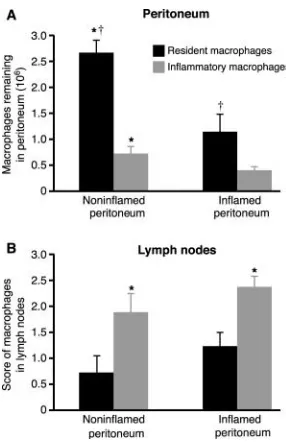
into the peritoneum in the presence/absence of resolving peritonitis and determining the number remaining after 3 (Fig. 1 A) and 10 d. Recovery of resident macrophages al-ways exceeded that of inflammatory macrophages with over three and a half times more resident than inflamma-tory macrophages recovered from the noninflamed
perito-neum (2.65 0.25 106 vs. 0.72 0.14 106, P
0.005) and nearly three times more from the inflamed
peri-toneum (1.14 0.3 106 vs. 0.4 0.07 106, P
0.05). Resolving peritonitis dramatically augmented the clearance of all macrophages with significantly more
resi-dent (P 0.005) and inflammatory (P 0.05)
macro-phages recovered from the noninflamed than the inflamed peritoneum after 3 d. Differences between resident and in-flammatory macrophage recovery persisted when examined
after 10 d (0.58 0.2 106 vs. 0.03 0.03 106, P
0.005 from noninflamed and 0.26 0.07 106 vs. 0.013
0.007 106, P 0.01 from inflamed peritoneum; n
6/group). These differences were maintained when
five-fold fewer cells were transferred (n 3/group,
unpub-lished data).
Fig. 1 B demonstrates that reduced peritoneal recovery of inflammatory macrophages was due to faster emigration into the draining lymphatics as there were always signifi-cantly more inflammatory than resident macrophages in the
parathymic nodes. Analysis of the peritoneal lining of the
abdominal wall, spleen, liver, kidney, and para-aortic lymph nodes for fluorescent cells verified that differences were not due to widespread nonspecific intra-abdominal adhesion. Consistent with reduced clearance rates in vivo, resident macrophage adhesion to noninflamed
mesothe-lium was only 71 4.1% (n 4, P 0.05) that of
inflam-matory macrophage adhesion to the inflamed mesothelium when examined ex vivo.
Macrophage–Mesothelial Adhesion Localizes to Areas
Overly-ing DrainOverly-ing Lymphatics. Macrophage–mesothelial
adher-ence was patchy and markedly reduced over the mesothe-lium lining the tendinous compared with the muscular portion of the diaphragm, both when adhesion was exam-ined in vivo or ex vivo. Adherence was localized to the In-dia ink–positive areas suggesting focal adhesion to particu-lar mesothelial cells or structures associated with the draining lymphatics. Macrophage binding to India ink–free
areas was minimal (n 3, Fig. 2 A). India ink outlined
lymphatic vessels under the diaphragmatic mesothelial sur-face that contained emigrating macrophages by 1 h (Fig. 2 B). India ink stained multiple small patchy areas to which macrophages commonly adhered as well as classic milky spot structures to which macrophages always adhered strongly (Fig. 2, C and D). Macrophage adhesion was simi-larly patchy in the absence of India ink, which suggested this was not an artifact induced by the India ink particles.
Very Late Antigen (VLA)-4 and VLA-5 Integrins Regulate
Macrophage–Mesothelial Adhesion Ex Vivo. Fig. 3 A(i)
dem-onstrates that inflammatory macrophage adhesion was
reduced by 70.6 6% in the absence of divalent cations
(P 0.0004), 50.5 5.4% by RGD (P 0.05), 29.2
8% by blocking the 4 chain of VLA-4 (PS/2; P 0.05),
and confirmed by another combination of anti-4
antibod-Figure 1. Macrophage emigration is regulated. A total of 12.5 106
fluorescent green–labeled resident and red-labeled inflammatory mac-rophages were instilled into the peritoneal cavity of mice in the absence of peritonitis (noninflamed peritoneum) or mice with resolving peritonitis (inflamed peritoneum). (A) The number of resident (black bars) and in-flammatory (gray bars) macrophages recovered from the peritoneal cavity after 3 d is shown. † significantly greater resident than inflammatory mac-rophage recovery (P 0.05, n 8/group). *, significantly greater recov-ery from noninflamed than inflamed peritoneum (P 0.05). (B) Fluores-cent-labeled macrophages in draining parathymic lymph nodes from the same mice. *, significantly more inflammatory than resident macrophages (P 0.05, n 8/group).
Figure 2. Macrophage–mesothelial adhesion is localized to regions overlying the draining lymphatics. (A) Ex vivo preparation of mesothelial surface of diaphragm with lymphatics localized by India ink. Adherence of inflammatory macrophages localizes to areas of mesothelium stained with India ink whereas virtually no macrophages adhere to India ink–free areas. Similar colocalization is seen when adhesion occurs in vivo. (B) Macrophages emigrating into draining lymphatics within 1 h of instilla-tion. (C) India ink labeling of a milky spot and (D) the same section un-der fluorescent microscopy where fluorescent macrophages are seen to adhere (80).
on December 5, 2008
jem.rupress.org
Downloaded from
ies (R1-2 with 9C10, 20 g/ml each), which decreased
adhesion by 27.2 6%. Blocking VLA-5 (5H10-27)
sig-nificantly inhibited binding by 31.2 11.8% (P 0.05).
In contrast, blocking v, another RGD-inhibitable
inte-grin, had no effect. The combination of blocking VLA-4 (PS/2) and VLA-5 (5H10-27) significantly increased
inhi-bition of adhesion (46.8 5.7%) over either PS/2 or
5H10-27 alone (P 0.01). In addition, the combination of RGD with VLA-4 blocking (PS/2) appeared to increase inhibition over RGD alone, although this was not
signifi-cant at the P 0.05 level (56.9 9 vs. 51.2 6.2).
Ad-hesion in the presence of RGD plus VLA-5 blocking
(5H10-27) was not different to RGD alone (51.1 6.8
vs. 49.8 6).
Importantly, Fig. 3 A(ii) shows that blocking 2 integrin
function with CD11a (M17/4), CD11b (5C6, M1/70), and CD18 (2E6, GAME-46) blocking antibodies had no effect on adhesion. However, the combination of VLA-4 (PS/2) with either CD11a or CD11b blocking antibodies resulted in significantly greater inhibition than with PS/2
alone (57.7 8.2 and 58.5 7.9 vs. 30.9 7.5,
respec-tively; P 0.05). Adhesion was compared with equivalent
concentrations of isotype control antibody, protein alone, and with F4/80 (used to determine whether there were any Fc-mediated effects). There was no difference between these controls. Silver nitrate staining confirmed mesothelial surface integrity (Fig. 3 B) and adhesion was easily quantifi-able by fluorescent microscopy (Fig. 3, C and D). Macro-phage adhesion to the mesothelium lining the abdominal
wall was reduced by 50% compared with the
subdia-phragmatic mesothelium, confirming the specificity of this
region for emigration (P 0.05, n 3). The difference in
adhesion induced by RGD peptide was not due to any proapoptotic effect of RGD on macrophages altering their ability to adhere, as there was no difference in the number
of apoptotic cells when peritoneal lavage cells (90%
mac-rophages) were exposed to 0.5mg/ml RGD or controls for up to 48 h in vitro.
Macrophage Emigration In Vivo Is Regulated by VLA-4 and
VLA-5 Integrins. Inflammatory macrophages express
sig-nificant levels of CD11b, CD49d, and CD49e (5.1 0.85,
2.23 0.31, and 2.27 0.48-fold greater than isotype
Figure 3. 1 integrins mediate
macrophage–meso-thelial adhesion. (A) (i) Macrophage–mesomacrophage–meso-thelial ad-hesion ex vivo is significantly reduced (P 0.05) by the following: Ca2/Mg2-free media (control: Ca2/
Mg2-containing media, n 5), 0.5 mg/ml RGD in
standard media (control: RGE, n 5), blocking: VLA-4 (PS/2 10 g/ml, n 6), VLA-5 (5H10–27 10 g/ml,
n 9), VLA-4 VLA-5 (n 7), VLA-4 RGD (n 9), and VLA-5 RGD (n 8), but not by block-ing v (H9.2B8, 10 g/ml, n 6). Controls (isotype
monoclonals, F4/80 and/or RGE) represent 100% ad-hesion. Adhesion significantly reduced compared to the following: controls (*), blocking VLA-4 (†), and blocking VLA-5 (#). (ii) Blocking 2 integrins: CD11a
(M17/4, n 8), CD11b (M1/70, 5C6 both n 6), and CD18 (GAME-46, 2E6 both n 9) do not inhibit adhesion, however blocking both 1 and
2 integrins, VLA-4 CD11a (n 6) and VLA-4 CD11b (M1/70,
n 6), inhibits adhesion significantly more than blocking 1 integrins alone
(PS/2, n 6). Adhesion was significantly reduced (P 0.05) compared to the following: controls (*) and blocking VLA-4 (†). (B) En face silver staining demonstrating the integrity of mesothelial surface after 6 h is shown (300). (C and D) Adhesion of fluorescent-labeled macrophages to mesothelium ex vivo in the presence of RGD or RGE, which shows the ease of visual quantification of assay (100).
on December 5, 2008
jem.rupress.org
Downloaded from
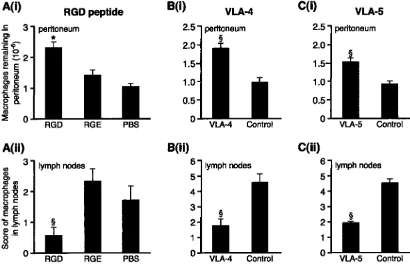
controls, respectively; all P 0.05, n 8 mice). Repeated intraperitoneal injection of RGD significantly delayed macrophage clearance in vivo as evidenced by increased numbers of fluorescent macrophages remaining in the
peri-toneum compared with RGE and medium controls (P
0.05, Fig. 4). In addition, RGD significantly reduced the number of labeled macrophages in the parathymic nodes in the RGD-treated mice compared with either control (P 0.01). Controls were similar for both peritoneal recovery and lymph node scores. There were no differences be-tween the groups in the number of fluorescent macro-phages adherent to bowel, spleen, or liver and the mesothelial surfaces of these organs remained intact. Corroborating the ex vivo data, macrophage emigration into the draining lymphatics was also significantly inhibited by blocking VLA-4 (PS/2) and VLA-5 (5H10-27), which increased the number of macrophages remaining in the
peritoneum (P 0.01 for blocking both 4 and
VLA-5) and reduced emigration into the lymphatics (P 0.01
for blocking both VLA-4 and VLA-5). They did not alter the distribution of macrophages on the mesothelial surface of spleen, bowel, or liver. Blocking CD11b had no effect on macrophage clearance in vivo, there being no signifi-cant difference in macrophage recovery from the
perito-neum for M1/70, isotype, or F4/80 controls (0.89 0.2
vs. 1.18 0.19 and 0.96 0.09 106, respectively; n
12). Likewise, lymphatic emigration was equally unaffected by blocking CD11b. Antibody was used at 100 g for each
injection and increasing to 225 g had no additional effect
(n 5, unpublished data).
Discussion
This study demonstrates for the first time that macro-phage clearance from the inflamed peritoneum can be reg-ulated, a finding that may have significant implications for our understanding of mechanisms leading to mononuclear persistence in chronic inflammation. Macrophages are
ca-pable of undergoing apoptosis, however they are generally resistant to this and during acute resolving peritoneal in-flammation are cleared by emigration, not apoptosis (2, 11). Hence, mechanisms regulating emigration may have pow-erful effects on inflammation.
Here, we demonstrate a dramatic difference in clearance between activated and nonactivated macrophages. Nearly seven times more nonactivated macrophages remained in the noninflamed peritoneum after 3 d compared with acti-vated macrophages in the inflamed peritoneum. These ob-servations, along with the demonstration that adhesion lo-calizes to the mesothelium overlying lymphatics, suggested specific adhesion molecule regulation that could be func-tionally up-regulated by inflammation. Adhesion was diva-lent cation and largely RGD sensitive and partially inhib-ited by blocking VLA-4 and VLA-5 but not by blocking CD11a, CD11b, or CD18, which suggests a predominant
role for 1 integrins. Ex vivo results were validated by in
vivo studies in which RGD and blocking VLA-4 and VLA-5 significantly delayed macrophage clearance, whereas blocking CD11b was ineffective. Alterations in peritoneal macrophage recovery was always mirrored by al-terations in recovery from draining lymph nodes. Controls confirmed that differences in recovery were not due to nonspecific widespread adhesion. These data thus confirm a role for VLA-4 and VLA-5 in regulating inflammatory macrophage clearance from the inflamed peritoneum. They do not exclude the possibility that resident cells may depend on distinct molecular mechanisms for clearance.
Mesothelial cells express intercellular adhesion molecule 1 and vascular cell adhesion molecule 1 and associated fi-bronectin strands can be demonstrated with inflammation, hence the ligands for VLA-4 and VLA-5 are present (12).
VLA-5 and v binding is RGD inhibitable whereas
VLA-4 binding is normally through LDV-inhibitable not RGD-inhibitable mechanisms (9, 13). The increased inhi-bition from the combination of VLA-4 and VLA-5 block-ing suggests that these two integrins are actblock-ing
indepen-Figure 4. VLA-4 and VLA-5 regulate macrophage clearance from resolving peritonitis. (A) (i) After fluo-rescently labeling macrophages in vivo, 0.4 mg RGD or controls, RGE (0.4 mg) or PBS, were injected 2 and 26 h later and macrophage recovery from peritoneum was determined 24 h later. This was significantly greater for RGD than controls. *, P 0.05. n 13 RGD, 8 RGE, and 10 PBS. Controls were not differ-ent. (ii) RGD significantly reduced the number of macrophages reaching draining lymph nodes compared with RGE or PBS (§, P 0.01). (B) (i) Blocking VLA-4
(PS/2) led to significantly more macrophages remain-ing in the peritoneum (§, P 0.01; n 10 VLA-4; 9
controls) and (ii) significantly less in the lymph nodes (§, P 0.01) than isotype control. (C) (i) Blocking
VLA-5 (5H10–27) led to significantly more macro-phages remaining in the peritoneum (§, P 0.01; n
10 VLA-5; 10 controls) and (ii) significantly less in the lymph nodes (§, P 0.01) than isotype control.
on December 5, 2008
jem.rupress.org
Downloaded from
dently. When combined with RGD, VLA-4 further decreased adhesion but not significantly, and VLA-5 had no added effect over that of RGD. Whether vascular cell adhesion molecule 1 or fibronectin are the ligands respon-sible for VLA-4–mediated binding is currently unknown. Both VLA-4 and VLA-5 are expressed on monocytes and implicated in leukocyte influx into inflamed sites, in par-ticular for migration through connective tissue (14). Given mesothelial intercellular adhesion molecule 1
ex-pression, the lack of effect from blocking 2 integrins was
surprising, however monocyte–fibronectin binding has
been shown to be VLA-5 rather than 2 integrin
depen-dent (15). In addition, the combination of 1 and 2
blocking had significantly greater inhibitory effect than
VLA-4 alone, suggesting 2 integrins are functional but
that 1 function is predominant.
VLA-4 inhibitors can attenuate inflammation, prevent-ing monocyte and lymphocyte influx and thus potentially preventing the detrimental effects of excessive leukocyte accumulation (16, 17). However, we demonstrate a cau-tionary note about using such inhibitors in established in-flammation as they can also prevent efflux. If the signal for leukocyte influx is waning, the balance of inflammatory cells at the inflamed site depends more on clearance. Hence the stage and nature of the inflammatory response need to be considered before modulating the function of these ad-hesion molecules.
Subdiaphragmatic macrophage–mesothelial adhesion, in close relationship with the lymphatics, has an anatomical explanation. Lymphatic drainage of the peritoneal cavity arises from this surface and the omentum where milky spots are found (8, 18). Electron microscopy demonstrates that these consist of dome-shaped mesothelial cells sur-rounding narrow pores overlying lymphatic vessels. This may have relevance for malignant cells that also adhere preferentially to milky spots, however the role of adhesion molecules in lymphatic metastases has received surprisingly little attention (19).
In summary, we propose that macrophages bind the me-sothelium overlying draining lymphatics by integrin-medi-ated mechanisms involving VLA-4 and VLA-5, and regula-tion of these interacregula-tions affects macrophage emigraregula-tion into the lymphatic circulation. As other coelomic cavities are mesothelial lined, these mechanisms might be impor-tant after pleural and pericardial inflammation. Moreover, as macrophage emigration to the lymphatics also occurs with inflammation in other organs such as the kidney and lung, an understanding of their clearance mechanisms is likely to have widespread significance (20, 21). Rapid and effective macrophage clearance may dictate the time course of inflammation and be a pivotal event determining the velopment of chronic inflammation. This work thus de-scribes a newly recognized process that offers potential novel therapeutic approaches to promote the effective res-olution of inflammation.
Medical Research Council UK funded G. Bellingan, P. Xu, and H. Cooksley.
Submitted: 24 October 2001 Revised: 27 September 2002 Accepted: 8 October 2002
References
1. Haslett, C. 1999. Granulocyte apoptosis and its role in the resolution and control of lung inflammation. Am. J. Respir. Crit. Care Med. 160:S5–S11.
2. Bellingan, G.J., H. Caldwell, S.E. Howie, I. Dransfield, and C. Haslett. 1996. In vivo fate of the inflammatory macro-phage during the resolution of inflammation: inflammatory macrophages do not die locally, but emigrate to the draining lymph nodes. J. Immunol. 157:2577–2585.
3. Van Furth, R. 1992. Development and distribution of mono-nuclear phagocytes. In Inflammation: Basic Principles and Clinical Correlates. Second Edition. J.I. Gallin, I.M. Gold-stein, and R. Snyderman, editors. Raven Press, New York. 325–341.
4. Leibovich, S.J., and R. Ross. 1975. The role of the macro-phage in wound repair. A study with hydrocortisone and anti-macrophage serum. Am. J. Pathol. 78:71–100.
5. Adams, D.O., and T.A. Hamilton. 1992. Macrophages as de-structive cells in host defence. In Inflammation: Basic Princi-ples and Clinical Correlates. Second Edition. J.I. Gallin, I.M. Goldstein, and R. Snyderman, editors. Raven Press, New York. 637–643.
6. Thepen, T., A.J. van Vuuren, R.C. Kieken, C.A. Damen, W.C. Vooijs, and J.G. van De Winkel. 2000. Resolution of cutaneous inflammation after local elimination of macro-phages. Nat. Biotechnol. 18:48–51.
7. Jonjic, N., G. Peri, S. Bernasconi, F.L. Sciacca, F. Colotta, P. Pelicci, L. Lanfrancone, and A. Mantovani. 1992. Expression of adhesion molecules and chemotactic cytokines in cultured human mesothelial cells. J. Exp. Med. 176:1165–1174. 8. Beelen, R.H., D.M. Fluitsma, and C.M. Hoefsmit. 1980.
The cellular composition of omentum milky spots and the ultrastructure of milky spot macrophages and reticulum. J. Retic. Endothel. Soc. 28:585–599.
9. Ruoslahti, E. 1996. RGD and other recognition sequences for integrins. Annu. Rev. Cell Dev. Biol. 12:697–715. 10. Ryan, G.B., J. Grobety, and G. Majno. 1973. Mesothelial
in-jury and recovery. Am. J. Pathol. 71:93–112.
11. Navarre, W.W., and A. Zychlinsky. 2000. Pathogen-induced apoptosis of macrophages: a common end for different pathogenic strategies. Cell Microbiol. 2:265–273.
12. Liang, Y., K. Jyoukura, N. Ogiwara, and K. Sasaki. 2001. Expression of adhesion molecules and fibronectin of activated peritoneal surface with lipopolysaccharide (LPS) analysed with immuno SEM. Ann. Anat. 183:353–356.
13. Lobb, R.R., and M.E. Hemler. 1994. The pathophysiologic role of alpha 4 integrins in vivo. J. Clin. Invest. 94:1722– 1728.
14. Shang, X.Z., B.J. Lang, and A.C. Issekutz. 1998. Adhesion molecule mechanisms mediating monocyte migration through synovial fibroblast and endothelium barriers: role for CD11/CD18, very late antigen-4 (CD49d/CD29), very late antigen-5 (CD49e/CD29), and vascular cell adhesion mole-cule-1 (CD106). J. Immunol. 160:467–474.
15. Werr, J., X. Xie, P. Hedqvist, E. Ruoslahti, and L. Lindbom. 1998. 1 integrins are critically involved in neutrophil loco-motion in extravascular tissue in vivo. J. Exp. Med. 187: 2091–2096.
on December 5, 2008
jem.rupress.org
Downloaded from
16. Allen, A.R., J. McHale, J. Smith, H.T. Cook, A. Karkar, D.O. Haskard, R.R. Lobb, and C.D. Pusey. 1999. Endothe-lial expression of VCAM-1 in experimental crescentic ne-phritis and effect of antibodies to very late antigen-4 or VCAM-1 on glomerular injury. J. Immunol. 162:5519–5527. 17. Abraham, W.M., A. Gill, A. Ahmed, M.W. Sielczak, I.T.
Lauredo, Y. Botinnikova, K.C. Lin, B. Pepinsky, D.R. Le-one, R.R. Lobb, et al. 2000. A small-molecule, tight-binding inhibitor of the integrin alpha(4)beta(1) blocks antigen-induced airway responses and inflammation in experimental asthma in sheep. Am. J. Respir. Crit. Care Med. 162:603–611. 18. Leak, L.V. 1983. Interaction of mesothelium to intraperito-neal stimulation. I. Aggregation of peritointraperito-neal cells. Lab.
In-vest. 48:479–491.
19. Tsujimoto, H., A. Hagiwara, M. Shimotsuma, C. Sakakura, K. Osaki, S. Sasaki, T. Ohyama, M. Ohgaki, T. Imanishi, J. Yamazaki, et al. 1996. Role of milky spots as selective im-plantation sites for malignant cells in peritoneal dissemination in mice. J. Cancer Res. Clin. Oncol. 122:590–595.
20. Lan, H.Y., D.J. Nikolic-Paterson, and R.C. Atkins. 1993. Trafficking of inflammatory macrophages from the kidney to draining lymph nodes during experimental glomerulonephri-tis. Clin. Exp. Immunol. 92:336–341.
21. Harmsen, A.G., B.A. Muggenburg, M.B. Snipes, and D.E. Bice. 1985. The role of macrophages in particle translocation from lungs to lymph nodes. Science. 230:1277–1280.
on December 5, 2008
jem.rupress.org
Downloaded from