_____________________________________________________________________________________________________
*Corresponding author: E-mail: [email protected];
(Past name: British Journal of Medicine and Medical Research, Past ISSN: 2231-0614, NLM ID: 101570965)
Evaluation of the Effect of Melatonin on Periodontal
Tissues in Rats with Periodontitis Induced
Experimentally
Bianca Caroline Custodio dos Santos
1*, Jossinelma Camargo Gomes
1,
Angela Esmeralda Zaparolli Miola
2, Simone Karine Rothen
2,
Célia Patricia Muller Rodrigues
2, Rose Meire Costa
1,
Patricia Oehlmeyer Nassar
3and Carlos Augusto Nassar
31Graduate Program in Biosciences and Health, Western Parana State University, Cascavel, Brazil.
2Western Paraná State University (UNIOESTE), Cascavel, Paraná, Brazil.
3
Department of Dentistry, Graduate Program in Biosciences and Health, Western Paraná State University (UNIOESTE), Cascavel, Paraná, Brazil.
Authors’ contributions
This work was carried out in collaboration among all authors. All authors read and approved the final manuscript.
Article Information
DOI: 10.9734/JAMMR/2020/v32i730457 Editor(s): (1)Dr. Emin Umit Bagriacik, Gazi University, Turkey. Reviewers: (1) Chahat Puri Periodontics, Himachal Pradesh University, India. (2)Jammula Surya Prasanna, Panineeya Institute of Dental Sciences and Research Center, India. Complete Peer review History:http://www.sdiarticle4.com/review-history/56171
Received 02 March 2020 Accepted 09 May 2020 Published 01 June 2020
ABSTRACT
Objective: To analyze the effects of melatonin administration on the periodontal tissues of rats, linked or not with ligature-induced periodontal disease.
Materials and Methods: 40 male Wistar rats aged eight weeks, divided into Control Group (GCON), Ligature Group (GLIG), Melatonin Group (GMEL) and Ligature and Melatonin Group (GLIGMEL). GLIG and GLIGMEL were induced to experimental periodontitis by placing a ligature on the lower molars for 30 days. During the experiment, after 16 days with the ligature, melatonin was administered orally for 10 mg/kg for 14 days in GMEL and GLIGMEL. In the end, euthanasia was performed and the hemi-mandibles were collected for the respective histological and radiographic analyzes; for the results, Shapiro-Wilk, ANOVA-One-Way and Tukey's multiple comparison tests were used.
Results: A significantly lower alveolar bone loss (p<0.05) was demonstrated in the animals that received the administration of melatonin, in which it had a prophylactic function against the effects of the disease, evidenced in radiographic, histomorphometric and histological analyzes in the bone cell count.
Conclusion: Results show that the therapy with administration of melatonin promotes a protective effect on the alveolar bone tissue of rats with induced experimental periodontitis.
Keywords: Melatonin; periodontitis; bone; alveolar.
1. INTRODUCTION
The relationship between oral health and general health has been grounded in the literature for a long time, especially highlighting the possibility that oral infection may expose individuals to other systemic diseases [1].
Periodontal disease is an inflammatory disease that affects adjacent structures of the oral cavity, which is the dental support tissue, in which the immune response is deregulated, generating oxidative stress and, subsequently, tissue destruction [2]. According to Lundmark et al. [3], there is a loss of connective tissue fixation, destruction of the alveolar bone, and loss of the tooth.
This oral pathology has been classically described as progressive, going
through several phases, that is, the initial and advanced phases [4], expressing itself as an inflammatory process with the presence of edema, redness, and bleeding gums. Duplat et al. [1] demonstrated a varied number of cytokines produced in response to systemic exposure of lipopolysaccharides from Gram-negative bacteria. The central cytokines involved in this pro-inflammatory response are tumor necrosis factor-alpha (TNF-α) and interleukins 1, 6, and 8 (IL-1; IL-6; IL-8). According to Montero et al. [5], the use of topical melatonin was associated with a significant improvement in the gingival index and the depth of the pocket, in addition to the concentrations of interleukin-1β, IL-6, and prostaglandin E2 (PGE2) in the crevicular gingival fluid.
Melatonin, known as N-acetyl-5-methoxy-tryptamine, is an indolaminergic hormone naturally produced and secreted by the pineal gland; it is synthesized from tryptophan and
derived from serotonin after two enzymatic transformations, in addition to the new
pineal synthesis that occurs in the retina, skin, gastrointestinal tract, stem cells and
lymphocytes. The circadian production of this by the pineal gland explains its
chronobiological influence on the activity of the organism, including the endocrine and non-endocrine rhythms [6].
The administration of melatonin can inhibit the activation of brain defense cells (microglia) and decrease the production of substances that activate inflammation (cytokines) in neuroinflammatory models in rats [7]. Thus, it can present applications in Dentistry, as, for example, in periodontal disease, due to its antioxidant activities, stimulating the proliferation of fibroblasts, anti-inflammation, and bone remodeling [6].
According to Cutando et al. [8], patients with periodontal disease showed changes in
cytokine levels, in which TNF-α, IL-6, and CRP are significantly higher than healthy
individuals, thus confirming that melatonin can modulate the inflammatory action of these molecules. In addition to its potent
antioxidant activity, melatonin has anti-inflammatory properties, preventing excessive expression of pro-inflammatory mediators and inhibiting the effects of various pro- inflammatory cytokines. The anti- inflammatory activity can protect the oral cavity against free radicals produced by
inflammatory diseases, such as periodontitis [5].
This study aimed to evaluate the effect of melatonin administration on the periodontal tissues of rats submitted or not to ligature-induced periodontal disease.
2. METHODOLOGY
average from 250 to 400 g, from the Central Vivarium of the Center for Biological and Health Sciences (CCBS) of the Western Paraná State University (UNIOESTE). They were kept in a sectoral vivarium of the Physiology and Endocrinology Laboratory of UNIOESTE (LAFEM), under controlled conditions of temperature (23 ± 2ºC) and light (12-hour light-dark cycle). All animals were separated in individual boxes in groups of 3-5 per box (41 cm long X 34 cm wide X 17 cm high) and fed a standard diet (commercial feed) and water at will for the entire experimental period.
The project was approved by the Ethics Committee on the Use of Animals (CEUA) of UNIOESTE (APPENDIX A), following the Ethical Principles in Animal Experimentation, adopted by the National Council for the Control of Animal Experimentation (CONCEA).
2.1 Experimental Groups
The entire sample calculation of the study was based on the ANOVA test to calculate the size of samples, with a power of 90% and a significance level of 5%, as well as in previous studies by groups of researchers [9-11]. This explanation is necessary to demonstrate the number of animals used for the experiment. The number of animals per group was based on this sample calculation and the other studies [9-11].
The animals were randomly divided into four groups of 10 animals each, totaling 40 male Wistar rats of 8 weeks of age for the experiment.
1- Control Group (GCON): No disease and no treatment;
2- Ligature Group (GLIG): In which periodontal disease by ligature was induced; 3- Melatonin Group (GMEL): Rats treated
with oral melatonin;
4- Melatonin and Ligature Group (GLIG-MEL): Rats that underwent oral melatonin and induction of periodontal disease by ligature.
2.2 Protocol of Periodontal Disease Induction
The animals in the LIG and LIG-MEL groups were anesthetized with 0.08mL/100g Ketamine Chloridate (Dopalen, Sespo Indústria e Comércio, Paulínia-SP) and 0.04mL/100g Xylazine Hydrochloride (Anasedan, Sespo Indústria e Comércio, Paulínia-SP) via intraperitoneal, and
positioned on an appropriate operating table, which allowed the maintenance of the mouth opening, facilitating access to the teeth of the posterior region of the mandible. With the aid of a modified clamp and an explorer probe, a size 40 cotton thread was placed around the lower right and left first molars. This ligature acted as a gingival irritant for 30 days, favoring the accumulation of bacterial plaque and, consequently, the development of PD [12].
2.3 Melatonin Administration
The animals in the groups MEL and LIGMEL received a dose of 10 mg/kg of body weight of melatonin orally after 16 days from the beginning of the experiment with a 1mL dosing plastic syringe; the melatonin was administered for 14 days, in the evening, for maintenance of melatonin production [13]. In Brazil, the melatonin is approved by the competent organs of the Brazilian government as a supplement, so this melatonin was obtained in a manipulated manner Active Pharmaceutical industry/Paraná/ Brazil. Lot:ML/80301, so that it could be administered at a dose of 10mg / Kg of body weight.
2.4 Euthanasia of Animals
On the last day of the experiment (30th), all animals were weighed and then euthanized. Subsequently, the right and left hemi-mandibles were collected for histological and histomorphometric analysis, in addition to radiographic analysis, respectively. This is a method followed by the group of researchers responsible for the experiment, as performed in other animal experiments [9-11].
2.5 Histological Processing
After the euthanasia of the animals, the right hemi-mandibles were collected, dissected, and fixed in a 10% formaldehyde solution for 48 hours. After this period, they were washed in running water for 24 hours and immersed in a 5% trichloroacetic acid (TCA) solution, prepared in the proportion of 100mL of distilled water to 5 grams of acid. The pieces were kept in the descaling solution for approximately twenty days and evaluated daily in order to verify the expected degree of descaling, with the TCA solution being renewed every five days.
histological processing was done for inclusion in paraffin, being dehydrated in series of alcoholic crescents and diaphanized in N-Butyl alcohol (Purified Paraffin, code 1228, lot 1008459, Vetec Química Fina, Rio de Janeiro, Brazil). The blocks were cut in a manual microtome (Olympus, CUT 4055 - Charleston, South Carolina, USA), and 7 µm thick cuts were obtained, which were mounted on histological slides per group and stained using the Hematoxylin and Eosin (H&E) technique [14].
2.6 Histological Analyses
Microscopic analysis was performed by a single examiner through the evaluation of stained histological sections. The slides were analyzed with the aid of a light microscope (Leica Microsystems, Switzerland). They were photomicrographed with an Olympus® DP71 microscope (USA), commonly transmitted for morphological observations of the gingival tissue, alveolar process, and osteocyte, osteoblast, and osteoclast count of the animals' hemi-mandibles.
2.7 Gingival Morphometry
Morphometric measurements were made on the marginal, buccal and right lingual gingiva in all groups, using an image analyzer program (Image Tool 3.0), coupled to a light microscope with a 10x objective, at intervals of 10 cuts between one
count and another in the severity of the cuts (approximately 50 μm) [10].
The measurements were made from pre-determined morphological points in the marginal gingiva, as shown in Fig. 1. The results were expressed in μm. Besides, the total area of the gingiva and connective tissue was checked.
2.8 Bone Morphometry
The quantification of osteoblasts, osteocytes, and osteoclasts present in five consecutive fields of the vestibular alveolar bone crest (ABC) starting from the highest point of the crest was performed. For observation, a 400x magnification was used. Two observations were made per field and then the average of the values for each animal and group was obtained [10].
The measurement of the ABC was performed using a microscope coupled to a computer, which allowed capturing the images, using the LazEz® software. A measure of the shortest distance between the apex of the buccal ABC and the cementoenamel junction was measured using a 40x magnification, with Image Tools 3.0 software (University of Texas Health Science Center, San Antonio, TX, USA). The measurements (in pixels) were repeated once a day on three different days to obtain an average between the values [10].
Fig. 1. Scheme of the rat's marginal gingiva, showing the reference points used for the morphometric measurements of the oral epithelium, epithelial crest, and connective tissue
2.9 Radiographic Analysis
Soon after euthanasia, the left hemi-mandible of each animal was removed and placed in buffered formaldehyde (pH 7.2) for 48 hours. The hemi-mandibles were placed with the lingual face on the Kodak RVG 6100 digital radiographic sensor with 20 pl/mm image resolution, theoretical sensor resolution of 27.03 pl/mm, optical fiber 1, 22 x 30 active surface dimensions mm, and matrix dimensions (pixels) 1200 x 1600 (1.92 million). Such positioning occurred so that the buccal and lingual cusps of the first molars were in the same vertical plane. A GE-1000 X-ray machine set to 15mA, 65Vp, 18 pulses, 50 cm focus/film distance with an X-ray incidence perpendicular to the pieces was used. The digitized images were analyzed with Image Tools 3.0 (University of Texas Health Science Center, San Antonio, TX, USA). Linear measurements were made from the cementoenamel junction to the ABC on the mesial side of the first lower left molar; these measurements were repeated once a day on three different days. An average value was carried out between the values and measurements were made in pixels [12].
2.10 Statistical Analysis
For statistical analysis, all numerical values were expressed as mean ± standard deviation. At first, using the Bioestat 5.3 program (Mamirauá Institute for Sustainable Development, Amazonas, Brazil), the Shapiro-Wilk test was performed to assess the normal distribution of data. Subsequently, parametric ANOVA - One-Way tests were performed, followed by Tukey's multiple comparison test to assess the observed differences considered significant when p <0.05 (5%). The data were presented in the form of tables and graphs.
3. RESULTS
3.1 Histological Analysis of the Right Hemi-Mandible
3.1.1 Control group
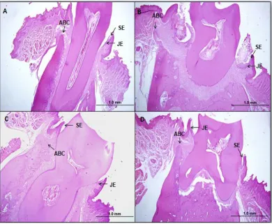
In this group, the evaluation of the parameters was within the normal range for the tissues of the oral, junctional, sulcular epithelium and connective tissue, without the presence of an inflammatory process. The alveolar bone was intact, compact and regular, with the bony crest showing height in the portion of the cervical third of the root. The presence of osteoclasts,
osteocytes, and osteoblasts was observed, incremental lines in the pattern of normal bone remodeling. Still, the cementoenamel junction, cementum, and periodontal ligament were observed, without particularities (Figs. 2-A and 3-A).
3.1.2 Ligature group
In the group in which periodontal disease was induced by ligature, abnormalities were observed in the morphology of the oral, junctional, and sulcular epithelia, with migration to the apical region and connective tissue with a predominance of an acute inflammatory state. As for bone cells, there was a higher concentration of osteoclasts when compared to the other groups (Figs. 2-B and 3-B).
3.1.3 Melatonin group
In the group in which only melatonin was administered, the oral, junctional, and sulcular epithelial tissue was healthy, and of the connective tissue, as well as in the control group, without the presence of phlogistic signs. The alveolar bone remained intact, and the presence of bone cells was observed, both osteoclasts and osteocytes, as well as osteoblasts (Figs. 2-C and 3-C).
3.1.4 Ligature-melatonin group
In this group, a bone retraction pattern was observed; however, the combination with melatonin treatment had less marked bone resorption, and, related to bone cells, it presented a lower concentration of osteoclasts
and a higher concentration of
osteoblasts/osteocytes, suggesting the prophylactic effect of melatonin on bone tissue. The oral junctional, sulcular, and connective tissues maintained the different morphological patterns of GCON, GMEL, and GLIGMEL (Figs. 2-D and 3-D), with collagen inhibition and destruction in the connective tissue spaces.
3.2 Histomorphometric Measurement of Gingival Tissue
Also, the groups that had periodontitis experimentally induced, both in the area of the epithelium and in the area of the connective tissue, had significant measures (p <0.05) when compared with the other groups (Table 2).
3.3 Radiographic and Histomorphome-tric Analysis of the Alveolar Bone
As shown in Table 3, GLIG had significantly fewer osteocytes and osteoblasts, as well as significantly higher osteoclastic activity compared to the other groups (p<0.05). GLIGMEL, however, showed less osteoclastic activity and a significantly higher number of osteocytes and
osteoblasts, when compared to groups GCON and GMEL (p <0.05).
The radiographic and histomorphometric analysis of the alveolar bone of the lower first molars (Tables 4 and 5) demonstrated a greater significant alveolar bone loss (p<0.05) in the groups in which periodontitis was induced by ligature, thus demonstrating the action of periodontitis induced on bone tissue. It is also noteworthy that in the GLIG group, alveolar bone loss was more statistically significant than in the GLIGMEL group. Thus, the group that received treatment with melatonin had less bone loss than the group without treatment.
Fig. 2. Photomicrographs of the alveolar bone crest of Wistar rats. A: GCON; B: GLIG; C: MEL; D: GLIGMEL. Bone matrix (BM), osteocytes (Ot), osteoblasts (Ob), osteoclasts (Oc), erythrocytes (hollow arrow) inside the blood vessel (VS), periodontal ligament area (Pd),
incremental lines (IL). Hematoxylin and Eosin (H&E) staining, sagittal section
Fig. 3. Photomicrographs of the teeth of Wistar rats. A: GCON; B: GLIG; C: MEL; D: GLIGMEL. Alveolar Bone Crest (ABC); Junctional epithelium (JE); Sulcular epithelium (SE). Hematoxylin
and Eosin (H&E) staining, 4x, sagittal section
Source: Author
Table 1. Histomorphometric analysis of the gingiva of the right hemimandible of animals of all groups. The values represent mean ± standard deviation and are expressed in pixels
Gingival crest epithelium height (C)
Epithelial width/ buccal base (E)
Connective tissue height (H)
Connective tissue width/base (L)
Control 535.88 ± 57.45 A 295.25 ± 47.36A 430.49 ± 49.35A 119.49 ± 36.73 A Ligature 639.42 ± 35.05 B 490.75 ±77.82B 538.26 ± 28.56B 247.50±24.40B Melatonin 517.64 ± 64.36 A 313.29 ± 74.85A 411.23 ± 55.16A 145.40±29.55ª Ligature
+Melatonin
594.66 ± 48.77C 403.81 ± 68.37C 503.48 ± 39.76C 193.20±58.43C
*The letters correspond to the comparisons within the same parameter. Different letters, statistically different groups with p <0.05, after the ANOVA test and Tukey's test
Table 2. Histomorphometric analysis of the gingiva of the right hemi-mandible of animals of all groups, the values represent mean ± standard deviation and are expressed in pixels
Epithelium area Connective tissue área
Control 57067.5 ± 15043.7A 27372.6 ± 7343.27ª
Ligature 103427 ± 26224.8 B 66648.7 ± 9380.57B
Melatonin 59407.8 ± 14117.6 A 30951.1 ± 10359.5ª
Ligature +Melatonin 80353.1 ± 19639.0 C 50769.2 ± 11471.1C
Table 3. Morphometric analysis of the right hemi-mandible of rats from the experimental groups to quantify osteocytes, osteoblasts and osteoclasts. The values represent mean ±
standard deviation and are expressed in units
Osteoblast Osteocyte Osteoclast
GCON 20.97±2.75 A 101.87±17.26A 0.10±0.08A
GLIG 13.3±1.79 B 71.86±5.86 B 0.64±0.37B
GMEL 21.6±0.78 A 94.12±8.21A 0.15±0.05A
GLIGMEL 16.15±2.95 C 83.1±4.49 C 0.30±0.18C
*Different letters on the same line indicate statistically significant differences (p<0.05) between groups, within the same parameter, after the ANOVA test and Tukey's test
Table 4. Radiographic analysis of the distance from the cementoenamel junction to the alveolar bone crest on the mesial side of the left first molar of rats of all groups. The values
represent the mean ± standard deviation and are expressed in pixels
Mean ± standard deviation
Control 63.70±17.40 A
Ligature 102.43±11.19 B
Melatonin 59.10±16.80 A
Ligature+Melatonin 87.38±18.27 C
*Different letters indicate statistically significant differences (p <0.05) between the groups after the ANOVA test and Tukey's test
Table 5. Histomorphometric analysis of the distance from the cementoenamel junction to the alveolar bone crest on the mesial side of the left first molar of rats of all groups. The values
represent the mean ± standard deviation and are expressed in pixels
Mean ± standard deviation
Control 865.96±251.78 A
Ligature 1413.36±230.05 B
Melatonin 825.85±322.97 A
Ligature +Melatonin 1142.03±271.71 C
*Different letters indicate statistically significant differences (p <0.05) between the groups after the ANOVA test and Tukey's test
4. DISCUSSION
PD is characterized as an inflammation in the oral cavity that is directly influenced by the action of inflammatory cytokines, especially TNF-α IL-1, IL-6, among others; besides, these cytokines have a direct effect on bone loss [1]. Studies with periodontal therapy in humans turn out to be very limited due to ethical issues and the risks involved in the experiment, so the use of experimental animal models [10,12,15].
The literature establishes an increase in inflammatory mediators in PD, such as TNF-α, [15-17], which accentuate the inflammatory process causing damage to the tissues of the periodontium. Still, it is worth noting that, in the histological parameters analyzed (Figs. 2 and 3), animals with PD presented marked alveolar bone resorption, as well as other changes in other periodontal tissues, thus highlighting the effectiveness of the experimental model, as well
as the consequences of the development of periodontitis. Several studies demonstrate that groups with PD have more significant alveolar bone resorption [16,18-20].
Thus, in an inflammatory process, the high concentrations of TNF-α associated with the increase of 46 reactive oxygen species are the main factors that accentuate this inflammatory process leading to the destruction of bone tissues [21].
The use of melatonin, with therapeutic action in this study, was related to its various uses within experimental therapy [22,23]. According to the literature, the most commonly used routes for administration of melatonin in laboratory animals are intraperitoneally, orally, and through gavage [15,24].
periodontitis have appeared, with several types of research in progress [15,25]. Therefore, this study sought to collaborate to understand the use of melatonin and its influence on periodontal tissues.
Similar to the radiographic analysis (Table 4), histomorphometric analysis of bone tissue (Tables 3 and 5) showed less alveolar bone loss (p<0.05) in GLIGMEL compared to GLIG, but with significantly higher bone destruction when compared the other groups (p<0.05). These results collaborate with the study by Marani (2018), in which the animals that were submitted to melatonin therapy presented protection against bone resorption, thus favoring the alveolar bone formation.
In general, there is evidence that melatonin can benefit bone tissue. In a study by Arabaci et al. [18], who investigated the effects of systemic administration of melatonin (10 mg/kg, intraperitoneally for 15 days) on alveolar bone
resorption in an experimental periodontitis model in rats, it was demonstrated that there was less osteoclastic activity in the treated group, characterized by the reduction of RANKL.
In a model of femur fracture in mice, Histint et al. [26] highlighted that this hormone helps in the formation of calluses and the bone remodeling process, and the expression of RANKL and OPG during the healing of the fracture. Corroborating the results, Muñoz et al. [27] highlighted that the topical use of melatonin associated with growth hormone accelerated bone healing around implants in a study carried out with dental implants in dogs. Cutando et al. highlighted a significant increase in bone density and formation, postulating that melatonin increases osseointegration in dental implants in dogs.
Evidence indicates that melatonin also influences by inhibiting osteoclast differentiation; this occurs by suppressing the expression of the activating ligand of the NFkB receptor (RANKL), which, through its stimulating actions in the differentiation of osteoblasts, activates melatonin 2 (MT2) receptors in osteoblasts. Such activation increases the activity of MAPKs and beta-catenin signaling pathways, which stimulates osteoblastogenesis by increasing the gene expression of runt-related transcription factor 2 (Runx2), which induces osteogenic expression including osterix, bone morphogenetic proteins-2 (Bmp-2) [25].
The literature shows that salivary and gingival melatonin levels decrease in individuals with periodontitis compared to clinically healthy subjects, indicating that melatonin may play a
protective role against PD [25]. The decrease in the levels of salivary melatonin in
periodontal diseases can occur in response to increased oxidative stress and bacterial attack, depleting it in the oral cavity [28] and reinforcing its antioxidant and free radical
scavenger action. Therefore, melatonin favors bone formation, promoting osteoblastic differentiation, and acting as
scavengers of free radicals with antioxidant properties [25].
Studies show the benefits of treatment with melatonin in controlling an inflammatory process due to the attenuation of TNF-α production [25,29, 30]. This effect of the hormone on TNF-α levels involves some mechanisms pointed out in the literature, among them, it is highlighted that melatonin has anti-inflammatory effects through the inhibition of the bearing and the adhesion of neutrophils [29].
Therefore, melatonin attenuates the production of inflammatory mediators such as TNF-α by mitigating the initial stage of an inflammatory process [25]. Studies point out its actions in inflammatory processes, such as PD, which contribute to the increase in the levels of local and systemic inflammatory mediators, including TNF-α and IL-6 [14,16,17].
5. CONCLUSION
Thus, the results here presented indicate that the therapy with administration of melatonin promotes a protective effect on the alveolar bone tissue of rats with experimental periodontitis induced.
6. LIMITATIONS
Thus, it can be concluded, even with the limitations of the study, such as time of administration and lack of serum melatonin dosage, that therapy with administration of melatonin promotes a protective effect on the alveolar bone tissue of rats with experimentally induced periodontitis.
CONSENT
ETHICAL APPROVAL
As authors, we declare that all experiences have been reviewed and approved by the appropriate ethics committee and, therefore, have been carried out by the ethical standards set out in the 1964 Helsinki Declaration.
COMPETING INTERESTS
Authors have declared that no competing interests exist.
REFERENCES
1. Duplat CB, et al. Associação entre doenças cardiovasculares e periodontite: Revisão de literatura. Revista. 2013;9 (2):60-66.
Available:Saúde.Com
2. Balaji TM, Vasanthi HR, Rao SR. Gingival, plasma and salivary levels of melatonin in periodontally healthy individuals and chronic periodontitis patients: A pilot study. Journal of Clinical and Diagnostic Research. 2015;9(3):ZC23-ZC25.
3. Lundmark AH, et al. Transcriptome analysis reveals mucin 4 to be highly associated with periodontitis and identifies pleckstrin as a link to systemic diseases, Scientific Reports. 2015;21(5).
4. Nath S, Raveendran R. What is there in a name?”: A literature review on chronic and aggressive periodontitis. Journal of Indian Society of Periodontology. 2011;15(4):318. 5. Montero J, et al. Changes in crevicular
cytokines after application of melatonin in patients with periodontal disease. Journal of Clinical and Experimental Dentistry. 2017;9(9):e1081–e1087.
6. Acuña-castroviejo D, et al. Extrapineal melatonin: Sources, regulation and potential functions. Cellular and Molecular Life Sciences. 2014;71(16):2997–3025. 7. Benítez-King G, et al. La melatonina como
un factor promotor de la diferenciación neuronal: Implicaciones en el tratamiento de las demencias. Salud Mental. 2013;36 (3):193–199.
8. Cutando A, et al. Effect of topical application of melatonin on serum levels of C-reactive protein (CRP), interleukin-6 (IL-6) and tumor necrosis factor-alpha (TNF-α) in patients with type 1 or type 2 diabetes and periodontal disease. Journal of Clinical and Experimental Dentistry. 2015;7(5): e628–e633.
9. Nascimento CM, et al. Radiographic evaluation of the effect of obesity on alveolar bone in rats with ligature-induced periodontal disease. Diabetes, Metabolic Syndrome and Obesity: Targets and Therapy. 2013;6:365–370.
10. Mattia TM. et al. The Influence of Obesity Induced by Monosodium Glutamate in Periodontal Tissues of Female Wister Rats with Experimental Periodontitis. Dissertação (Mestrado) – Universidade Estadual do Oeste do Parná, Cascavel. 2017;7(3):28–40.
11. Pedrotti S, Nassar PO, Sagae SC, Costa KF, Beu CL, Nassar CA. Evaluation of the influence of experimental periodontitis on the sexual behavior of male wistar rats, British Journal of Medicine & Medical Research. 2016;15(9):1-8.
12. Nassar PO, et al. Simvastatin therapy in cyclosporine A-induced alveolar bone loss in rats. Journal of Periodontal Research. 2009;44(4):479–488.
13. Aranda M, et al. In vivo hepatic oxidative stress because of carbon tetrachloride toxicity: Protection by melatonin and pinoline. Journal of Pineal Research. 2010; 49(1):78–85.
14. Junqueira LCU, Carneiro J. Histologia Básica. 12. ed. Rio de Janeiro: Guanabara Koogan; 2013.
15. Kara A, Akman S, Ozkanlar S, Tozoglu U, Kalkan Y, Canakci CF, Tozoglu S. Immune modulatory and antioxidant effects of melatonin in experimental periodontitis in rats. Free Radic Biol Med. 2013;55:21-6. 16. Colombo NH, Shirakashi DJ, Chiba FY,
Coutinho MS, Ervolino E, Garbin CA, Machado UF, Sumida DH. Periodontal disease decreases insulin sensitivity and insulin signaling. J. Periodontol. 2012; 83(7):864-70.
17. Hernandez M, et al. Host-pathogen interactions in progressive chronic periodontitis. J. Dent. Res. 2011;90(10): 1164-1170.
18. Arabaci T, et al. Therapeutic effects of melatonin on alveolar bone resorption after experimental periodontitis in rats: A biochemical and immunohistochemical study. J. Periodontol. 2015;86:874-881. 19. Mattera MSLC. et al. Maternal periodontitis
decreases plasma membrane GLUT4 content in skeletal muscle of adult offspring. Life Sciences. 2016;148:194-200. 20. Ricoldi MST, et al. Effects of the probiotic
the non-surgical treatment of periodontitis. A histomorphometric, Microtomographic and Immunohistochemical Study in Rats. PLoS ONE. 2017;12(6):1-15.
21. Blaser H, Dostert C, Mak TW, Brenner D. TNF and ROS crosstalk in inflammation. Trends Cell Biol. 2016;26(4):249-261. 22. Ghallab NA, Hamdy E, Shaker OG.
Malondialdehyde, superoxide dismutase and melatonin levels in gingival crevicular fluid of aggressive and chronic periodontitis patients. Australian Dental Journal. 2016; 61(1):53–61.
23. Lodhi K, et al. Evaluation of melatonin levels in saliva in gingivitis and periodontitis cases: A pilot study. Contemporary Clinical Dentistry. 2016;7(4):519.
24. Kose O, Arabaci T, Kara A, Yemenoglu H, Kermen E, Kizildag A, Gedikli S, Ozkanlar S. Effects of melatonin on oxidative stress index and alveolar bone loss in diabetic rats with periodontitis. J. Periodontol. 2016;87 (5):e82-90.
25. Marani F. Efeitos da suplementação com melatonina na remodelaçõao óssea
alveolar em ratos pinealectomizados com doença periodontal. 2018. Dissertação (Mestrado) - Universidade Estadual Paulista, Araçatuba; 2018.
26. Histing T, et al. Melatonin impairs fracture healing by suppressing RANKL mediated bone remodeling. J. Surgical Research. 2012;173:83–90.
27. Muñoz F, et al. Topical application of melatonin and growth hormone accelerates bone healing around dental implants in dogs. Clin. Implant Dent Relat Res. 2012; 14(2):226-235.
28. Bertl K, Schoiber A, Haririan, H. et al. Non-surgical periodontal therapy influences salivary melatonin levels. Clin. Oral Invest. 2013;17:1219–1225.
29. Markus RP, Ferreira ZS, Fernandes PACM. Cecon E. The immune-pineal axis: A shuttle between endocrine and paracrine melatonin sources. Neuroimmuno-modulation. 2007;14:126-133.
30. Cutando A, et al. Melatonin stimulates osteointegration of dental
APPENDIX A
_________________________________________________________________________________
© 2020 Santos et al.; This is an Open Access article distributed under the terms of the Creative Commons Attribution License (http://creativecommons.org/licenses/by/4.0), which permits unrestricted use, distribution, and reproduction in any medium, provided the original work is properly cited.
Peer-review history: