Open Access
Research article
Adhesive capsulitis and dynamic splinting: a controlled, cohort
study
Paul D Gaspar
1and F Buck Willis*
2Address: 1Gaspar Doctors of Physical Therapy, 981 Lomas Santa Fe, Ste. A Solana Beach, CA 92075, USA and 2Texas State University, HPER Adjunct Faculty (at the time of this study), Dynasplint Systems, Inc., Clinical Research, PO Box 92135, Austin, TX 78709, USA
Email: Paul D Gaspar - [email protected] ; F Buck Willis* - [email protected] * Corresponding author
Abstract
Background: Adhesive Capsulitis (AC) affects patient of all ages, and stretching protocols are commonly prescribed for this condition. Dynamic splinting has been shown effective in contracture reduction from pathologies including Trismus to plantar fasciitis. The purpose of this study was to examine the efficacy of dynamic splinting on patients with AC.
Methods: This controlled, cohort study, was conducted at four physical therapy, sports medicine clinics in Texas and California. Sixty-two patients diagnosed with Stage II Adhesive Capsulitis were grouped by intervention. The intervention categories were as follows: Group I (Control); Group II (Physical Therapy exclusively with standardized protocols); Group III; (Shoulder Dynasplint system exclusively); Group IV (Combined treatment with Shoulder Dynasplint and standardized Physical Therapy). The duration of this study was 90 days for all groups, and the main outcome measures were change in active, external rotation.
Results: Significant difference was found for all treatment groups (p < 0.001) following a one-way ANOVA. The greatest change with the smallest standard deviation was for the combined treatment group IV, (mean change of 29°).
Conclusion: The difference for the combined treatment group was attributed to patients' receiving the best PT combined with structured "home therapy" that contributed an additional 90 hours of end-range stretching. This adjunct should be included in the standard of care for adhesive Capsulitis.
Trial Registration: Trial Number: NCT00873158
Background
Adhesive capsulitis (AC) is an idiopathic disease that affects an estimated 2-6% of the American population (6 to 18 million Americans) [1-6]. It is characterized by fibrosis, decreased volume of the glenoid capsule, pain, and progressive pain with loss of both active and passive Range of Motion (ROM). The direct cost of treating this
pathology in the United States in the year 2000 was $7 bil-lion [7] and it affects patients predominantly over 50 years of age.
This condition therefore is a serious pathology, which is also known as "Frozen Shoulder" with three phases: 1) The Painful stage is characterized by the gradual onset of
Published: 7 September 2009
BMC Musculoskeletal Disorders 2009, 10:111 doi:10.1186/1471-2474-10-111
Received: 24 January 2009 Accepted: 7 September 2009
This article is available from: http://www.biomedcentral.com/1471-2474/10/111 © 2009 Gaspar and Willis; licensee BioMed Central Ltd.
diffuse shoulder pain and which usually lasts one to two months; 2) The Frozen stage is characterized by progres-sive loss of motion (particularly glenohumeral external rotation) which lasts several months to a year or longer [8]. This stage also exhibits decreased capsular volume which can be visualized with MRI, for differential diagno-sis; 3) The Thawing stage is the final stage during which range of motion gradually improves over several months to years. Range of motion deficits may continue to be unresolved for more than 3-5 years following the onset of AC.
Contracture is defined as shortening of connective tissue (ligaments, tendons, and cartilage) caused by excessive arthrofibrosis, immobilization, inactivation, adhesions, or excessive neuromuscular tone [9-11]. Contracture in the shoulder is primarily seen in decreased capsular vol-ume, and is measured with MRI for differential diagnosis. There are many treatment methods for adhesive capsulitis including physical therapy, corticosteroid injections (intra-articular), hydroplasty, manipulation of the joint while under anesthetics and surgery [1-3,5-8,11-20]. The conservative primary treatment for adhesive capsulitis are intra-articular corticosteroid injections and physical ther-apy, which was examined by Dudkiewicz et al. [8]. They conducted a long-term follow-up (mean 9.2 years) of 54 patients suffering from idiopathic adhesive capsulitis, and their results showed that conservative treatment alone (physical therapy and non-steroidal anti-inflammatory medications) was an effective, long-term treatment method.
Current Treatments for AC range from surgical interven-tion or manipulainterven-tion under anesthetics [21], stretching protocols combined with glenohumeral intra-articular corticosteroid injections [22], and continuous passive motion devices [23]. Studies of often report benefits from early intervention [7,12,22] which Earley and Shannon said may help prevent the "downward spiral of forced dis-use" leading to contracture [7]. Joint mobilization and flexibility training are common features in treatment of this condition.
Griggs, et al. revealed in a prospective study that showed a significant benefit from participating in a "Four-direction shoulder-stretching exercise program"[12]. In their study, 75 patients diagnosed with Stage II idiopathic adhesive capsulitis participated, and 90% of the patients were satis-fied with the outcome of the four-direction shoulder-stretching exercise program. As a long-term study, (ROM measurements were taken at 3, 6, 12,18, and 22 months) ninety percent of the patients gained significant increases in ROM of external rotation, internal rotation, flexion and abduction in the first few months and maintained the
ROM through a daily four-direction shoulder-stretching exercise program.
The protocol of using low-load prolonged-duration stretch, combined with the therapeutic principle of increased time at end range allows the patient to reduce contracture by achieving permanent elongation of con-nective tissue [9,10,13,14,24]. The protocol of increasing total end range time has been shown to be beneficial, despite the cause of contracture in the shoulder joint [7,9,10,25,26]. This is the protocol used with the Dynas-plint Systems, (DynasDynas-plint Systems, Inc., Severna Park, MD) and twenty-five years ago a biomechanically correct device was developed to utilize a low-load prolonged-duration stretch with dynamic tension to reduce contrac-ture of the elbow and knee joints [10,24]. This stretching protocol was subsequently included in the Shoulder Dynasplint systems (SDS) modality which allows patients to stretch in flexion, abduction, external, or internal rota-tion.
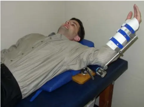
The SDS is often prescribed as home therapy because it designed to help the patient stretch the shoulder in multi-ple planes. (See figure 1.) The purpose of this study was to examine the efficacy of dynamic splinting with low-load, prolonged-duration of stretching on adhesive capsulitis. This study used a three-month duration which measured change in the active external rotation of the shoulder, (supine position with humerus abducted to 90°) which is the most common ROM deficit in AC [2,3,5].
Methods
Subjects
Sixty-two patients between the ages of 36 and 75 with Stage II adhesive capsulitis were prospectively recruited by
[image:2.612.313.554.511.690.2]Dynasplint Shoulder System in External Rotation Figure 1
referral from clinicians in California and Texas (Mean age 55.6 ± 7.9) and all patients had deficits in external rota-tion. All patients enrolled had been previously treated with cortical steroid injection(s) but no patients had pre-viously undergone manipulation or surgery. Patients were give informed consent and all patients' rights, protection, and privacy have been ensured in this study as required by the Gaspar-PT Biomed IRB and ethical approval was received from this IRB. Patients were independently pre-scribed treatment in one of the following groups:
• Group I was the control group, and these patients were only treated with cortical steroid injections, (n = 15).
• Group II patients were treated exclusively with stand-ardized physical therapy, twice per week, (n = 15). • Group III patients were treated exclusively with the SDS as "home therapy," (n = 16).
• Group IV patients were treated with both physical therapy (twice weekly) and the SDS for daily end-range stretching, (n = 16).
When enrolled, patients were instructed that if they required additional treatment such as additional cortical steroid injections then their participation in this study would be completed but only two patients required such additional treatment methods. Standardized Training and reporting was used for all patients in all groups. All subject data was transmitted in confidential documents without jeopardizing the patients' privacy according to the federal health information privacy protection act.
Clinical Protocols
All treatment categories were prescribed by the attending physicians rather than being randomized, which may reflect current treatments in use. Physical therapy was standardized, based on the protocols of Vermuelen, Hsu, and Mulligan.[4,6,16,17] These methods included moist heat, patient education and re-evaluation of symptoms, joint mobilization (limited to progressive end-range joint mobilization), passive range of motion, AROM and PNF, and therapeutic exercise. Group II and Group IV patients participated in physical therapy for two or more times per week, and the SDS was worn twice a day, seven days per week.
Group III and Group IV patients who wore the SDS received a standardized treatment protocol and wearing schedule. These patients were instructed on the use by the by the physical therapist and a Dynasplint consultant who accomplished a customized fitting of the unit and taught the standardized protocol regarding how to increase
ten-sion in the direction of external rotation, with humeral abduction to 90 degrees. Each subject was instructed to fax a weekly tracking form to investigators which reported daily duration(s) in the SDS and tension settings used. Patients were instructed to begin the dynamic splinting with only the tension setting of #1 for the first week for accommodation, and then they increase the tension set-ting to #2 which equals 3.0 foot pounds of force. During this period, patients were instructed to increase the dura-tion in the SDS unit for 20-30 minutes, twice each day (with the goal of stretching 60 total minutes per day). If the patient had post-wear discomfort or stiffness lasting more than one hour after removing the splint, the dura-tion of the treatment was then reduced for the next two scheduled stretching bouts. After the patient was able to tolerate 60 minutes of stretching, (30 min, bid) then the patient was then instructed to increase the tension every two weeks as tolerated, without discomfort lasting more than one hour following each stretching session.
After 90 consecutive days in the SDS the patients' Active External Rotation was measured again by the same, pre-scribing clinician. While treatment with the SDS may be performed in multiple planes, this study chose to only evaluate ER rotation because it is the most common restricted ROM from Adhesive Capsulitis [5-7]. Analysis of "Intention to Treat" include the data from patients who were non-compliant (less than 90% PT attendance and/or less than 90% scheduled use of SDS) or did not complete the study duration. All patients' data was included in this analysis.
Data Analysis
The dependent variable was the change in Active Range of Motion, Supine External Rotation (Humerus abducted to 90°), and the independent variables were groups (Con-trol vs. Physical therapy vs. SDS vs. Combined SDS and Physical Therapy). One-Way Analyses Of Variance (ANOVA) was performed using the Graph-Pad, InStat software, and post-hoc T-tests were performed to measure difference between groups. (An alpha level of 0.05 was used for all tests.) All other calculations were made with the Microsoft Excel program.
Equipment Used
Dynasplint® Shoulder System
800-638-6771
Dynasplint® and Dynasplint Shoulder System
Are registered trademarks of Dynasplint Systems, Inc. In-Stat Software
2055 Gateway Place, Suite 150 San Jose CA 95110
408-345-4495
Results
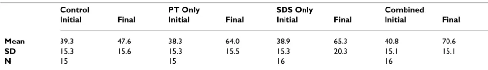
There was a significant difference for each treatment groups (PT Only: T = 4.441, P < 0.001; SDS Only: T = 4.887, P < 0.001; Combined: T = 5.318, P < 0.001). Due to the low power there was not a significant difference between treatment groups but the greatest change and the smallest Standard Deviation was seen for the Combined Treatment Group PT + SDS (mean 29.8° of change and SD = 12.36; see Figure 2.) This suggests that the SDS is a measurably effective adjunct to physical therapy as a struc-tured home therapy. (See Table 1.)
Discussion
The purpose of this study was to examine the efficacy of dynamic splinting on adhesive capsulitis in a prospective, cohort study. Although dynamic splinting for other extremity joints have been studied [10,24], this is the first controlled study investigating the effects of the dynasplint shoulder system. The results showed the efficacy of dynamic splinting as an effective "home therapy" adjunct to physical therapy. The additional 80 to 90 hours of end-range stretching as home therapy combined with stand-ardized physical therapy is considered to be responsible for the greatest change in AROM of external rotation. The results were in agreement with the study by Griggs et. al. [9], which demonstrated that a conservative treatment protocol of four-direction shoulder-stretching exercise program would benefit shoulder flexibility. This experi-ment also confirmed the findings of Dudkiewicz et al.[6] which described the efficacy of "conservative protocols." Because ROM deficits frequently exist in external rotation, this experiment chose to examine only that plane follow-ing treatment with physical therapy and/or the SDS.
Conclusion
Use of the SDS may be an effective adjunct "home ther-apy" for adhesive capsulitis, and the additional 60 min-utes per day of low-load, prolonged-stretch was beneficial. (The mean time recorded was 85 hours in this 90 days study.) Earley and Shannon [7] proposed that conserva-tive interventions of adhesive capsulitis would be the most beneficial when initiated as soon as the diagnosis is made, and DS could be an effective initial modality of conservative treatment.
Confounding variables in this study included lack of ran-domization. Grouping was done by the prescribing clini-cian who may have been biased, based on patient history. The total duration that each patient endured Stage II of adhesive capsulitis was not differentiated. Limitations of this study also included that it was only performed on a small number of patients and was limited to examining the Active Range of Motion, Supine External Rotation (Humerus abducted to 90°).
A future study comparing the duration of treatment to dis-charge between groups would be greatly beneficial in
[image:4.612.59.293.97.256.2]Graph displaying the results of the study Figure 2
Graph displaying the results of the study.
Table 1: Active Range of Motion in External Rotation
Control PT Only SDS Only Combined
Initial Final Initial Final Initial Final Initial Final
Mean 39.3 47.6 38.3 64.0 38.9 65.3 40.8 70.6
SD 15.3 15.6 15.3 15.5 15.3 20.3 15.1 15.1
[image:4.612.54.554.657.729.2]measuring the benefits that the SDS has in treating adhe-sive capsulitis. A larger subject population would reduce the chance for type two error, and a randomized, control-led trial would eliminate most of the limitations discussed regarding this study. A pain scale should also be used in the next study.
List of Abbreviations
(AC): Adhesive capsulitis; (ROM): Range of Motion; (SDS): Shoulder Dynasplint systems; (ER): External Rota-tion; (ANOVA): Analyses Of Variance; (PT): Physical Ther-apy.
Competing interests
PDG has no competing interest and has received no finan-cial compensation for this study or manuscript. FBW is employed by the parent company of Dynasplint systems but he has no stock or vested interest in either company.
Authors' contributions
PDG was responsible for experimental design, patient recruitment and treatment, and development of this man-uscript. FBW contributed in experimental design, study coordination, data analysis, and development of this manuscript. Both authors read and approved the final manuscript.
Acknowledgements
We would like to convey our thanks to Craig Neffendorf, PT who helped recruit patients for this study from his practice at Eagle Rehab in Austin Texas. This study was completed without extramural funding.
References
1. Baums MH, Spahn G, Nozaki M, Steckel H, Schultz W, Klinger HM:
Functional outcome and general health status in patients after arthroscopic release in adhesive capsulitis. Knee Surg Sports Traumatol Arthrosc 2007, 15(5):638-44.
2. Castellarin G, Ricci M, Vedovi E, Vecchini E, Sembenini P, Marangon A, Vangelista A: Manipulation and arthroscopy under general anesthesia and early rehabilitative treatment for frozen shoulders. Arch Phys Med Rehabil 2004, 85(8):1236-40.
3. Farrell CM, Sperling JW, Cofield RH: Manipulation for frozen shoulder: long-term results. J Shoulder Elbow Surg 2005,
14(5):480-4.
4. Hsu AT, Hedman T, Chang JH: Changes in abduction and rota-tion range of morota-tion in response to simulated dorsal and ventral translational mobilization of the glenohumeral joint.
Phys Ther 2002, 82(6):544-56.
5. Rundquist PJ, Anderson DD, Guanche CA, Ludewig PM: Shoulder kinematics in subjects with frozen shoulder. Arch Phys Med Rehabil 2003, 84(10):1473-9.
6. Vermeulen HM, Rozing PM, Obermann WR, le Cessie S, Vliet Vlieland TP: Comparison of high-grade and low-grade mobilization techniques in the management of adhesive capsulitis of the shoulder: randomized controlled trial. Phys Ther 2006,
86(3):355-68.
7. Earley D, Shannon M: The use of occupation-based treatment with a person who has shoulder adhesive capsulitis: a case report. Am J Occup Ther 2006, 60(4):397-403.
8. Dudkiewicz I, Oran A, Salai M, Palti R, Pritsch M: Idiopathic adhe-sive capsulitis: long-term results of conservative treatment.
Isr Med Assoc J 2004, 6(9):524-6.
9. Ada L, Goddard E, McCully J, Stavrinos T, Bampton J: Thirty min-utes of positioning reduces the development of shoulder
external rotation contracture after stroke: a randomized controlled trial. Arch Phys Med Rehabil 2005, 86(2):230-4. 10. Lai JM, Francisco GE, Willis FB: Dynamic Splinting on Range of
Motion and Spasticity after Treatment with Botulinum Toxin-A: a Randomized, Controlled Pilot Study. Advances in Therapy 2009, 26(2):241-8.
11. Karduna AR, Williams GR, Iannotti JP, Williams JL: Kinematics of the glenohumeral joint: Influences of muscle forces, ligamen-tous constraints, and articular geometry. Journal of Orthopaedic Research 1996, 4(6):986-993.
12. Griggs SM, Ahn A, Green A: Idiopathic adhesive capsulitis. A prospective functional outcome study of nonoperative treat-ment. J Bone Joint Surg Am 2000, 82-A(10):1398-407.
13. Adrian MJ, Cooper JM: Biomechanics of Human Movement.
2nd edition. Wisconsin: Brown & Benchmark; 1989.
14. Olensek A, Matjacic Z, Bajd T: Muscle contracture emulating system for studying artificially induced pathological gait in intact individuals. J Appl Biomech 2005, 21(4):348-58.
15. Hamdan TA, Al-Essa KA: Manipulation under anaesthesia for the treatment of frozen shoulder. Int Orthop 2003, 27(2):107-9. 16. Hsu AT, Ho L, Ho S, Hedman T: Joint position during anterior-posterior glide mobilization: its effect on glenohumeral abduction range of motion. Arch Phys Med Rehabil 2000,
81(2):210-4.
17. Hsu AT, Ho L, Ho S, Hedman T: Immediate response of gleno-humeral abduction range of motion to a caudally directed translational. Arch Phys Med Rehabil 2000, 81(11):1511-6. 18. Meislin RJ, Sperling JW, Stitik TP: Persistent shoulder pain:
epide-miology, pathophysiology, and diagnosis. Am J Orthop 2005,
34(12 Suppl):5-9.
19. Moskowitz RW, Blaine TA: An overview of treatment options for persistent shoulder pain. Am J Orthop 2005, 34(12 Suppl):10-5.
20. Placzek JD, Roubal PJ, Freeman DC, Kulig K, Nasser S, Pagett BT:
Long-term effectiveness of translational manipulation for adhesive capsulitis. Clin Orthop Relat Res 1998:181-91.
21. Ng CY, Amin AK, Narborough S, McMullan L, Cook R, Brenkel IJ:
Manipulation under anaesthesia and early physiotherapy facilitate recovery of patients with frozen shoulder syn-drome. Scott Med J 2009, 54(1):29-31.
22. Kelley MJ, McClure PW, Leggin BG: Frozen shoulder: evidence and a proposed model guiding rehabilitation. J Orthop Sports Phys Ther 2009, 39(2):135-48.
23. Dundar U, Toktas H, Cakir T, Evcik D, Kavuncu V: Continuous pas-sive motion provides good pain control in patients with adhesive capsulitis. Int J Rehabil Res 2009, 32(3):193-8.
24. Finger E, Willis FR: Dynamic splinting for knee flexion contrac-ture following total knee arthroplasty: a case report. Cases J
2008, 1(1):421.
25. Uhl TL: Motion Analysis: Shoulder mechanics mobilize joint movement. BioMechanic 2000:5-10.
26. Boakes JL, Foran J, Ward SR, Lieber RL: Muscle Adaptation by Serial Sarcomere Addition 1 Year after Femoral Lengthen-ing. Clin Orthop Relat Res 2007, 456:250-3.
Pre-publication history
The pre-publication history for this paper can be accessed here: