INTRODUCTION
The mechanisms employed to segregate chromosomes reflect the specific requirements of a cell. Although many features of spindle structure and function remain unchanged, some aspects are dependent upon cell type and developmental stage. Well known examples include the absence of centrioles in oocytes and the reduction in spindle size during early embryonic cleavages. The challenge therefore is to identify which features of spindle function remain unchanged and which are developmentally regulated.
In most eukaryotic cells spindle microtubules (MTs) continually move towards the spindle poles as a result of balanced MT disassembly at spindle poles and assembly in the spindle midzone, a process known as poleward MT flux (Mitchison et al., 1986; Mitchison, 1989). Poleward flux occurs in prophase, prometaphase and metaphase in mitotic cells, and participates in several aspects of spindle function, including controlling spindle length, promoting correct MT-kinetochore attachments, and contributing to the forces that drive chromosomes to opposing spindle poles in anaphase (Ferenz and Wadsworth, 2007; Ganem and Compton, 2006; Rogers et al., 2005). However, the molecular details of how poleward flux is regulated and therefore how steady-state spindle length is established remain poorly understood.
Kinesin 5 has long been considered as a candidate MT motor that might participate in poleward flux. Kinesin 5 is bipolar, possessing two heads, each of which move towards MT plus ends (Kashina et al., 1996). In vitro, therefore, kinesin 5 has the capacity to slide adjacent anti-parallel MTs, thereby pushing their minus ends apart (Kapitein et al., 2005; van den Wildenberg et al.,
2008). Because spindle MTs are normally oriented with their minus ends towards spindle poles, kinesin 5 has the potential to participate in poleward flux by pushing overlapping anti-parrallel MTs in the spindle midzone towards spindle poles. Kinesin 5 might also contribute to spindle pole focussing by binding and crosslinking parallel MTs near the pole (Gaglio et al., 1996; Sawin et al., 1992; Uteng et al., 2008; Yang et al., 2008). However, studies of the role of kinesin 5 in poleward flux in intact spindles have yielded contrasting results. In the Xenopus egg extract system, inhibition of kinesin 5 causes metaphase spindles to shorten and collapse (Kapoor et al., 2000), and, under experimental conditions that prevent spindle collapse, kinesin 5 inhibiton or immunodepletion was also found to inhibit poleward MT flux (Groen et al., 2008; Miyamoto et al., 2004; Yang et al., 2008; Yang et al., 2007). The inhibition of the kinesin 5 KLP61F in syncitial Drosophila embryos caused a concentration-dependent range of defects, including spindle collapse and a reduction of poleward flux (Brust-Mascher et al., 2009). By contrast, although kinesin 5 plays essential roles in spindle assembly, kinesin 5 inhibition does not collapse spindles that have already formed in cultured vertebrate cells (Blangy et al., 1995; Cameron et al., 2006; Kapoor et al., 2000), and has only a minor affect upon the rate of poleward flux (Cameron et al., 2006; Ferenz and Wadsworth, 2007). Demonstration of an essential role for kinesin 5 in poleward flux in an intact vertebrate cell has so far proven elusive, and the reason for these differences is unknown.
Here, an assay for monitoring poleward flux in live mouse eggs has been established and used to demonstrate that kinesin 5 is essential for poleward flux in this system. The inhibition of poleward flux leads to spindle collapse as a result of persistent MT disassembly at the poles. Kinesin 5 is shown to be essential for spindle bipolarity in eggs and early embryos, but spindles become resistant to kinesin 5-inhibition in morulae and blastocysts. This switch in spindle function occurs irrespective of the fate or clonal
A shift from kinesin 5-dependent metaphase spindle
function during preimplantation development in mouse
Greg FitzHarrisMicrotubules within meiotic and mitotic spindles continually move towards spindle poles in a process termed poleward flux, which is essential for spindle integrity and faithful chromosome segregation. Kinesin 5 is a longstanding candidate for a molecular motor that might drive poleward flux, and has been shown to drive flux and to be necessary for spindle bipolarity in Xenopusegg extracts. However, kinesin 5 is not necessary for poleward flux or for maintaining metaphase spindle bipolarity in intact mammalian cells, and the reason for the different results in these systems is unknown. The experiments presented here test the hypothesis that these results might reflect developmental differences in spindle function by examining the role of kinesin 5 in mouse eggs and
preimplantation embryos. In contrast to cultured somatic cells, poleward flux in mouse eggs is critically dependent upon kinesin 5. Inhibition of poleward flux leads to spindle shortening as a result of continued microtubule depolymerisation at the pole, and eventual loss of spindle bipolarity. Spindle bipolarity is also dependent upon kinesin 5 during the first three embryonic cleavages, but becomes kinesin 5-independent in the majority of spindles by the blastocyst stage. This switch occurs asynchronously in different blastomeres but is independent of clonal cell heritage and of whether the blastomere is within the inner cell mass or the trophoectoderm. These experiments reveal a novel developmental switch in the requirements for spindle function and
chromosome segregation during preimplantation development.
KEY WORDS: Spindle, Poleward flux, Kinesin 5, Mouse
Development 136, 2111-2119 (2009) doi:10.1242/dev.035089
University College London Institute for Women’s Health, Gower Street, London WC1E 6BT, UK.
e-mail: [email protected]
Accepted 14 April 2009
D
E
V
E
LO
P
M
E
N
lineage of the blastomeres. The data presented here therefore establish a novel developmental transition in the requirements for spindle bipolarity, which might underpin the different results obtained from Xenopus egg extracts and mammalian cultured cells.
MATERIALS AND METHODS
Egg and embryo handling
Metaphase II eggs were obtained from MF1 mice (Harlan, UK) previously administered with pregnant mares serum gonadotropin (PMSG; 7 IU) and human chorionic gonadotropin (hCG; 5 IU) at a 48-hour interval. Eggs were collected 13-14 hours after hCG administration and cumulus cells were removed using hyaluronidase. All live culture and imaging was carried out at 37°C. To collect embryos, mice were mated at the time of hCG administration and embryos collected 28 hours (one-cell embryos) or 48 hours (two-cell embryos) later. Four-cell embryos, morulae and blastocysts, were obtained by culturing two-cell embryos in KSOM media (Lawitts and
Biggers, 1993) at 37°C, 5% CO2for 24, 48 and 72 hours, respectively.
Immunofluorescence
Eggs/embryos were briefly permeabilised with 0.25% Triton X-100 in
PHEM solution (10 mM EGTA, 2 mM MgCl2, 60 mM PIPES, 25 mM
HEPES, pH 6.9) for 5 seconds, then fixed using 3.7% paraformaldehyde in PHEM for 50 minutes. Eggs were subsequently permeabilised for 10 minutes, and blocked overnight at 4°C in 3% bovine serum albumin (BSA). The pre-permeabilisation step was omitted in experiments in which GFP and mCherry were expressed. Antibodies used were as follows: rabbit anti-kinesin 5 antibodies from Abcam (ab37009) or Duane Compton
(Dartmouth, USA) (Mountain et al., 1999); rat anti-α-tubulin (YL1/2;
Abcam); mouse anti-Oct4 (C10; Santa Cruz Biotechnology). Each was used at a dilution of 1:300 for 1 hour at 37°C. Following three washes in PBS with 1% BSA, cells were labelled with Alexa Fluor-labelled secondary antibodies as appropriate (Invitrogen). Fixed chromatin was labelled by a 5-minute
exposure to 5 μg/ml Hoechst 33342 (Invitrogen).
Expression of fluorescent proteins
PhotoActivatable-GFP::tubulin (PAGFP-tubulin) (Tulu et al., 2003) was purchased in the pIREShyg vector from Addgene. MAP7fragment::GFP (MAP7-GFP) and MAP7fragment::RFP (MAP7-RFP) were a gift from Alex McDougall (Villefranche, France). Plasmids were amplified and linearised, and mRNA made using Ambion mMessage Machine T7 Ultra, according to the manufacturer’s instructions. Polyadenylated RNA was delivered using a microinjection apparatus consisting of Narishige micromanipulators mounted on a Leica inverted microscope. The plasma membrane was breached using a brief pulse of negative capacitance provided by an intracellular electrometer, and a controlled volume of mRNA delivered through a fine glass pipette using a picopump (World Precision Instruments).
Imaging and analysis
Live epifluorescence microscopy was carried out on a Zeiss Axiovert microscope fitted with a cooled CCD camera (Micromax; Princeton Scientific Instruments), controlled using Metafluor software. GFP and Hoechst were imaged using a 510 nm dichroic mirror. GFP was excited using a 480/50-nm bandpass filter, and emitted light monitored with a
520/35-nm bandpass filter. Cells were loaded with 5 μg/ml Hoechst for 10
seconds prior to imaging. Microtubules were imaged using MAP7-GFP or MAP7-RFP, similar to the approach recently used to study MT dynamics in mouse meiosis I (Schuh and Ellenberg, 2007). BSA was omitted from the culture media to allow eggs to adhere to the coverslip thereby immobilising them for imaging where necessary. Rates of spindle collapse following monastrol addition were calculated by linear regression of spindle length during the first five minutes following monastrol addition, during which time the relationship between spindle length and time was approximately linear.
Confocal imaging was performed on a Zeiss LSM 510 confocal microscope. GFP was imaged using a 488-nm laser and a 505- to 550-nm bandpass emission filter. RFP and Alexa594-tubulin were imaged using a
546-nm laser and a 560-nm longpass emission filter. Hoechst was excited using a 405-nm blue diode, and emitted light collected with a 420- to 480-nm bandpass filter.
For PAGFP-tubulin experiments, photoactivation was performed by brief (<1 second) illumination by the 405-nm blue diode using the ‘bleach’ function. A 520-nm longpass filter was used to collect emitted light from photoactivated PAGFP. Eggs were co-injected with a small amount of
Alexa594-tubulin (estimated final concentration 75 μg/ml) to co-label the
spindle during photoactivation experiments (gift from Paul Chang, Harvard, USA). The velocity of poleward MT flux was calculated from the location of peak PAGFP-tubulin fluorescence. To ensure that the determined velocity was not affected by noise, Gaussian curves were fitted to the peak region of each curve, and the mean value used as the position of the PAGFP-tubulin bar. Velocity was calculated as the mean speed during the first 6 minutes after photoactivation, during which time velocity was constant.
Fluorescence dissipation from the area of photoactivation was used as a measure of MT turnover, and calculated according to the method of Cimini et al. (Cimini et al., 2006). Background subtraction was performed using a corresponding area on the opposite side of the spindle. Each individual data point was corrected for photobleaching. The extent of photobleaching was calculated by imaging photoactivated PAGFP-tubulin in taxol-treated eggs
(2 μM, 1 hour). Photobleaching in these experiments was not excessive;
when all three fluorophores (Alexa594-tubulin, PAGFP-tubulin and Hoechst) were imaged simultaneously in taxol-treated eggs, five iterations cumulatively resulted in ~17% PAGFP-tubulin fluorescence reduction. In
order to correlate clonal lineage with spindle morphology, χ2values were
calculated for each individual embryo using Yates’ correction for small
numbers. χ2values were added to provide a χ2statistic with ndegrees of
freedom.
RESULTS
Measurement of poleward microtubule motion in live eggs
The mouse egg provides an excellent model in which to study spindle dynamics as it has a large meiotic spindle (~25 μm in length) that remains arrested at metaphase until the egg is fertilised. Although poleward MT flux has been examined in a number of somatic cell types, spermatocytes, Drosophilaembryos and Xenopus egg extracts (Rogers et al., 2005), flux has not previously been demonstrated in a live mammalian egg. To establish a method of monitoring MT dynamics in eggs, a fusion protein consisting of tubulin and the photoactivatable form of green fluorescent protein was employed (PAGFP-tubulin) (Patterson and Lippincott-Schwartz, 2002; Tulu et al., 2003), which has been used previously to measure poleward flux in cultured somatic cells (Cimini et al., 2006; Ganem et al., 2005; Ferenz and Wadsworth, 2007; Cameron et al., 2006). PAGFP-tubulin is incorporated into the spindle MTs but is only weakly fluorescent under 488-nm excitation until activated by 405-nm light (Patterson and Lippincott-Schwartz, 2002). A subpopulation of MTs can therefore be selectively labelled by illuminating a specific area of the cell with 405-nm light, and their position subsequently monitored. PAGFP-tubulin was introduced by microinjecting mRNA encoding the fusion protein. Spindles were co-labelled with Alexa594-tubulin in order to position eggs such that the spindle was parallel with the coverslip, thereby eliminating any effect of spindle orientation. A line of PAGFP-tubulin was activated close to the metaphase chromosomes by brief illumination with the 405-nm laser. The line of photoactivated MTs moved away from the chromosomes towards the spindle pole in all cases (Fig. 1A), confirming that egg spindle MTs undergo poleward flux. Tubulin dynamics were quantified using linescan analyses (Fig. 1B; see Materials and methods for more details). Peak tubulin fluorescence diminished and the distribution of fluorescence broadened as the PAGFP bar moved towards the pole, which was
D
E
V
E
LO
P
M
E
N
most likely the combined result of MT turnover and non-uniform flux velocities of MTs. Fluorescence photobleaching was relatively minor (see Materials and methods for details). By monitoring the location of peak fluorescence, the initial velocity of poleward movement was calculated as 0.52±0.04 μm minute (n=15), which is similar to flux rates in mammalian cell lines (Rogers et al., 2005).
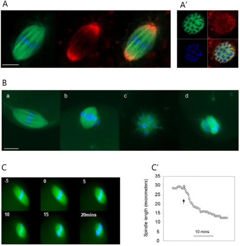
Kinesin 5 drives poleward flux and maintains spindle bipolarity in metaphase-arrested eggs Next the role of kinesin 5 in the MII spindle was examined. Immunofluorescence revealed that kinesin 5 is highly enriched at the spindle poles (Fig. 2A), as has been shown previously (Mountain et al., 1999), and transverse confocal sections through the spindle midzone confirmed that kinesin 5 is also detectable on MTs throughout the spindle (Fig. 2A⬘). This pattern is similar to that in other systems (Kapoor et al., 2000; Kapoor and Mitchison, 2001; Sawin and Mitchison, 1995), and might reflect poleward transport of kinesin 5 by dynein/dynactin (Kapoor and Mitchison, 2001; Uteng et al., 2008). Next the function of kinesin 5 was addressed using monastrol, a small-molecule inhibitor that has been extensively characterised in a number of systems and shown to be specific for kinesin 5 (Maliga and Mitchison, 2006; Mayer et al., 1999) (see Discussion for further details). Eggs incubated in 100 μM monastrol for 1 hour (Fig. 2B, part b) had spindles that were much shorter than those in untreated controls (Fig. 2B, part a), such that
[image:3.612.315.561.62.315.2]monastrol-treated spindles typically looked rounded. Two hours after treatment the majority of eggs had monopolar spindles (Fig. 2B, part c) or a disorganised array of MTs surrounding the chromatin (Fig. 2B, part d). To more closely examine spindle shortening, live epifluorescence imaging of spindles was carried out during monastrol addition (Fig. 2C). Eggs were immobilised by adherence to the coverslip in the absence of BSA, thereby allowing live imaging of the spindle during the addition of monastrol. The velocity of spindle collapse was calculated by plotting the length of the spindle against time (Fig. 2C⬘). The velocity of monastrol-induced spindle shortening was concentration dependent with a maximal concentration in the range 100-200 μM (see Fig. S1 in the supplementary material), which is similar to the concentration dependency found for poleward flux inhibition by monastrol in Fig. 1. Poleward microtubule flux in mouse eggs.(A)
PAGFP-tubulin was photoactivated in eggs using a 405-nm blue diode focussed as a bar across the spindle (top row). Spindles were co-labelled with Alexa594-tubulin and only eggs that were oriented with the spindle parallel to the coverslip were examined
[image:3.612.48.293.62.284.2](PAGFP-tubulin/Alexa594-tubulin overlay, bottom row). An image of the Alexa594-labelled spindle and Hoechst-labelled chromosomes (blue) taken at the end of the experiment is shown to confirm that the egg was in metaphase (bottom row, right; n=15). Scale bar: 5μm. (B) Linescan analysis of PAGFP-tubulin fluorescence across the same spindle. The dotted line across the inset spindle indicates the location of the linescan. Linescans shown are as follows: blue, 0 minutes; green, 4 minutes; red, 8 minutes; black, 12 minutes; purple, 16 minutes; brown, 20 minutes. See Materials and methods for further details.
Fig. 2. Kinesin 5 maintains metaphase spindle length in the mouse egg.(A) Confocal image of a fixed MII spindle. MAP7-GFP-labelled microtubules (green), Hoechst-MAP7-GFP-labelled chromosomes (blue) and kinesin 5 immuofluorescence (red) are shown. Note that kinesin 5 colocalises with microtubules throughout the spindle and is enriched at the spindle poles. (A⬘) A single confocal section through the metaphase plate illustrating that kinesin 5 (red) is detectable on microtubules in the centre of the spindle. Similar results were obtained using two different kinesin 5 antibodies (see Materials and methods). Scale bar: 10μm. (B) Microtubule configurations in mouse eggs exposed to 100μM monastrol. Microtubules are labelled green, chromosomes are labelled blue. (a) Normal untreated bipolar MII spindle; (b) short MII spindle with a relatively coherent metaphase plate; (c) monoaster; (d) disorganised array of chromosomes and dense microtubules. Following a 1-hour exposure to monastrol, most eggs had short spindles (83%; b). Following 2 hours of monastrol exposure, most spindles had collapsed to form monoasters (29%; c) or a disorganised array (43%; d). Scale bar: 10μm. Normal bipolar spindles were never seen following 100μM monastrol treatment. (C) Live epifluorescence images of the spindle (MAP7-GFP, green) and chromosomes (Hoechst, blue) during monastrol-induced spindle collapse. Monastrol was added at t=0 minutes. (C⬘) Typical plot of spindle length following monastrol addition. Arrowhead indicates time of monastrol addition.
D
E
V
E
LO
P
M
E
N
Xenopusextracts (Miyamoto et al., 2004). At maximal concentration (200 μM), monastrol caused spindles to shorten at a rate of 1.16±0.05 μm/minute (i.e. spindle poles approached the midzone at ~0.58 μm/minute). Spindles became rounded within ~20 minutes, during which time chromosome movement at the metaphase plate was minimal. These experiments indicate that, similar to inXenopus egg extracts, but unlike in somatic cells, kinesin 5 activity is necessary to maintain spindle length and bipolarity during metaphase in the mouse egg.
Next it was important to understand how kinesin 5 inhibition causes spindle shortening, and whether kinesin 5 is necessary for poleward flux. I therefore set out to directly examine MT dynamics during monastrol-induced spindle shortening. PAGFP-tubulin was activated midway between the chromosomes and spindle poles at the same time as monastrol addition during live imaging. Spindle MTs, chromosomes and the PAGFP-tubulin bar were simultaneously imaged, and the metaphase chromosomes were used as a reference point for the centre of the spindle. Strikingly, the PAGFP bar remained a constant distance from the metaphase chromosomes during spindle shortening (n=8; Fig. 3A). Linescan analysis of pixel intensities across the spindle confirmed that, following monastrol addition, the PAGFP-tubulin bar moved minimally compared with in controls eggs (Fig. 3A⬘; 0.24±0.08 μm away from the chromosomes in the first 6 minutes after photoactivation, compared with 3.12±0.19 μm in controls;
P<0.0001). Reduced movement of MTs away from the spindle midzone was also confirmed by calculating the rate of dissipation of PAGFP signal from the initial activation area (Fig. 3A⬙). Thus kinesin 5 inhibition prevents the poleward movement of MTs away from the spindle midzone (i.e. poleward flux). However, the spindle pole moved towards the PAGFP line and the chromosomes in all cases. Thus, kinesin 5 is necessary for poleward flux and in its absence continued MT disassembly at the poles causes the spindle to shorten.
[image:4.612.54.358.358.739.2]One possible interpretation of these data is that kinesin 5 inhibition might prevent MT polymerisation at plus ends in the spindle midzone, thereby allowing continued depolymerisation at the pole to shorten the spindle. It was therefore necessary to determine whether MT addition in the spindle midzone continues during monastrol-induced spindle collapse, by carrying out high magnification confocal microscopy of spindle MTs during monastrol-induced collapse (Fig. 3B). If monastrol prevented MT polymerisation then MT density would be expected to remain constant as depolymerisation at the pole causes the spindle to shorten, whereas continued polymerization should cause an increase in MT density in the shortening spindle. To test this, spindle collapse was triggered by monastrol addition while MTs were visualised using MAP7-RFP and confocal microscopy (see Materials and methods). Linescan analysis of collapsing spindles revealed that spindle shortening was accompanied by substantial increases in the
Fig. 3. Poleward microtubule flux is kinesin 5 dependent in the mouse egg.(A) Monastrol (200μM) was added immediately after creation of the PAGFP-tubulin bar (t=0), and confocal images were taken at the timepoints indicated. The plane of focus was quickly checked between images to ensure that the confocal plane bisected the spindle, to confirm that the observed spindle shortening was not attributable to focus shift. Colour scheme is the same as in Fig. 1. (A⬘) Linescans of PAGFP-tubulin (solid line) and Hoechst fluorescence (dotted line) from the same experiment. Note that the PAGFP-tubulin bar remains at a constant distance from the metaphase plate during spindle collapse (n=8). (A⬙) Analysis of fluorescence dissipation after photoactivation similarly confirmed that microtubule dissipation from the site of photoactivation was significantly reduced by kinesin 5 inhibition (see Materials and methods). (B) Confocal images of monastrol-induced spindle collapse at the times indicated following monastrol addition. Microtubules are labelled with MAP7-RFP. (B⬘) Linescan analyses of microtubule fluorescence intensity at different times, as indicated by line colour: blue, 0 minutes; grey, 4 minutes; red, 8 minutes. Dotted lines in B illustrate position of linescan. Note that fluorescence intensity increases markedly predominantly within the central portion of the spindle. Scale bar: 10μm.
D
E
V
E
LO
P
M
E
N
fluorescence of MT bundles in the central region of the spindle, such that after four minutes the fluorescence intensity of MTs had approximately doubled (Fig. 3B⬘, n=7). Therefore, MTs accumulate in the spindle midzone during monastrol-induced spindle collapse, indicating that MT polymerisation in the midzone continues despite kinesin 5 inhibition.
The experiments described this far indicate that kinesin 5 inhibition does not prevent MT depolymerisation at spindle poles, nor prevent MT addition in the midzone, but that it does stop the movement of MTs away from the midzone towards the poles. Kinesin 5 is thus essential for driving MTs polewards during poleward flux in the mouse egg.
Loss of kinesin 5 dependence in metaphase during preimplantation embryo development The dependence of mouse egg and Xenopus egg extract spindles upon kinesin 5 contrasts with that of mammalian somatic (mitotic) cells, in which kinesin 5 inhibition has been reported to have a minimal effect upon poleward flux and metaphase spindle bipolarity (Blangy et al., 1995; Cameron et al., 2006; Ferenz and Wadsworth, 2007; Kapoor et al., 2000). One possible explanation for these contrasting results was that there might be a difference in the requirement for kinesin 5 between meiosis and mitosis. To test this,
the role of kinesin 5 in spindles from the first mitotic embryonic division was examined. One-cell embryos were allowed to undergo nuclear envelope breakdown (observed under the light microscope), then were treated with the proteasome inhibitor MG132 to prevent anaphase, thereby ensuring that all spindles examined were in metaphase. Kinesin 5 inhibition by the addition of monastrol caused spindle collapse within 30 minutes in all embryos examined (Fig. 4A). Live epifluorescence imaging revealed that spindle collapse occurred as a result of progressive spindle shortening, as had occurred in eggs (see Fig. S2 in the supplementary material). Therefore, the different requirement for kinesin 5 in eggs and somatic cells is not simply due to a difference between meiosis and mitosis.
[image:5.612.49.360.332.738.2]Another possibility was that spindles might undergo a transition from kinesin 5 dependence to kinesin 5 independence during early embryonic development. To address this, the role of kinesin 5 in metaphase spindle length maintenance was examined during the second mitotic division (two-cell to four-cell transition) and the third mitotic division (four-cell to eight-cell transition), by the same method that had been applied to one-cell embryos. Kinesin 5 inhibition caused the loss of spindle bipolarity and the formation of monoasters in embryos arrested in metaphase of the second and third mitotic divisions in almost all cases (over 90%; Fig. 4A). Thus
Fig. 4. Metaphase spindle length becomes insensitive to kinesin 5 inhibition during late preimplantation development.(A) Embryos were treated with 25μM MG132 to cause metaphase arrest, and were subsequently treated with monastrol for 30 minutes before fixation. Control embryos (no monastrol treatment) are shown for comparison (top). Typical images of α-tubulin (red) and chromosomes (Hoechst, blue) are shown. Note that the monastrol-treated morula shown in the figure possesses both an apparently normal bipolar spindle and a collapsed spindle. Scale bar: 25μm. (B) Analysis of spindle morphology in
preimplantation embryos. Eighty spindles were examined in morulae and 195 spindles examined in blastocysts over the course of three separate experiments. A minimum of 17 spindles were examined for each group at the one-, two- and four-cell stage. C, control; M, monastrol.
D
E
V
E
LO
P
M
E
N
metaphase spindles are dependent upon kinesin 5 for the maintenance of bipolarity in unfertilised eggs and also in the first three mitotic embryonic divisions.
Finally, the dependence upon kinesin 5 was examined in morulae and blastocysts. Morulae and blastocysts were treated with MG132 for 2 hours prior to monastrol exposure to ensure that a proportion of spindles were in metaphase, and that no spindles that had entered anaphase would be examined. By using this approach, I was able to examine an average of ~3 spindles per morula and ~5 spindles per blastocyst. Strikingly, 38% of morulae spindles and 56% of blastocyst spindles remained as normal bipolar spindles following kinesin 5 inhibiton (Fig. 4B). Approximately 10% of spindles were bipolar, but short and rounded in appearance, suggesting that those spindles that do collapse at this stage of development do so by shortening, similar to as occurs in eggs and one-cell embryos.
These results suggest that metaphase spindle bipolarity becomes independent of kinesin 5 in morulae and blastocysts. However, it was important to exclude the possibility that these results might instead reflect a change in permeability of the embryos, or an unexpected breakdown of monastrol, such that there was reduced access of the drug to spindles in later embryos. To address this, the ability of monastrol to prevent spindle formation in blastocysts was investigated. Embryos were treated with nocodazole for 2 hours to allow a population of blastomeres to enter mitosis with depolymerised MTs. The nocodazole was subsequently removed, and the embryos transferred for 1 hour either to media containing MG132 to allow spindles to be built that would then arrest in metaphase, or to a co-treatment of MG132 and monastrol. Following this regimen, 97% of mitotic cells possessed normal bipolar spindles in control blastocysts (MG132 only), whereas 94% of monastrol-treated blastomeres contained monopolar MT arrays, revealing that kinesin 5 is essential for spindle formation in blastocysts and confirming that monastrol can access the mitotic machinery in later preimplantation embryos (see Fig. S3 in the supplementary material). In addition, a doubling in concentration of monastrol to 400 μM did not increase the proportion of metaphase spindles that collapsed, which might have been expected had the heterogeneous phenotype observed in blastocysts been attributable to partially reduced access of the drug (see Fig. S4 in the supplementary material). Thus, whereas the metaphase spindle is critically dependent upon kinesin 5 in eggs and during the first three mitotic divisions, the majority of spindles in later preimplantation embryos remain bipolar despite kinesin 5 inhibition.
The switch to kinesin 5 independence is not related to blastomere fate or cell lineage
An interesting feature of the above result is that the switch to kinesin 5 independence does not occur simultaneously in all cells. Rather, ~60% of spindles continue to require kinesin 5 in morulae, and ~40% in blastocysts, revealing that there are two populations of cells at these stages with spindles that behave differently when kinesin 5 is inhibited. The transition from morula to blastocyst represents the establishment of the first two major cell lineages in the developing mammalian embryo: the inner cell mass (ICM), which gives rise to the embryo proper; and the trophoectoderm (TE), which results in the trophoblast. It was therefore possible that the heterogeneity of the spindle response at morula and blastocyst stage might correspond to these two different cell lineages.
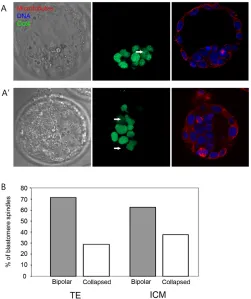
To test this, the effect of kinesin 5 inhibition in the TE and ICM of blastocysts was compared. To reliably identify cells of the inner cell mass, blastocysts were labelled with antibodies for the ICM-specific transcription factor Oct4 (Fig. 5A). As previously, kinesin
5 inhibition in blastocysts caused spindle collapse in approximately one third of all spindles in blastocysts (306 spindles from a total of 61 blastocysts were examined from four separate experiments). A comparison of spindles in the TE and the Oct4-labelled inner cell mass revealed no difference in the proportion of spindles that collapsed following kinesin 5 inhibition in the two cell lines (Fig. 5B; 62% of spindles remained bipolar in the ICM, 71% remained bipolar in the TE; P>0.2). Thus, the different requirement for kinesin 5 observed in blastocysts was not attributable to differences between the inner cell mass and the trophoectoderm.
[image:6.612.313.564.60.359.2]An alternative possibility was that the unexpected heterogeneity of spindle function in morulae and blastocysts might relate to the clonal heritage of blastomeres. Although the progeny of the two blastomeres at the two-cell stage might be preferentially predisposed towards the ICM or TE, the two cell types in the blastocyst are not exclusively the progeny of each of the two-cell stage blastomeres (Piotrowska et al., 2001). Therefore it was possible that the two populations of blastomeres with different spindles might relate to the two lines of Fig. 5. The switch to kinesin 5 independence occurs in
trophoectoderm and inner cell mass cells.(A,A⬘) Blastocysts were fixed following monastrol treatment and labelled with Oct4 antibodies (green) to label the inner cell mass, tubulin antibodies to label the spindle (red), and Hoechst to label DNA (blue). Two examples of this experiment are shown. Note that example A shows one spindle that is in an Oct4-labelled cell and which has collapsed following monastrol addition (marked with arrow). A⬘shows two spindles each of which are intact and bipolar, one lying within an Oct4-labelled cell, the other within an Oct4-negative cell (marked with arrows). (B) Spindle morphology was assessed in the trophoectoderm (TE) and the inner cell mass (ICM) and the proportion of spindles that had collapsed
quantified. There was no significant difference in the proportion of intact and collapsed spindles in Oct4-positive (ICM) and -negative (TE) cells (P>0.2).
D
E
V
E
LO
P
M
E
N
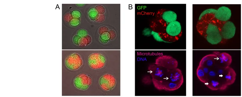
clonal progeny rather than to the eventual fate of the blastomere (ICM or TE). To test this possibility, different coloured fluorescent proteins were expressed in each of the two blastomeres at the two-cell stage. Embryos were cultured for a further two days to morula stage, at which time the two clonal lines were easily distinguishable by using fluorescence microscopy (Fig. 6A). This approach is similar to that which has been used previously to determine the relative contributions of the two blastomeres of the two-cell embryo to the inner cell mass and trophoectoderm of the blastocyst (Piotrowska et al., 2001). Fluorophore expression had no effect upon embryonic development to blastocyst or upon the proportion of spindles that collapsed following monastrol treatment. Having labelled the two cell lineages, the effect of monastrol upon spindles was examined in morulae as described above (Fig. 6B). If the different spindle phenotypes following kinesin 5 inhibition were indeed related to cell lineage, then within any given embryo collapsed spindles should be largely confined to one clone (indicated by either mCherry or GFP), whereas bipolar spindles should be confined to the other. Collapsed and bipolar spindles were confined to opposite lineages in this manner in only 2 out of the 34 embryos examined. Spindles with similar phenotypes (collapsed or bipolar) were found in different cell lineages in 22 out of 34 embryos examined, and different spindle phenotypes were observed within the same cell lineage in 27 of 34 embryos. Figure 6B shows two examples of such embryos; the embryo on the left has two collapsed spindles in the plane of focus that are in opposite clonal lineages; the embryo on the right shows both collapsed and bipolar spindles residing within the same clonal lineage. Correlation between blastomere lineage and spindle morphology was highly non-significant (P>0.95). Therefore, the switch from kinesin 5 dependence to independence in metaphase of mitosis occurs both in the ICM and TE, irrespective of the clonal cell heritage of the blastomere.
DISCUSSION
Mammalian eggs and early embryos are vulnerable to chromosome segregation errors, which are rare in normal somatic cells. Identification of mechanistic differences in spindle function between eggs and early embryos, and somatic cells, will probably be
important in understanding the unusual susceptibility during early development. The experiments presented here reveal a shift in the requirements for spindle bipolarity during early development, whereby an essential dependence upon kinesin 5 in metaphase is lost in the majority of cells by the blastocyst stage, irrespective of the lineage or eventual fate of the blastomere. The discussion that follows will focus on the insights these results provide into the mechanism of action of kinesin 5 in eggs and early embryos, and on the nature of the switch to kinesin 5 independence.
The role of kinesin 5 in poleward flux and determination of spindle length in early mouse development
[image:7.612.59.462.488.650.2]Mitotic spindles are normally considered to be composed of two interdigitating half spindles, with MT minus ends oriented towards spindle poles, and plus ends that terminate at kinetochores (kinetochore microtubules, kMTs), or elsewhere within the spindle (interpolar microtubules, ipMTs). Poleward flux in mitotic cells is therefore typically viewed as a conveyorbelt-like motion of MTs underpinned by balanced rates of MT disassembly at the poles and assembly at the MT plus-ends (Margolis and Wilson, 1981; Rogers et al., 2005). The in vitro capacity of kinesin 5 to slide MTs apart supports the hypothesis that this property might participate in flux by generating a poleward force upon spindle MTs. However, direct evidence for a role for kinesin 5 in metaphase poleward MT flux has previously only come from the Xenopus egg extract system (Miyamoto et al., 2004) and syncitial Drosophilaembryos (Brust-Mascher et al., 2009), and similar experiments in mammalian cells in culture revealed only a minor role for kinesin 5 (Cameron et al., 2006; Ferenz and Wadsworth, 2007). The experiments presented here show that kinesin 5 drives poleward flux in the mouse egg, and that kinesin 5 inhibition causes spindle collapse. Spindle collapse is a direct consequence of poleward flux inhibition because the collapse is a result of continued MT depolymerisation at the pole in the absence of poleward MT movement. The experiments also indicate that MT polymerisation in the midzone continues during spindle collapse. Together, the data are consistent with a model in
Fig. 6. The switch to kinesin 5 independence is independent of clonal lineage.(A) Blastomeres of two-cell embryos were labelled with either GFP or mCherry such that the clonal descendants of the two cells could be identified at the morula stage. Typical epifluorescence images of embryos labelled in this manner at the four-cell stage (24 hours after labelling, top) and morula (48 hours after labelling, bottom) are shown. (B) Morulae were treated with monastrol as previously, fixed, and microtubules labelled with a far-red secondary antibody (Alexa633) in order to visualise mCherry, GFP, microtubules and DNA (Hoechst) simultaneously. Typical examples of this experiment are shown. Note that in the first example (left), two metaphase blastomeres can be seen, each of which contains a collapsed spindle (indicated by an arrow), one blastomere being labelled with mCherry, the other with GFP. In the second example (right), three metaphase cells can be seen, each labelled with GFP, two of which
are intact and bipolar (broad arrows), one of which has collapsed (arrow).
D
E
V
E
LO
P
M
E
N
which poleward flux in metaphase mouse eggs occurs as a result of balanced rates of MT assembly and disassembly in the midzone and spindle poles, respectively, while kinesin 5 provides the force to drive MTs polewards. The data thus provide a clear demonstration of an essential role for kinesin 5 in poleward flux in an intact vertebrate cell.
The current experiments were made possible by the use of monastrol as a specific cell-permeable inhibitor of kinesin 5. Monastrol was first identified in a chemical genetic screen as being capable of inhibiting kinesin 5-dependent MT motility in vitro (Mayer et al., 1999). Monastrol exerts an allosteric inhibition upon the ATPase activity of the motor (Maliga et al., 2002) at a site conserved amongst vertebrate kinesin 5s, but poorly conserved in other kinesins (Maliga and Mitchison, 2006). Although specificity has not been explicitly addressed in the current study, the spindle phenotypes and poleward flux inhibition caused by monastrol in mouse eggs are consistent with the effect of monastrol in other systems (Kapoor et al., 2000; Miyamoto et al., 2004), with other approaches to inhibiting kinesin 5 in other systems (Brust-Mascher et al., 2009; Miyamoto et al., 2004), and with the in vitro function of kinesin 5 (Kapitein et al., 2005; van den Wildenberg et al., 2008). In addition, monastrol has been employed previously in meiosis-I mouse oocytes and was found to produce phenotypes consistent with kinesin 5 inhibition in other systems and with microinjection of kinesin 5 antibodies (Mailhes et al., 2004; Mountain et al., 1999; Schuh and Ellenberg, 2007).
Recent Xenopusegg extract experiments have led to new models of spindle architecture, in which minus ends of non-kinetochore MTs (non-kMTs) are located throughout the spindle, such that MTs of various lengths are distributed in a so-called ‘tiled-array’ of spindle MTs. Within this model, poleward movement of non-kMTs is proposed to occur as a result of the formation of MT polymers in the spindle midzone that slide towards and then depolymerise at the pole (Burbank et al., 2006; Burbank et al., 2007; Ohi et al., 2007; Yang et al., 2007). One variation of this model questions the need for minus-end depolymerisation in fluxing meiotic spindles in that system, instead proposing that the clustering of fluxing MTs provides a braking force that reduces flux speed to essentially zero at the pole, allowing the balance of MTs within the spindle to be maintained by MT plus-end turnover (the slide and cluster model) (Burbank et al., 2007; Ohi et al., 2007). By contrast, the PAGFP-tubulin experiment presented here points to the presence of a pole-resident depolymerising activity in mouse eggs that has the capacity to shorten the spindle when kinesin 5 is inhibited, and the direction of this shortening (i.e. from the pole towards the spindle midzone) is consistent with minus end depolymerisation. Other recent experiments in Xenopusegg extracts revealed a relatively minor change in flux speed at the pole, which also implies the need for minus end depolymerisation (Yang et al., 2008). Although the experiments presented here do not explicitly distinguish between the classical mitotic model and more recent models of MT layout in this system, I saw no evidence of MT bending or buckling within collapsing spindles in any experiments, which might be expected within the classical model of spindle structure if nucleation were to continue within kinetochore MTs as the spindle shortens.
Developmental switch in spindle function during preimplantation development
In contrast to the Xenopus egg system and to mouse eggs, a dependence upon kinesin 5 in metaphase is apparently not a general feature of cultured mammalian somatic cells, as inhibition fails to collapse bipolar spindles in BCC1 (Kapoor et al., 2000), PtK1
(Cameron et al., 2006) or HeLa (Blangy et al., 1995) (G.F., unpublished) cells. This disparity has previously been suggested to be a difference between higher and lower vertebrates, meiosis and mitosis, or artefacts attributable to a cell-free system. However, the experiments here suggest that in mouse a high level of dependence upon kinesin 5 in metaphase is a feature of eggs and early preimplantation embryos, and that a switch occurs whereby this requirement for kinesin 5 has been lost in ~40-50% of morulae spindles and ~50-70% of blastocyst spindles. As metaphase spindle bipolarity has been found not to be kinesin 5 dependent in the few mammalian cell lines studied so far, this might reflect a gradual shift towards complete independence from kinesin 5. It is not yet known whether a similar developmental shift will be found in other species, although it is interesting that syncitial Drosophilaembryo spindles are very sensitive to kinesin 5 inhibition in mitosis (Sharp et al., 1999; Brust-Mascher et al., 2009), whereas spindle length is relatively insensitive to the depletion of kinesin 5 in metaphase in S2 cells (Goshima et al., 2005).
Although the mechanism of the transition has not been directly examined here, its timing would appear inconsistent with a number of otherwise appealing hypotheses. The kinesin 5 dependence of the first three mitotic metaphases clearly precludes the influence of meiosis-specific factors, such as cytostatic factor (CSF), and the direct involvement of embryonic genome activation (EGA) appears unlikely because EGA occurs predominantly at the two-cell stage in mice. Mouse eggs and cleavage-stage embryos possess acentriolar centrosomes, and centrioles first begin to appear in blastomeres during late preimplantation development (Abumuslimov et al., 1994; Calarco-Gillam et al., 1983; Gueth-Hallonet et al., 1993). It is unlikely that there is a direct correlation between the appearance of centrioles/conventional centrosomes and resistance to spindle collapse, as centrioles are first detected by electron microscopy in late blastocysts, and not in morulae (Abumuslimov et al., 1994; Gueth-Hallonet et al., 1993). However, changes in centrosomal appearance have been observed by immunofluorescence somewhat earlier in 32-cell embryos (Calarco-Gillam et al., 1983; Gueth-Hallonet et al., 1993), making changes to the spindle pole that might influence the behaviour of kinetochore, interpolar or astral MTs an attractive possibility. Finally, one speculative possibility is that the switch from kinesin 5 dependence to independence in metaphase might coincide with a switch from a tiled array of MTs to a classical mitotic spindle of two interdigitating half spindles. Testing this hypothesis would require the development of single fluorophore speckle imaging (Yang et al., 2007) in live eggs and embryos, or of tools to localise MT minus ends.
Conclusion
The experiments here show that poleward flux in the mouse egg spindle occurs as a result of balanced MT depolymerisation and polymerisation at spindle poles and midzone, and the simultaneous kinesin 5-dependent translocation of MTs towards the poles. In addition, they reveal a novel developmental transition prior to implantation, whereby mitotic spindles no longer require kinesin 5 for metaphase spindle bipolarity in morulae and blastocysts. Given the stability of mammalian metaphase II and its amenability to imaging, the mouse egg and early embryo will probably prove to be a valuable model for future studies of spindle MT dynamics in early development.
I thank Guillaume Halet, Guillaume Charras and Carolyn Moores for critical reading of the manuscript, and John Carroll for advice and encouragement. Thanks also to Alex McDougall (Villefeanche, France), Duane Compton
(Dartmouth, NH, USA) and Paul Chang (Harvard, USA) for gifts of plasmids,
D
E
V
E
LO
P
M
E
N
antibodies and tubulin, respectively, and Tony Gardner-Medwin for discussions regarding statistics. This work was funded by an MRC NIRG to G.F. Deposited in PMC for release after 6 months.
Supplementary material
Supplementary material for this article is available at http://dev.biologists.org/cgi/content/full/136/12/2111/DC1
References
Abumuslimov, S. S., Nadezhdina, E. S. and Chentsov, I.(1994). An electron microscopic study of centriole and centrosome morphogenesis in the early development of the mouse. Tsitologiia36, 1054-1061.
Blangy, A., Lane, H. A., d’Herin, P., Harper, M., Kress, M. and Nigg, E. A.
(1995). Phosphorylation by p34cdc2 regulates spindle association of human Eg5, a kinesin-related motor essential for bipolar spindle formation in vivo. Cell83, 1159-1169.
Brust-Mascher, I., Sommi, P., Cheerambathur, D. K. and Scholey, J. M.(2009). Kinesin-5-dependent poleward flux and spindle length control in Drosophila embryo mitosis. Mol. Biol. Cell20, 1749-1762.
Burbank, K. S., Groen, A. C., Perlman, Z. E., Fisher, D. S. and Mitchison, T. J.
(2006). A new method reveals microtubule minus ends throughout the meiotic spindle. J. Cell Biol. 175, 369-375.
Burbank, K. S., Mitchison, T. J. and Fisher, D. S.(2007). Slide-and-cluster models for spindle assembly. Curr. Biol. 17, 1373-1383.
Calarco-Gillam, P. D., Siebert, M. C., Hubble, R., Mitchison, T. and Kirschner, M.(1983). Centrosome development in early mouse embryos as defined by an autoantibody against pericentriolar material. Cell35, 621-629.
Cameron, L. A., Yang, G., Cimini, D., Canman, J. C., Kisurina-Evgenieva, O., Khodjakov, A., Danuser, G. and Salmon, E. D.(2006). Kinesin 5-independent poleward flux of kinetochore microtubules in PtK1 cells. J. Cell Biol. 173, 173-179.
Cimini, D., Wan, X., Hirel, C. B. and Salmon, E. D.(2006) Aurora kinase promotes turnover of kinetochore microtubules to reduce chromosome segregation errors.Curr. Biol.16, 1711-1718.
Ferenz, N. P. and Wadsworth, P.(2007). Prophase microtubule arrays undergo flux-like behavior in mammalian cells. Mol. Biol. Cell18, 3993-4002.
Gaglio, T., Saredi, A., Bingham, J. B., Hasbani, M. J., Gill, S. R., Schroer, T. A. and Compton, D. A.(1996). Opposing motor activities are required for the organization of the mammalian mitotic spindle pole. J. Cell Biol. 135, 399-414.
Ganem, N. J. and Compton, D. A.(2006). Functional roles of poleward microtubule flux during mitosis. Cell Cycle5, 481-485.
Ganem, N. J., Upton, K. and Compton, D. A.(2005) Efficient mitosis in human cells lacking poleward microtubule flux.Curr. Biol.15, 1827-1832.
Goshima, G., Wollman, R., Stuurman, N., Scholey, J. M. and Vale, R. D.
(2005). Length control of the metaphase spindle. Curr. Biol. 15, 1979-1988.
Groen, A. C., Needleman, D., Brangwynne, C., Gradinaru, C., Fowler, B., Mazitschek, R. and Mitchison, T. J.(2008). A novel small-molecule inhibitor reveals a possible role of kinesin-5 in anastral spindle-pole assembly. J. Cell Sci.
121, 2293-2300.
Gueth-Hallonet, C., Antony, C., Aghion, J., Santa-Maria, A., Lajoie-Mazenc, I., Wright, M. and Maro, B.(1993). gamma-Tubulin is present in acentriolar MTOCs during early mouse development. J. Cell Sci. 105, 157-166.
Kapitein, L. C., Peterman, E. J., Kwok, B. H., Kim, J. H., Kapoor, T. M. and Schmidt, C. F.(2005). The bipolar mitotic kinesin Eg5 moves on both microtubules that it crosslinks. Nature435, 114-118.
Kapoor, T. M. and Mitchison, T. J.(2001). Eg5 is static in bipolar spindles relative to tubulin: evidence for a static spindle matrix. J. Cell Biol. 154, 1125-1133.
Kapoor, T. M., Mayer, T. U., Coughlin, M. L. and Mitchison, T. J.(2000). Probing spindle assembly mechanisms with monastrol, a small molecule inhibitor of the mitotic kinesin, Eg5. J. Cell Biol. 150, 975-988.
Kashina, A. S., Baskin, R. J., Cole, D. G., Wedaman, K. P., Saxton, W. M. and Scholey, J. M.(1996). A bipolar kinesin. Nature379, 270-272.
Lawitts, J. A. and Biggers, J. D.(1993) Culture of preimplantation embryos. Methods Enzymol.225, 153-164.
Mailhes, J. B., Mastromatteo, C. and Fuseler, J. W.(2004). Transient exposure to the Eg5 kinesin inhibitor monastrol leads to syntelic orientation of chromosomes and aneuploidy in mouse oocytes. Mutat. Res. 559, 153-167.
Maliga, Z. and Mitchison, T. J.(2006). Small-molecule and mutational analysis of allosteric Eg5 inhibition by monastrol. BMC Chem. Biol. 6, 2.
Maliga, Z., Kapoor, T. M. and Mitchison, T. J.(2002). Evidence that monastrol is an allosteric inhibitor of the mitotic kinesin Eg5. Chem. Biol. 9, 989-996.
Margolis, R. L. and Wilson, L.(1981). Microtubule treadmills-possible molecular machinery. Nature293, 705-711.
Mayer, T. U., Kapoor, T. M., Haggarty, S. J., King, R. W., Schreiber, S. L. and Mitchison, T. J.(1999). Small molecule inhibitor of mitotic spindle bipolarity identified in a phenotype-based screen. Science286, 971-974.
Mitchison, T. J.(1989). Polewards microtubule flux in the mitotic spindle: evidence from photoactivation of fluorescence. J. Cell Biol. 109, 637-652.
Mitchison,T., Evans, L., Schulze, E. and Kirschner, M.(1986). Sites of microtubule assembly and disassembly in the mitotic spindle. Cell45, 515-527.
Miyamoto, D. T., Perlman, Z. E., Burbank, K. S., Groen, A. C. and Mitchison, T. J.(2004). The kinesin Eg5 drives poleward microtubule flux in Xenopus laevis egg extract spindles. J. Cell Biol. 167, 813-818.
Mountain, V., Simerly, C., Howard, L., Ando, A., Schatten, G. and Compton, D. A.(1999). The kinesin-related protein, HSET, opposes the activity of Eg5 and cross-links microtubules in the mammalian mitotic spindle. J. Cell Biol. 147, 351-366.
Ohi, R., Burbank, K., Liu, Q. and Mitchison, T. J.(2007). Nonredundant functions of Kinesin-13s during meiotic spindle assembly. Curr. Biol. 17, 953-959.
Patterson, G. H. and Lippincott-Schwartz, J.(2002). A photoactivatable GFP for selective photolabeling of proteins and cells. Science297, 1873-1877.
Piotrowska, K., Wianny, F., Pedersen, R. A. and Zernicka-Goetz, M.(2001). Blastomeres arising from the first cleavage division have distinguishable fates in normal mouse development. Development128, 3739-3748.
Rogers, G. C., Rogers, S. L. and Sharp, D. J.(2005). Spindle microtubules in flux. J. Cell Sci. 118, 1105-1116.
Sawin, K. E. and Mitchison, T. J.(1995). Mutations in the kinesin-like protein Eg5 disrupting localization to the mitotic spindle. Proc. Natl. Acad. Sci. USA 92, 4289-4293.
Sawin, K. E., LeGuellec, K., Philippe, M. and Mitchison, T. J.(1992). Mitotic spindle organization by a plus-end-directed microtubule motor. Nature359, 540-543.
Schuh, M. and Ellenberg, J.(2007). Self-organization of MTOCs replaces centrosome function during acentrosomal spindle assembly in live mouse oocytes. Cell130, 484-498.
Sharp, D. J., McDonald, K. L., Brown, H. M., Matthies, H. J., Walczak, C., Vale, R. D., Mitchison, T. J. and Scholey, J. M.(1999). The bipolar kinesin, KLP61F, cross-links microtubules within interpolar microtubule bundles of Drosophila embryonic mitotic spindles. J. Cell Biol. 144, 125-138.
Tulu, U. S., Rusan, N. M. and Wadsworth, P.(2003). Peripheral, non-centrosome-associated microtubules contribute to spindle formation in centrosome-containing cells. Curr. Biol. 13, 1894-1899.
Uteng, M., Hentrich, C., Miura, K., Bieling, P. and Surrey, T.(2008). Poleward transport of Eg5 by dynein-dynactin in Xenopus laevis egg extract spindles. J. Cell Biol. 182, 715-726.
van den Wildenberg, S. M., Tao, L., Kapitein, L. C., Schmidt, C. F., Scholey, J. M. and Peterman, E. J.(2008). The homotetrameric kinesin-5 KLP61F preferentially crosslinks microtubules into antiparallel orientations. Curr. Biol. 18, 1860-1864.
Yang, G., Houghtaling, B. R., Gaetz, J., Liu, J. Z., Danuser, G. and Kapoor, T. M.(2007). Architectural dynamics of the meiotic spindle revealed by single-fluorophore imaging. Nat. Cell Biol. 9, 1233-1242.
Yang, G., Cameron, L. A., Maddox, P. S., Salmon, E. D. and Danuser, G.
(2008). Regional variation of microtubule flux reveals microtubule organization in the metaphase meiotic spindle. J. Cell Biol. 182, 631-639.