R E S E A R C H
Open Access
Evaluation of left ventricular ejection fraction
using through-time radial GRAPPA
Gunhild Aandal
1,2, Vidya Nadig
3, Victoria Yeh
4, Prabhakar Rajiah
1, Trevor Jenkins
5, Abdus Sattar
6, Mark Griswold
1,7,
Vikas Gulani
1,7, Robert C Gilkeson
1and Nicole Seiberlich
7*Abstract
Background:The determination of left ventricular ejection fraction using cardiovascular magnetic resonance (CMR) requires a steady cardiac rhythm for electrocardiogram (ECG) gating and multiple breathholds to minimize
respiratory motion artifacts, which often leads to scan times of several minutes. The need for gating and
breathholding can be eliminated by employing real-time CMR methods such as through-time radial GRAPPA. The aim of this study is to compare left ventricular cardiac functional parameters obtained using current gold-standard breathhold ECG-gated functional scans with non-gated free-breathing real-time imaging using radial GRAPPA, and to determine whether scan time or the occurrence of artifacts are reduced when using this real-time approach. Methods:63 patients were scanned on a 1.5T CMR scanner using both the standard cardiac functional examination with gating and breathholding and the real-time method. Total scan durations were noted. Through-time radial GRAPPA was employed to reconstruct images from the highly accelerated real-time data. The blood volume in the left ventricle was assessed to determine the end systolic volume (ESV), end diastolic volume (EDV), and ejection fraction (EF) for both methods, and images were rated for the presence of artifacts and quality of specific image features by two cardiac readers. Linear regression analysis, Bland-Altman plots and two-sided t-tests were performed to compare the quantitative parameters. A two-sample t-test was performed to compare the scan durations, and a two-sample test of proportion was used to analyze the presence of artifacts. For the reviewers´ ratings the Wilcoxon test for the equality of the scores’distributions was employed.
Results:The differences in EF, EDV, and ESV between the gold-standard and real-time methods were not statistically significant (p-values of 0.77, 0.82, and 0.97, respectively). Additionally, the scan time was significantly shorter for the real-time data collection (p<0.001) and fewer artifacts were reported in the real-time images (p<0.01). In the qualitative image analysis, reviewers marginally preferred the standard images although some features including cardiac motion were equivalently rated.
Conclusion:Real-time functional CMR with through-time radial GRAPPA performed without ECG-gating under free-breathing can be considered as an alternative to gold-standard breathhold cine imaging for the evaluation of ejection fraction in patients.
Keywords:Real-time imaging, Left ventricular ejection fraction, Cardiovascular magnetic resonance, Cardiac function
* Correspondence:[email protected]
7Biomedical Engineering, Case Western Reserve University, Room 309 Wickenden Building 2071 Martin Luther King Jr. Drive, Cleveland, OH 44106-7207, USA
Full list of author information is available at the end of the article
Background
Cardiovascular Magnetic Resonance (CMR) is consid-ered to be the current gold-standard for the assessment of cardiac functional parameters, including left ventricu-lar function [1,2]. While CMR has significant advantages over other imaging modalities, it can only be reliably used if the patient has a steady cardiac rhythm and the ability to perform the requisite breath-holds. These limi-tations restrict the patient populations that can be im-aged with CMR, and can result in time-consuming and artifact-prone CMR examinations.
Real-time CMR has recently emerged as an alternative to standard CMR. In real-time CMR, imaging data are collected rapidly enough to effectively eliminate artifacts from cardiac or respiratory motion. Several real-time, non-breath-hold and non-ECG-gated imaging methods have been shown to be similar to the standard CMR methods in terms of image quality, and superior to echo-cardiography [3,4]. In order to achieve the high temporal resolution required (i.e. less than 50 ms per frame), many techniques have been investigated [5-15]. These tech-niques rely on data undersampling in conjunction with image reconstruction methods such as parallel imaging, compressed sensing, view-sharing, and retrospective navi-gation and/or registration. Real-time methods, while po-tentially effective for CMR without breath-holding or gating, are often hindered by challenges including low ac-celeration factors, the potential for temporal blurring, long reconstruction times, and the continued need for breath-holding to avoid motion artifacts. Additionally, many real-time methods have not been studied in large patient populations, and thus practical applicability remains to be determined. However, based on early studies, it has been shown that real-time cardiac imaging methods can pro-vide significant new information for physicians, such as beat-to-beat or respiratory-dependent variations in motion or ventricular function [16].
Through-time radial GRAPPA is a real-time CMR technique that has been shown to provide robust image quality with temporal resolutions of less than 50 ms per frame [17]. This technique has been previously reported to allow high quality non-gated and free-breathing car-diac images in healthy volunteers by employing a radial data collection scheme in conjunction with a parallel im-aging method based on GRAPPA [18]. Through-time radial GRAPPA offers several advantages over other real-time imaging techniques, including the ability to use high acceleration factors without relying on view-sharing or temporal regularization, which can lead to temporal blur-ring. Additionally, it has been shown that through-time radial GRAPPA reconstruction times can be less than 40 ms per frame using a Graphics Processing Unit (GPU) [19], which could allow for true real-time image collec-tion and visualizacollec-tion at the CMR scanner. A previous
study showed no significant differences between cardiac functional parameters when using standard breath-hold cine and highly accelerated through-time radial GRAPPA performed during a breath-hold in 20 patients [20].
While real-time free-breathing CMR using radial GRAPPA may prove useful for the assessment of functional parame-ters in patients who cannot currently be imaged with CMR, this technique cannot be employed without first validating the quantitative parameters obtained using this approach against gold-standard methods in patients who can be imaged with CMR. Thus, the primary purpose of this study is to compare quantitative left ventricular func-tional values determined using gold-standard breath-held and gated CMR images with those parameters obtained using free-breathing and ungated images reconstructed with through-time radial GRAPPA in a population of pa-tients referred for CMR. The main hypothesis is that the functional parameters determined using free-breathing real-time imaging with through-time radial GRAPPA will be equivalent to those collected using gold-standard aging. Additionally, the scan times needed to obtain im-ages using the gold-standard method and the proposed real-time method were compared. Finally, the presence of artifacts and qualitative image ratings were compared in order to determine whether the use of the radial GRAPPA imaging technique leads to either fewer artifacts or a sig-nificant loss in visibility of image features.
Methods Study population
This is a single-center, IRB-compliant prospective study. The study population included 63 consecutive patients undergoing routine CMR. At least 12 of the 63 patients were known to have experienced arrhythmias, at least 13 were known to have difficulty breath-holding, and at least two experienced both difficulties, although further information regarding the exact type of arrhythmia or poor breath-holding during the CMR scans was not available. Informed consent was obtained after the na-ture of the procedure had been fully explained. No pa-tients above the age of 18 were excluded for reasons besides generally accepted contraindications for CMR in order to assure that the population was representative of patients undergoing CMR at our institution.
CMR scanning
left ventricle was covered in twelve to sixteen slices with separate breath-holds. In order to ensure that a fair com-parison to the clinical standard was performed for each subject, parameters were optimized (as per the clinical protocol at our institution) for each subject according to their breath-holding capabilities and heart rate. The following ranges of parameters were employed: TR = 1.84-3.30 ms, TE = 0.92-1.65 ms, BW = 930-1502Hz/ px, flip angle = 68-82°, read FoV = 230-450 mm, phase FoV = 228-366 mm, effective temporal resolution = 31-62 ms, in-plane resolution = 1.4-2.6 mm2, slice thick-ness = 6-8 mm, number of slices = 12-16, slice gap = 0-20%, cardiac phases = 13-33. The average temporal resolution (± standard deviation) was 40.3 ± 4.5 ms, and the average spatial resolutions in the phase and read directions were 1.81 ± 0.19 mm and 1.80 ± 0.19 mm.
The real-time, free-breathing scans were performed im-mediately following the standard scan with no ECG-gating or breath-holding. A radial bSSFP sequence was employed with the following sequence parameters: TR = 2.74 ms, TE = 1.37 ms, BW = 1115 Hz/px, flip angle = 70°, FoV = 300 mm2, temporal resolution = 43.8 ms, in-plane reso-lution = 2.3 mm2, acceleration factor of 8 with respect to Cartesian bSSFP (16 projections for 1282 matrix). The slice thicknesses, number of slices, and slice gaps were matched to the gold-standard scans used for each subject in order to provide the same coverage for both imaging methods. A total of 60 accelerated short-axis images were acquired for each slice in order to ensure capture of a complete cardiac cycle (approximately 2.5 seconds of im-aging per slice).
The through-time radial GRAPPA reconstruction requires additional calibration data in the form of fully-sampled ra-dial datasets. Thus, a total of either six (40 patients) or 26 (23 patients) fully-sampled calibration datasets for each slice were also acquired directly following the acquisition of the real-time data. These calibration scans had se-quence parameters as described above, with the exception of the number of projections, which was set to 144 to form a fully-sampled radial dataset. The slice thicknesses, number of slices, and slice gaps were matched to the real-time data and gold-standard data. This calibration phase lasted an additional 2.4 seconds (six calibration frames) or 10.3 seconds (26 calibration frames) per slice. All calibra-tion and accelerated radial data were acquired without cardiac gating during free-breathing. The start and end times of the standard breath-hold scans and the real-time free-breathing scans (calibration times included) were noted for each subject.
Image reconstruction
The gold-standard cardiac images were collected from the scanner in DICOM format, with no further post-processing. The real-time radial data were exported to an off-line
computer and reconstructed into images using through-time radial GRAPPA (Matlab R2011b, The Mathworks, Natick, MA). The radial GRAPPA reconstruction kernel size was set to 2×3 (projection × read) for all reconstruc-tions, as described in [17]. Similarly, segment sizes of 8×4 (read × projection) were used for the reconstruction when 26 calibration frames were available, and this segment size was increased to 16×8 when only six calibration frames were collected. The segment size for the longer calibration scan was determined based on results from the original through-time radial GRAPPA paper [17]. The shorter cali-bration scan was selected to be 6 frames to equalize the calibration and accelerated imaging times. The recon-struction parameters for the shorter calibration time were chosen to keep the number of equations for the GRAPPA weight determination approximately equal for both the long and the short calibration schemes.
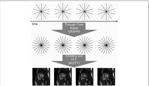
A schematic overview of the through-time radial GRAPPA reconstruction is shown in Figure 1. The GRAPPA weights generated from the fully-sampled data are applied to the undersampled radial data (Figure 1, top) to reconstruct fully-sampled radial data (Figure 1, middle), then trans-formed to the image domain (Figure 1, bottom) using the NUFFT [21]. The reconstruction required approximately three minutes per slice in the off-line implementation.
Quantitative parameter determination
The blood volume in the left ventricle for each of the datasets was assessed by a single physician to determine the end-diastolic volume (EDV), end-systolic volume (ESV) and ejection fraction (EF) for both imaging tech-niques. For the standard gated CMR images, only one composite heartbeat is available for analysis. The end-diastolic frame for the gold-standard images was deter-mined as the frame with the largest blood pool volume, and the end-systolic frame was determined as the frame with the smallest blood pool volume, automatically using Argus Ventricular Function software (Siemens Medical Solutions). For the real-time images, sixty cardiac images spanning several cardiac cycles were collected for each slice. The first full cardiac cycle acquired for each slice was chosen and EDV and ESV were determined for each individual slice using Argus as described above. No con-trol for breath-hold state or image registration was performed.
Qualitative image review
visualization of myocardium, blood pool contrast, and car-diac motion. Images were also reviewed for the presence of artifacts (including artifacts due to respiration or mis-gating, as well as parallel imaging reconstruction and ra-dial streak artifacts). Artifacts were graded on a 5 point scale: no artifact (1); minimal artifact not affecting volu-metric analysis (2), mild artifact affecting voluvolu-metric ana-lysis (3), moderate artifact affecting volumetric anaana-lysis (4); extensive artifact affecting volumetric analysis (5).
Statistical analysis
Statistical analysis was performed using commercially available software (Excel 12.3.6, Stata 11.2). Linear re-gression analyses were performed and Bland-Altman plots [22] were generated to evaluate the agreement between the two methods in the estimation of EDV, ESV and EF. Two-sided t-tests with unequal variance assumption were used in testing the equality of means of EDV, ESV and EF measures obtained from the two methods. A two-sample t-test was performed to compare the scan durations be-tween the gold-standard scans and the real-time scans. Average scores for each metric of reviewer ratings were computed and the distributions were compared using the Wilcoxon test for the equality of the scores’distributions. Finally, a two-sample test of proportion was used to analyze the presence and absence of artifacts.
Results
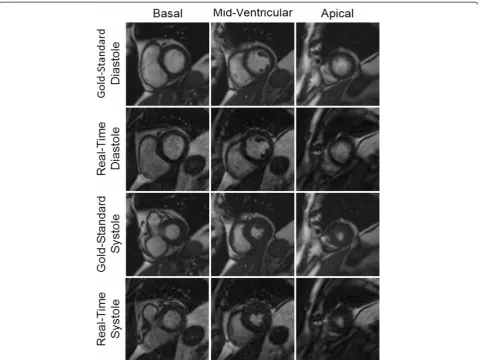
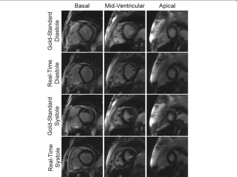
Example images collected using both the gold-standard breath-hold gated scan as well as the real-time, free-breathing and ungated images with radial GRAPPA are shown in Figures 2 and 3. For each patient, multiple slice positions in end-systole and end-diastole are shown in order to demonstrate the relative image quality.
[image:4.595.57.540.88.366.2]variance for the differences in the two EF measurements was 5.1%.
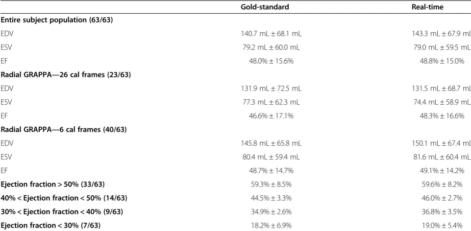
A total of 33 of the 63 subjects had EF values above 50%, 14 had EF values between 40-50%, 9 had EF values between 30-40%, and 7 had EF values of less than 30%. The average and standard deviations for the ESV, EDV, and EF values broken down into these EF ranges for both the gold-standard and real-time radial GRAPPA scans are also shown in Table 1 so that the two methods can be compared in patients with different levels of car-diac function. Of the 63 patients, two patients crossed the 35% EF threshold commonly used to determine EF dys-function. For the first of these patients, the breath-hold EF value was 34.0% and the real-time EF was 35.4%, and for the second patient, the breath-hold EF was 33.9% and the real-time EF was 35.2%. Please note that the images for this second patient are shown in Figure 3.
[image:5.595.59.538.88.448.2]and 0.98 for the EDV and ESV, respectively. The Bland-Altman analysis of the EDV data showed that the 95% limits of agreement (−29.0 mL, 23.9 mL) contains 93.6% (59/63) of the difference scores, and the mean difference of the measurements was−2.6 mL. Similarly, the 95% limits of agreement of ESV measurements (−17.0 mL, 17.6 mL) contains 93.6% (59/63) of the difference scores, and the mean difference was 0.3 mL. For all three quantitative pa-rameters, namely EF, EDV and ESV, the mean differences are small (approximately 1%) and limits of agreement are narrow, indicating that the two methods are systematically producing similar results. The hypothesis tests of equal mean show that the means of EF, EDV, and ESV obtained from the two methods are equivalent with p-values 0.77, 0.82, and 0.97, respectively.
The start and end times for both the gold-standard and radial GRAPPA scans were recorded for all subjects. The mean and standard deviation of the scan duration for the gold-standard functional imaging was 5.8 ± 1.9 minutes. For the real-time radial GRAPPA scans using 26 cali-bration frames, the mean and standard deviation of the scan duration was 4.6 ± 0.8 minutes, and for the radial GRAPPA scans using six calibration frames, the mean and standard deviation of the scan duration was 2.8 ± 0.4 minutes. Based on the two-sample t-test, the scan time was shortened by a statistically significant amount when using the real-time method (p < 0.001) either with 26 or six calibration frames.
[image:6.595.60.538.90.445.2]radial GRAPPA scans (1 ± 0) exhibited artifacts and re-viewer two indicated that 13 gold-standard (1.3 ± 0.6) and two real-time scans (with 26 calibration frames) (1.0 ± 0.1) contained such artifacts. The two-sample test of proportion showed that this is a statistically signifi-cant difference (p < 0.01), indicating that the use of real-time imaging in the form of radial GRAPPA reduces the number of image exhibiting artifacts.
[image:7.595.56.537.100.336.2]Results of the qualitative image feature ratings and statistical analysis are summarized in both Table 3 and Figure 7. As indicated in Table 3, the overall averages in each category were above 3, indicating that the feature visibility for both types of imaging methods was good to excellent. Figure 7 shows that the ratings were almost exclusively chosen to be 3 or 4 (“good”or“excellent”) al-though there were some exceptions. For instance, both
Figure 4Linear regression (left) and Bland-Altman plot (right) for the ejection fraction values.Values from all 63 subjects are included in both plots. Gray diamonds denote subjects for which only 6 calibration frames were used when performing the radial GRAPPA reconstruction, and black circles denote subjects for which 26 calibration frames were used. In the Bland-Altman plot, the mean of the two measurements is plotted on the x-axis, and the difference (gold-standard–radial GRAPPA) on the y-axis. The mean difference and 95% limits of agreement are noted on the plot.
Table 1 Summary of Quantitative Assessment
Gold-standard Real-time
Entire subject population (63/63)
EDV 140.7 mL ± 68.1 mL 143.3 mL ± 67.9 mL
ESV 79.2 mL ± 60.0 mL 79.0 mL ± 59.5 mL
EF 48.0% ± 15.6% 48.8% ± 15.0%
Radial GRAPPA—26 cal frames (23/63)
EDV 131.9 mL ± 72.5 mL 131.5 mL ± 68.7 mL
ESV 77.3 mL ± 62.3 mL 74.4 mL ± 58.9 mL
EF 46.6% ± 17.1% 48.3% ± 16.6%
Radial GRAPPA—6 cal frames (40/63)
EDV 145.8 mL ± 65.8 mL 150.1 mL ± 67.4 mL
ESV 80.4 mL ± 59.4 mL 81.6 mL ± 60.4 mL
EF 48.7% ± 14.7% 49.1% ± 14.2%
Ejection fraction > 50% (33/63) 59.3% ± 8.5% 59.6% ± 8.2%
40% < Ejection fraction < 50% (14/63) 44.5% ± 3.3% 46.0% ± 2.7%
30% < Ejection fraction < 40% (9/63) 34.9% ± 2.6% 36.8% ± 3.5%
Ejection fraction < 30% (7/63) 18.2% ± 6.9% 19.0% ± 5.4%
[image:7.595.57.545.487.675.2]reviewers found the mitral valves to be more poorly visu-alized in the radial GRAPPA images in comparison to the standard images. One reviewer indicated that papillary muscle visualization was “poor” for both imaging tech-niques in one subject, although the rest of the ratings were in the“good”and“excellent”categories. No other features
besides these two were rated as “poor” (or 2) for either type of scan, and no features for either type of image were rated as“not visible”(or 1) by either reader.
[image:8.595.57.540.100.363.2]Statistical analyses using the Wilcoxon test were per-formed using all 63 subjects together, and also on each subgroup with different numbers of calibration frames
Table 2 Summary of Results of Statistical Analysis of Quantitative Parameters
Ejection fraction End diastolic volume End systolic volume
For all subjects (63/63)
R2 0.98 0.96 0.98
Mean difference −0.9% −2.6 mL 0.3 mL
Lower 95% limit of agreement −5.3% −29.0 mL −17.0 mL
Upper 95% limit of agreement 3.6% 23.9 mL 17.6 mL
p-value 0.77 0.82 0.97
Radial GRAPPA with 26 calibration frames (23/63)
R2 0.98 0.98 0.99
Mean difference −1.7% 0.4 mL 2.9 mL
Lower 95% limit of agreement −6.1% −21.0 mL −10.1 mL
Upper 95% limit of agreement 2.7% 21.7 mL 15.9 mL
p-value 0.73 0.99 0.87
Radial GRAPPA with 6 calibration frames (40/63)
R2 0.98 0.95 0.97
Mean difference −0.4% −4.3 mL −1.2 mL
Lower 95% limit of agreement −4.7% −33.1 mL −20.1 mL
Upper 95% limit of agreement 3.9% 24.5 mL 17.7 mL
p-value 0.90 0.78 0.93
A summary of the R2
values from the regression analysis, the mean differences and upper and lower 95% limits of agreement from the Bland-Altman plots, and the p-values from the hypothesis tests of equal mean for the EF, ESV, and EDVs calculated using the two different imaging methods. The top third of the table shows values derived from all subjects, while the central and bottom thirds show the values when using only subsets of the subjects with the same number of calibration frames for the radial GRAPPA reconstructions.
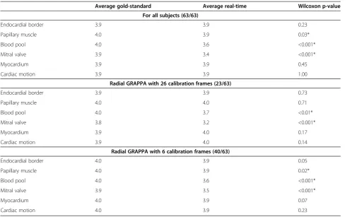
[image:8.595.58.545.503.684.2]for the radial GRAPPA reconstruction. The Wilcoxon tests showed that the image quality scores for the myocar-dium (p = 0.45 overall, p = 0.07 with 6 calibration frames, p = 0.17 with 26 calibration frames) and cardiac motion (p = 1.00 overall, p = 0.23 with 6 calibration frames, p = 0.14 with 26 calibration frames) follow the same distribution; this indicates that neither set of images was preferred
[image:9.595.58.541.89.245.2]for these features. The reviewers strongly preferred the standard images for blood pool contrast (p < 0.001 overall and with 6 calibration frames, and p < 0.01 with 26 cali-bration frames) and mitral valve visualization (p < 0.001 overall and for both subgroups). For the visualization of the endocardial border, there was no statistically signifi-cant difference between the gold-standard scans and the
Table 3 Summary of Reviewer Ratings
Average gold-standard Average real-time Wilcoxon p-value
For all subjects (63/63)
Endocardial border 3.9 3.9 0.23
Papillary muscle 4.0 3.9 0.03*
Blood pool 4.0 3.6 <0.001*
Mitral valve 3.9 3.4 <0.001*
Myocardium 3.9 3.9 0.45
Cardiac motion 3.9 3.9 1.00
Radial GRAPPA with 26 calibration frames (23/63)
Endocardial border 3.9 3.9 0.73
Papillary muscle 4.0 4.0 0.71
Blood pool 4.0 3.7 <0.01*
Mitral valve 3.8 3.2 <0.001*
Myocardium 3.9 4.0 0.17
Cardiac motion 3.9 4.0 0.14
Radial GRAPPA with 6 calibration frames (40/63)
Endocardial border 4.0 3.9 0.05
Papillary muscle 4.0 3.9 0.02*
Blood pool 4.0 3.6 <0.001*
Mitral valve 3.9 3.5 <0.001*
Myocardium 4.0 3.9 0.07
Cardiac motion 4.0 3.9 0.23
[image:9.595.56.555.411.718.2]Average results and p-values of the reviewer ratings for image features. The subject population was examined as a whole, and statistics were also calculated for the two subsets of images with different numbers of calibration frames for the radial GRAPPA reconstruction. Stars denote statistically significant differences.
radial GRAPPA reconstructions when 26 calibration frames were used (p = 0.73), However, when only six calibration frames were used for the radial GRAPPA, the p-value drops substantially (p = 0.05). Similar results are found for the visualization of the papillary muscles, where the image rat-ings show no statistically significant differences when 26 calibration frames are (p = 0.71), but significant differences when only six frames are used (p = 0.02).
Discussion
This study demonstrates that through-time radial GRAPPA used for real-time, free-breathing CMR imaging produces equivalent results for volumetric analysis of the left ven-tricle when compared to the current gold-standard CMR imaging technique. The high R2 values in the linear re-gressions, small mean differences and narrow 95% limits of agreement in the Bland-Altman plots, and large p-values for EF, EDV, and ESV indicate that the radial GRAPPA images can be used to obtain similar volumetric information as the standard scans. The two types of scans are equivalent regardless of the number of calibration frames used for the radial GRAPPA reconstruction;
reconstructions with 26 and six frames have high R2 and p-values. Prior studies [23-27] have indicated that ejection fraction measurements with a variance of 10% can be considered equivalent, and the data presented here have a smaller variance of only 5.1%. Additionally, a com-parison of average and standard deviations of the ESV, EDV, and EF values measured using the gold-standard methods and the real-time approach for subjects sorted into ranges of EF > 50%, 40% < EF < 50%, 30% < EF < 40%, and EF < 30% shows that these values are similar for all ranges. These results indicate that the real-time approach offers similar volumetric estimates for all levels of systolic dysfunction.
[image:10.595.59.539.88.415.2]COPD [28,29]. Additionally, slice positions can change dur-ing the collection of images when usdur-ing a free-breathdur-ing ap-proach. Such motion may introduce small changes in the orientation of the heart within the chest, which could also result in differences in calculated EF values. Finally, the presence of arrhythmias in several cases and the resulting artifacts may lead to errors in the volumetric analysis. How-ever, despite these potential sources of differences between the EF values as measured with free-breathing and breath-hold methods, only small deviations were seen between dif-ferent scans in the same subject.
The statistical analysis performed based on reviewer ratings of artifacts demonstrated that the use of the ra-dial GRAPPA approach results in fewer image artifacts (p < 0.01 based on the two-sample test of proportion that could adversely affect functional assessment of the left ventricle. The presence of fewer artifacts in the radial GRAPPA images is most likely due to the lack of gating and breath-holding in the real-time scans; no misgating or failed breath-holding can occur when no gating or breath-holding is used.
The average scan times were significantly shorter (p < 0.001) when using the real-time radial GRAPPA approach, despite the need for calibration data. The shorter scan times were due to the lack of a need for breath-holding and recovery periods. In cases where only an evaluation of EF is needed, the use of the real-time method may make CMR more rapid, accurate, and cost-effective when compared to other methods of meas-uring EF. While only EF evaluation was explored in this study, radial GRAPPA can be applied to accelerate other portions of the CMR examination [30,31]. If such a real-time method were employed for the entire CMR study, significant and meaningful reduction in scan time could be achieved. As other CMR researchers have shown, such a reduction in scan time has several advantages including, but not limited to, improved patient comfort and reduc-tion in CMR and overall costs [11]. The true impact of real-time CMR may be the removal of the requirement of a steady cardiac rhythm and the ability to breath-hold, which could potentially lead to improved image quality in patients experiencing arrhythmias and who cannot hold their breath. Based on the results of this study and other similar studies using real-time CMR for patients with arrhythmia [16], it may be possible to measure EF in these patients using such real-time techniques.
The reviewer ratings of the image features indicate that the gold-standard cine imaging method offers superior visualization of several specific image features than the ra-dial GRAPPA approach. Previous qualitative assessments of real-time CMR images reconstructed with other tech-niques have shown that real-time image quality is slightly [16] to moderately [12,13] worse than the standard func-tional images. The real-time radial GRAPPA images
shown here have similar image quality for some features, and had only a modest drop in ratings for other features, when compared to the standard images. Overall, the rat-ings for the real-time images generated in this study were high, averaging between“good”and“excellent”for all cat-egories. For features including myocardium, endocardial border, and cardiac motion, all related to ejection fraction, statistical tests showed that there was no significant differ-ence between the gold-standard method and real-time imaging with radial GRAPPA. However, the analysis indi-cates that standard cine scans should be performed when specific anatomical structures (i.e. the mitral valve) must be assessed, when patients can provide the requisite breath-holds.
One limitation of the through-time radial GRAPPA tech-nique is the need for a potentially lengthy calibration phase, although this data collection can occur without breath-holding or ECG gating. In the case of 26 calibration frames, the calibration requires approximately four times longer than the collection of the actual imaging data. While using only six calibration frames greatly reduces the scan time, the resulting images may be blurry due to the need for a larger k-space segment for calibration [17]. In-deed, the statistical analysis of the ratings has shown that the use of more calibration frames for the radial GRAPPA reconstruction (26 vs. 6) may lead to better image quality when looking at some specific image features, such as endocardial border definition and visualization of the pap-illary muscles. In cases where these features are to be assessed in addition to calculating the ejection fraction, the use of radial GRAPPA with more calibration frames may be preferred despite the longer scan time. However, for both types of real-time calibration (i.e. with 26 and 6 frames), the overall scan times were significantly shorter than the standard clinical scan, even with the collection of calibration data included. A comparison between through-time radial GRAPPA and other real-time car-diac imaging techniques which do not require large amounts of calibration data has not been performed. However, the potential advantages of through-time ra-dial GRAPPA, including the high temporal resolution without temporal regularization or view-sharing and capability for true real-time image reconstruction, make this technique attractive despite the need for calibration data.
Clinical implications
general anesthesia to facilitate acquisition of sequences that require breath-holding.
Conclusion
Real-time CMR using through-time radial GRAPPA method has been shown to yield quantitative left ventricu-lar functional parameters equivalent to the gold-standard technique in an overall shorter scan time. While reviewers overall preferred the standard images, the radial GRAPPA images were also generally rated to have “excellent” to
“good” image quality, and the radial GRAPPA images exhibited fewer artifacts as compared to gold-standard images. Based on this validation study, it may be possible to replace traditional cine techniques with the radial GRAPPA approach in cases where only EF assessment is required, and to use real-time imaging with radial GRAPPA for EF evaluation in patients with difficulties breath-holding or arrhythmias.
Abbreviations
CMR:Cardiovascular Magnetic Resonance; GRAPPA: GeneRalized Autocalibrating Partially Parallel Acquisitions; HIPAA: The Health Insurance Portability and Accountability Act of 1996; IRB: Institutional Review Board; bSSFP: Balanced steady-state free precession; DICOM: Digital Imaging and Communications in Medicine; NUFFT: Non-Uniform Fast Fourier Transform; EDV: End-Diastolic Volume; ESV: End-Systolic Volume; EF: Ejection fraction; GPU: Graphics Processing Unit.
Authors’contributions
GA; guarantor of integrity of entire study, data acquisition and data analysis/ interpretation, manuscript revision for important intellectual content, literature research, clinical studies. VN; study concepts/study design and data acquisition and data analysis/interpretation, manuscript revision for important intellectual content, literature research, clinical studies. VY; data acquisition and data analysis/interpretation, clinical studies. PR; data analysis/interpretation, manuscript revision for important intellectual content. TJ; data analysis/ interpretation. AS; data analysis/interpretation. MG; experimental studies. VG; experimental studies, manuscript revision for important intellectual content. RG; study concepts/study design. NS; guarantor of integrity of entire study, study concepts/study design and data acquisition and data analysis/interpretation, manuscript revision for important intellectual content, literature research, experimental studies. All authors read and approved the final manuscript.
Competing interest
Research Support from Siemens Medical Solutions: Aandal, Griswold, Gulani, Seiberlich.
Patent licenses with Siemens, GE, Bruker: Griswold. None: Nadig, Yeh, Rajiah, Jenkins, Sattar, Gilkeson.
Funding sources
This work was funded by the NIH (R00EB011527, 1RO1HL094557, and UL1 RR024989) and Siemens Medical Solutions (Erlangen, Germany).
Author details
1Radiology, Case Western Reserve University and University Hospitals Case Medical Center, Cleveland, OH, USA.2Haraldsplass Deaconess Hospital, Bergen, Norway.3Cardiology, MetroHealth Medical Center at Case Western University, Cleveland, OH, USA.4Case Western Reserve University School of Medicine, Cleveland, OH, USA.5Division of Cardiovascular Medicine, Harrington Heart & Vascular Institute, University Hospitals Case Medical Center, Case Western Reserve University, Cleveland, OH, USA.6Epidemiology and Biostatistics, Case Western Reserve University, Cleveland, OH, USA. 7Biomedical Engineering, Case Western Reserve University, Room 309 Wickenden Building 2071 Martin Luther King Jr. Drive, Cleveland, OH 44106-7207, USA.
Received: 3 March 2014 Accepted: 1 September 2014
References
1. Karamitsos TD, Francis JM, Myerson S, Selvanayagam JB, Neubauer S. The role of cardiovascular magnetic resonance imaging in heart failure.
J Am Coll Cardiol.2009;54:1407–24.
2. Grothues F, Smith GC, Moon JC, et al.Comparison of interstudy reproducibility of cardiovascular magnetic resonance and 2D echocardiography in normal subjects and in patients with heart failure or left ventricular hypertrophy.Am J Cardiol.2002;90:29–34.
3. Schalla S, Nagel E, Lehmkuhl H, et al.Comparison of magnetic resonance real-time imaging of left ventricular function with conventional magnetic resonance imaging and echocardiography.Am J Cardiol.2001;87:95–9. 4. Kühl HP, Spuentrup E, Wall A, et al.Assessment of myocardial function
with interactive non-breath-hold real-time MR imaging: comparison with echocardiography and breath-hold Cine MR imaging.Radiology.2004; 231:198–207.
5. Beer M, Stamm H, Machann W, et al.Free breathing cardiac real-time cine MR without ECG triggering.Int J Cardiol.2010;145:380–2.
6. Brinegar C, Zhang H, Wu YJ, et al.Real-time cardiac MRI using prior spatial-spectral information [abstr].Paper presented at Conf Proc IEEE Eng Med Biol Soc.2009;2009:4383–6.
7. Kellman P, Chefd’hotel C, Lorenz CH, Mancini C, Arai AE, McVeigh ER.High spatial and temporal resolution cardiac cine MRI from retrospective reconstruction of data acquired in real time using motion correction and resorting.Magn Reson Med.2009;62:1557–64.
8. Sümbül U, Santos JM, Pauly JM.A practical acceleration algorithm for real-time imaging.IEEE Trans Med Imaging.2009;28:2042–51.
9. Naegel B, Cernicanu A, Hyacinthe JN, Tognolini M, Vallée JP.SNR enhancement of highly-accelerated real-time cardiac MRI acquisitions based on non-local means algorithm.Med Image Anal.2009;13:598–608. 10. Ding Y, Chung YC, Raman SV, Simonetti OP.Application of the Karhunen-Loeve
transform temporal image filter to reduce noise in real-time cardiac cine MRI.
Phys Med Biol.2009;54:3909–22.
11. Lurz P, Muthurangu V, Schievano S, et al.Feasibility and reproducibility of biventricular volumetric assessment of cardiac function during exercise using real-time radial k-t SENSE magnetic resonance imaging.J Magn Reson Imaging.2009;29:1062–70.
12. Bauer RW, Radke I, Block KT, et al.True real-time cardiac MRI in free breathing without ECG synchronization using a novel sequence with radial k-space sampling and balanced SSFP contrast mode.Int J Cardiovasc Imaging.2013; [E-pub ahead of print], doi:10.1007/s10554-013-0183-0.
13. Feng L, Srichai MB, Lim RP, et al.Highly accelerated real-time cardiac cine MRI using k-t SPARSE-SENSE.Magn Reson Med.2012; [E-pub ahead of print], doi:10.1002/mrm.24440.
14. Hansen MS, Sørensen TS, Arai AE, Kellman P.Retrospective reconstruction of high temporal resolution cine images from real-time MRI using iterative motion correction.Magn Reson Med.2012;68:741–50. 15. Uecker M, Zhang S, Voit D, Karaus A, Merboldt KD, Frahm J.Real-time MRI
at a resolution of 20 ms.NMR Biomed.2010;23:986–94.
16. Voit D, Zhang S, Unterberg-Buchwald C, Sohns JM, Lotz J, Frahm J. Real-time cardiovascular magnetic resonance at 1.5 T using balanced SSFP and 40 ms resolution.J Cardiovasc Magn Reson.2013;15:79. doi:10.1186/1532-429X-15-79.
17. Seiberlich N, Ehses P, Duerk J, Gilkeson R, Griswold M.Improved radial GRAPPA calibration for real-time free-breathing cardiac imaging.
Magn Reson Med.2011;65:492–505.
18. Griswold MA, Jakob PM, Heidemann RM, et al.Generalized autocalibrating partially parallel acquisitions (GRAPPA).Magn Reson Med.2002;47:1202–10. 19. Saybasili H, Herzka DA, Seiberlich N, Griswold MA.Real-time imaging with
radial GRAPPA: Implementation on a heterogeneous architecture for low-latency reconstructions.Magn Reson Imaging.2014;32(6):747–58. doi:10.1016/j.mri.2014.02.022. Epub 2014 Mar 15.
20. Raju VM, Sussman M, Pellow A, Griswold M, Seiberlich N, Wintersperger BJ. Undersampled real time cine SSFP with through-time radial GRAPPA: analysis of global LV function and ventricular mass.Proc RSNA.2013; LL-CAS-TH6B.
21. Fessler JA.On NUFFT-based gridding for non-Cartesian MRI.J Magn Reson.
22. Bland JM, Altman DG.Statistical methods for assessing agreement between two methods of clinical measurement.Lancet.1986;1(8476):307–10. 23. Pattynama PM, Lamb HJ, Van der Velde EA, van der Wall EE, de Roos A.Left
ventricular measurements with cine and spin echo MR imaging: a study of reproducibility with variance component analysis.Radiology.1993; 187:261–8.
24. Cohn PF, Levine JA, Bergeron GA, Gorlin R.Reproducibility of the angiographic left ventricular ejection fraction in patients with coronary artery disease.Am Heart J.1974;88:713–20.
25. Gordon EP, Schnittger I, Fitzgerald PJ, Williams P, Popp RL.Reproducibility of left ventricular volumes by two-dimensional echocardiography.
J Am Coll Cardiol.1983;2:506–13.
26. Dorosz JL, Lezotte DC, Weitzenkamp DA, Allen LA, Salcedo EE.Performance of 3-dimensional echocardiography in measuring left ventricular volumes and ejection fraction: a systematic review and meta-analysis.J Am Coll Cardiol.2012;59:1799–808.
27. Malm S, Frigstad S, Sagberg E, Larsson H, Skjaerpe T.Accurate and reproducible measurement of left ventricular volume and ejection fraction by contrast echocardiography: a comparison with magnetic resonance imaging.J Am Coll Cardiol.2004;44:1030–5.
28. Buda AJ, Pinsky MR, Ingels NB Jr, Daughters GT, Stinson EB, Alderman EL. Effect of intrathoracic pressure on left ventricular performance.N Engl J Med.1979;301:453–9.
29. Yoshida S, Wu D, Fukumoto M, Akagi N, Seguchi H.Quantitative study of the difference in pulmonary perfusion in different respiratory phases in healthy volunteers.Ann Nucl Med.2002;16:533–9.
30. Hamilton JI, Barkauskas K, Seiberlich N.Accelerated 2D multi-slice first-pass contrast-enhanced myocardial perfusion using through-time radial GRAPPA.JCMR.2014;16(Suppl 1):378.
31. Sayin O, Saybasili H, Halperin H, Zviman M, Griswold MA, Seiberlich N, Herzka DA.Accelerated delayed enhancement imaging of myocardial infarction with through-time radial GRAPPA.JCMR.2014;16(Suppl 1):W6.
doi:10.1186/s12968-014-0079-8
Cite this article as:Aandalet al.:Evaluation of left ventricular ejection fraction using through-time radial GRAPPA.Journal of Cardiovascular Magnetic Resonance201416:79.
Submit your next manuscript to BioMed Central and take full advantage of:
• Convenient online submission
• Thorough peer review
• No space constraints or color figure charges
• Immediate publication on acceptance
• Inclusion in PubMed, CAS, Scopus and Google Scholar
• Research which is freely available for redistribution