Copyright © 2003, American Society for Microbiology. All Rights Reserved.
Multilocus Sequence Typing System for Group B Streptococcus
Nicola Jones,
1* John F. Bohnsack,
2Shinji Takahashi,
3Karen A. Oliver,
1Man-Suen Chan,
4Frank Kunst,
5Philippe Glaser,
5Christophe Rusniok,
5Derrick W. M. Crook,
1Rosalind M. Harding,
6Naiel Bisharat,
1and Brian G. Spratt
7Nuffield Department of Clinical Laboratory Sciences
1and Paediatric Molecular Medicine, Institute for Molecular Medicine,
4John Radcliffe Hospital, Oxford OX3 9DU, The Peter Medawar Building for Pathogen Research, University of Oxford,
Oxford OX1 3SY,
6and Department of Infectious Disease Epidemiology, Faculty of Medicine, Imperial College,
St. Mary’s Hospital, London W2 1PG,
7United Kingdom; Department of Pediatrics, University of Utah
Health Sciences Center, Salt Lake City, Utah 84132
2; Division of Microbiology, Joshi-Eiyoh
University, Chiyoda, Sakado, Saitama 350-0288, Japan
3; and Laboratoire de Ge´nomique
des Microorganismes Pathoge`nes, Institut Pasteur, 75724 Paris Cedex 15, France
5Received 29 January 2003/Returned for modification 7 March 2003/Accepted 17 March 2003
A multilocus sequence typing (MLST) system was developed for group B streptococcus (GBS). The system
was used to characterize a collection (
n
ⴝ
152) of globally and ecologically diverse human strains of GBS that
included representatives of capsular serotypes Ia, Ib, II, III, V, VI, and VIII. Fragments (459 to 519 bp) of seven
housekeeping genes were amplified by PCR for each strain and sequenced. The combination of alleles at the
seven loci provided an allelic profile or sequence type (ST) for each strain. A subset of the strains were
characterized by restriction digest patterning, and these results were highly congruent with those obtained with
MLST. There were 29 STs, but 66% of isolates were assigned to four major STs. ST-1 and ST-19 were
significantly associated with asymptomatic carriage, whereas ST-23 included both carried and invasive strains.
All 44 isolates of ST-17 were serotype III clones, and this ST appeared to define a homogeneous clone that was
strongly associated with neonatal invasive infections. The finding that isolates with different capsular serotypes
had the same ST suggests that recombination occurs at the capsular locus. A web site for GBS MLST was set up
and can be accessed at http://sagalactiae.mlst.net. The GBS MLST system offers investigators a valuable typing
tool that will promote further investigation of the population biology of this organism.
Streptococcus agalactiae
, group B streptococcus (GBS), is an
important human pathogen. It is the leading cause of neonatal
sepsis in the United Kingdom (18) and the United States (23).
It is regarded as an emerging pathogen in the elderly (13) and
is a frequent cause of maternal sepsis. However, GBS is usually
a commensal organism and can be isolated from the
genito-urinary and gastrointestinal tracts of up to 35% of healthy
adults (1).
Capsular serotyping has been one of the mainstays in the
descriptive epidemiology of GBS. Nine capsular serotypes
have been described (Ia, Ib, and II to VIII). Serotype III GBS
strains are of particular importance, as they are responsible for
the majority of infections, including meningitis, in neonates
worldwide (22). Diverse lineages of serotype III strains can be
distinguished with multilocus enzyme electrophoresis (12, 19),
pulsed-field gel electrophoresis (20), and restriction digest
pat-tern (RDP) analysis (2), and the lineages appear to vary in
pathogenic potential.
Multilocus sequence typing (MLST) is an unambiguous
se-quence-based typing method that involves sequencing
approx-imately 500-bp fragments of seven housekeeping genes and has
been used successfully to type strains and investigate the
pop-ulation structure of a number of human bacterial pathogens,
including
Neisseria meningitidis
(16) and
Streptococcus
pneu-moniae
(9). MLST is particularly suitable for epidemiological
studies because it provides data that can easily be compared
between laboratories over the Internet.
The primary aim of this study was to develop an MLST
system for GBS. Secondary aims were to show that the system
could be used on a diverse globally derived collection of strains
isolated from neonates and adults and that the system could
distinguish between strains that were different with capsular
serotyping and RDP typing.
MATERIALS AND METHODS
Strain collection.The study collection consisted of 152 isolates of GBS from North America, New Zealand, Thailand, Singapore, Israel, Japan, and the United Kingdom. In addition, two well-characterized strains were included, the
[image:1.603.302.542.582.715.2]* Corresponding author. Mailing address: Nuffield Department of
Clinical Laboratory Sciences, Level 7, John Radcliffe Hospital, Oxford
OX3 9DU, United Kingdom. Phone: 44 1865 220855. Fax: 44 1865
220890. E-mail: [email protected].
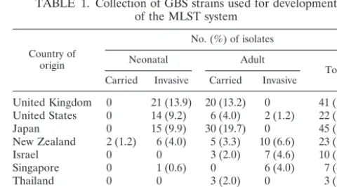
TABLE 1. Collection of GBS strains used for development
of the MLST system
Country of origin
No. (%) of isolates
Neonatal Adult
Total Carried Invasive Carried Invasive
United Kingdom 0 21 (13.9) 20 (13.2) 0 41 (27.1) United States 0 14 (9.2) 6 (4.0) 2 (1.2) 22 (14.4)
Japan 0 15 (9.9) 30 (19.7) 0 45 (29.6)
New Zealand 2 (1.2) 6 (4.0) 5 (3.3) 10 (6.6) 23 (15.1)
Israel 0 0 3 (2.0) 7 (4.6) 10 (6.6)
Singapore 0 1 (0.6) 0 6 (4.0) 7 (4.6)
Thailand 0 0 3 (2.0) 0 3 (2.0)
Unknown* 0 1 (0.6) 0 0 1 (0.6)
Total 2 (1.2) 58 (38.2) 67 (44.2) 25 (16.4) 152
*NEM316 (ATCC 12403).
2530
on May 15, 2020 by guest
http://jcm.asm.org/
NCTC-8541 strain (isolated from a vaginal carrier; Public Health Laboratory, United Kingdom) and the NEM316 strain (ATCC 12403, isolated from a case of fatal neonatal sepsis, country of origin unknown), whose genome has been fully sequenced (11). The collection was globally diverse and included strains from asymptomatic carriers as well as human infections. Most capsular serotypes of GBS were represented, although serotypes IV and VII, which are rarely associ-ated with disease in humans, were not included. Table 1 further describes the strain collection.
Identification of GBS.Isolates were grown on blood agar and identified as group B streptococcus by the following criteria (21):-hemolysis on a Columbia agar plate containing 5% horse blood (two strains of the 152 analyzed were nonhemolytic), Gram staining showing gram-positive cocci in pairs or short chains, negative reaction with catalase reagent, and Lancefield grouping with type B antisera (Oxoid, Basingstoke, United Kingdom).
Characterization of strains.Capsular serotyping was carried out on all strains in the laboratory of John Bohnsack. Capsular serotyping was repeated in a second laboratory with different method for 25 (16.5%) randomly selected iso-lates (Streptococcal Reference Laboratory, Central Public Health Laboratory, Colindale, United Kingdom [n⫽20], and Clinical Microbiology Laboratory, KK Women’s and Children’s Hospital, Singapore [26] [n⫽5]). Forty strains had previously been characterized by RDP (4).
DNA extraction.The DNeasy kit (Qiagen GmbH) was used to extract DNA, and the gram-positive bacterial protocol was followed. A single colony of each strain was streaked across a Columbia agar plate containing 5% horse blood. Several colonies were picked off into phosphate-buffered saline and centrifuged at 5,500⫻g. The cell pellet was resuspended in 180l of enzymatic lysis buffer containing lysozyme (20 mg/ml) and incubated for 30 min at 37°C. Then 25l of proteinase K (10 mg/ml) was added, and incubation was continued at 70°C for 30 min. The DNA in the clear viscous lysates was precipitated with 95% (vol/vol) ethanol and added to DNeasy minicolumns. Ethanol (70%, vol/vol)-based buffers AW1 and AW2 were added sequentially to the columns and centrifuged at 5,500 ⫻g. The supernatants were discarded, and the DNA was resuspended in sterile water and stored at⫺20°C.
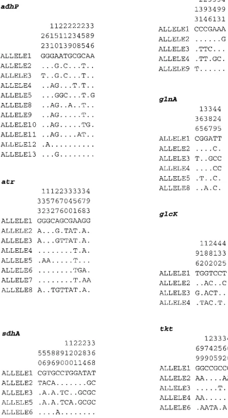
Choice of loci for MLST.Ten candidate loci, encoding enzymes involved in intermediary metabolism, were identified by searching the genome sequence of the GBS strain NEM316 (11) with homologous sequences from other bacteria. Suitable genes were then chosen on the basis of chromosomal location and sequence diversity observed in pilot studies with a restricted set of GBS strains. Three genes were excluded, two because they failed to distinguish between GBS strains and one which was not reliably amplified. The following seven loci were selected for the MLST scheme (Table 2): alcohol dehydrogenase gbs0054 (adhP), phenylalanyl tRNA synthetase (pheS), amino acid transporter gbs0538 (atr), glutamine synthetase (glnA), serine dehydratase gbs2105 (sdhA), glucose kinase gbs0518 (glcK), and transketolase gbs2105 (tkt). The chromosomal loca-tions of these housekeeping loci (Table 2) suggested that it was unlikely for any of them to be coinherited in the same recombination event, as the minimum distance between two loci was 20 kb.
Amplification and nucleotide sequence determination.PCR products were amplified with oligonucleotide primer pairs designed from the NEM316 GBS genome sequence (11). A range of primers were tested, with those shown in Table 3 providing reliable amplification from a diverse range of GBS isolates. Each 50-l amplification reaction mixture comprised 10 ng of GBS chromosomal DNA, 100 pmol of each PCR primer (MWG Biotech, Ebersberg, Germany), 1⫻ PCR buffer with 1.5 mM MgCl2(Qiagen GmbH), 0.5 U ofTaqDNA polymerase
(Qiagen GmbH), and 1.6 mM deoxynucleoside triphosphate mix (ABgene, Ep-som, United Kingdom). The reaction conditions were denaturation at 94°C for 1 min, primer annealing at 55°C for 45 s, and extension at 72°C for 1 min for 30 cycles.
[image:2.603.43.542.81.171.2]The amplification products were purified by precipitation with 20% polyeth-ylene glycol and 2.5 M NaCl (8), and their nucleotide sequences were determined at least once on each DNA strand with internal nested primers (Table 3) and ABI Prism BigDye Terminators version 3.0 reaction mix (Applied Biosystems, Foster City, Calif.) in accordance with the manufacturer’s instructions. Unincor-porated dye terminators were removed by precipitation of the termination prod-ucts with sodium acetate (3 M, pH 5.2) and 95% (vol/vol) ethanol, and the reaction products were separated and detected with an ABI Prism 3700 DNA
TABLE 2. Characteristics of loci included in the GBS MLST system
aLocus Putative function of gene Size of sequencedfragment (bp) No. of allelesidentified No. (%) of polymorphicnucleotide sites G%⫹C dn/ds Position in GBSgenomeb(bp)
adhP Alcohol dehydrogenase (gbs0054) 498 11 12 (2.4) 43.1 0.13 72286
pheS Phenylalanyl tRNA synthetase 501 5 7 (1.4) 37.1 0.17 912817
atr Amino acid transporter (gbs0538) 501 8 12 (2.4) 36.9 0.14 560085
glnA Glutamine synthetase 498 6 6 (1.2) 35.7 0.12 1868862
sdhA Serine dehydratase (gbs2105) 519 6 13 (2.5) 41.4 0.12 2179923
glcK Glucose kinase (gbs0518) 459 4 7 (1.5) 42.6 0.13 538770
tkt Transketolase (gbs0268) 480 5 8 (1.7) 38.9 0.42 287111
aGenes:adhP, alcohol dehydrogenase (gbs0054);pheS, phenylalanyl tRNA synthetase;atr, amino acid transporter (gbs0538);glnA, glutamine synthetase;sdhA, serine dehydratase (gbs2105);glcK, glucose kinase (gbs0518);tkt, transketolase (gbs2105). Alleles of the seven housekeeping loci can be obtained at http://sagalactiae.mlst.net.
[image:2.603.47.541.562.724.2]bFrom reference 11.
TABLE 3. Oligonucleotide primers for GBS MLST
Locus Use Name and sequence of primer Ampliconsize (bp)
Forward (5⬘to 3⬘) Reverse (5⬘to 3⬘)
adhP
Amplification
GTTGGTCATGGTGAAGCACT
ACTGTACCTCCAGCACGAAC
672
Sequencing
GGTGTGTGCCATACTGATTT
ACAGCAGTCACAACCACTCC
498
pheS
Amplification
GATTAAGGAGTAGTGGCACG
TTGAGATCGCCCATTGAAAT
723
Sequencing
ATATCAACTCAAGAAAAGCT
TGATGGAATTGATGGCTATG
501
atr
Amplification
CGATTCTCTCAGCTTTGTTA
AAGAAATCTCTTGTGCGGAT
627
Sequencing
ATGGTTGAGCCAATTATTTC
CCTTGCTCAACAATAATGCC
501
glnA
Amplification
CCGGCTACAGATGAACAATT
CTGATAATTGCCATTCCACG
589
Sequencing
AATAAAGCAATGTTTGATGG
GCATTGTTCCCTTCATTATC
498
sdhA
Amplification
AGAGCAAGCTAATAGCCAAC
ATATCAGCAGCAACAAGTGC
646
Sequencing
AACATAGCAGAGCTCATGAT
GGGACTTCAACTAAACCTGC
519
glcK
Amplification
CTCGGAGGAACGACCATTAA
CTTGTAACAGTATCACCGTT
607
Sequencing
GGTATCTTGACGCTTGAGGG
ATCGCTGCTTTAATGGCAGA
459
tkt
Amplification
CCAGGCTTTGATTTAGTTGA
AATAGCTTGTTGGCTTGAAA
859
Sequencing
ACACTTCATGGTGATGGTTG
TGACCTAGGTCATGAGCTTT
480
on May 15, 2020 by guest
http://jcm.asm.org/
analyzer (Applied Biosystems). Sequences were assembled from the resultant chromatograms with the Staden suite of computer programs and edited to resolve any ambiguities (24).
Allele and sequence type assignment.For each locus, every different sequence was assigned a distinct allele number in order of identification; these were internal fragments of the gene which contained an exact number of codons. Any change in the nucleotide sequence, whether or not the amino acid sequence was altered, was defined as a new allele. Each isolate was therefore designated by a seven-integer number, constituting its allelic profile. Isolates with the same allelic profile were assigned to the same sequence type (ST), which were numbered in the order of their identification (ST-1, ST-2, etc.). The data have been deposited in a database accessible on the Internet at http://sagalactiae.mlst.net.
Computational analyses.Determination of the number of polymorphic nucle-otide sites, calculation ofdn/ds, wherednis nonsynonymous substitutions andds is synonymous substitutions, and construction of dendrograms with the un-weighted pair group method with arithmetic mean (UPGMA) were performed with START (http://www.mlst.net) (14). STs were grouped into lineages or clonal complexes with BURST (START version 1.05, http://www.mlst.net [14]). The members of a BURST lineage were defined as groups of two or more indepen-dent isolates where each isolate had iindepen-dentical alleles at six or more loci with at least one other member of the group.
RESULTS
Variation at the seven MLST loci.
The sequences of the
seven chosen loci were determined for the 152 strains, and
allelic profiles were assigned. The alleles defined for the MLST
scheme were based on sequence lengths of between 459 (
glcK
)
and 519 bp (
sdhA
). Between four (
glcK
) and 11 alleles (
adhP
)
were present at each locus. The average number of alleles at
each locus was 6.4, providing the potential to distinguish 4.4
⫻
10
5different genotypes. The proportion of variable nucleotide
sites present in the selected housekeeping genes ranged from
1.2% (
glnA
) to 2.5% (
sdhA
) (Table 2 and Fig. 1). The
propor-tions of nucleotide alterapropor-tions that changed the amino acid
sequence (nonsynonymous substitutions,
dn
) and the
propor-tions of silent changes (synonymous substitupropor-tions,
ds
) were
calculated for each gene. With these data, the
dn
/
ds
ratios were
calculated for all seven loci and were all
⬍
1 (Table 2).
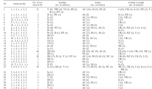
Relatedness of GBS isolates.
The 152 isolates were resolved
into 29 STs, 14 of which were identified only once (Table 4).
One hundred and one isolates (66.5% of the data set) were
represented by one of four STs, ST-1, ST-17, ST-19, and ST-23.
The most common ST (ST-17) was identified 44 times in the
data set, followed by ST-1 (21 isolates), ST-19 (20 isolates),
and ST-23 (16 isolates). ST-3 was not identified in this data set
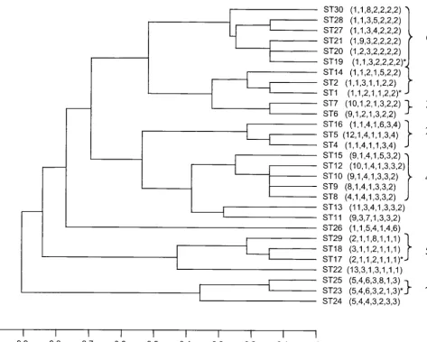
but had been identified in a pilot study. UPGMA was used to
construct a dendrogram from the matrix of pairwise allelic
differences between the 29 STs of all 152 isolates (Fig. 2).
BURST grouped the isolates into seven lineages (Fig. 2),
which approximated well with the clusters of STs obtained by
UPGMA.
Relationship between ST, capsular serotype, and restriction
digest patterns.
Capsular serotype was known for all 152
[image:3.603.32.543.81.380.2]strains (Table 5), and there was complete correlation
be-tween capsular serotyping results among three laboratories.
Five strains proved to be nontypeable. Serotype III was most
common (78 strains, 44 of which belonged to ST-17),
fol-lowed by serotypes Ia (19 strains), Ib (17 strains), V (15
strains), II (8 strains), VI (6 strains), and VIII (4 strains).
TABLE 4. Characteristics of GBS isolates according to ST
aST Allelic profile No. of iso-lates in ST (no. of isolates)Serotype (no. of isolates)Source Country of origin(no. of isolates)
1 1, 1, 2, 1, 1, 2, 2 21 V (9), VIII (4), VI (4), III (2),
Ib (1), NT (1) AC (16), AI (3), NI (2) J (10), UK (4), Is (3), NZ (3), T (1)
2 1, 1, 3, 1, 1, 2, 2 2 II (1), NT (1) AI (2) Is (1), NZ (1)
4 1, 1, 4, 1, 1, 3, 4 2 Ia (2) AC (1), NI (1) J (1), UK (1)
5 12, 1, 4, 1, 1, 3, 4 1 Ia (1) AC (1) J (1)
6 9, 1, 2, 1, 3, 2, 2 1 Ib (1) AC (1) UK (1)
7 10, 1, 2, 1, 3, 2, 2 3 Ia (3) AC (1), NI (2) J (3)
8 4, 1, 4, 1, 3, 3, 2 7 Ib (6), NT (1) AC (4), NI (1), AI (2) UK (3), NZ (2), J (1), S (1)
9 8, 1, 4, 1, 3, 3, 2 1 Ib (1) AC (1) Is (1)
10 9, 1, 4, 1, 3, 3, 2 5 Ib (3), II (1), NT (1) AC (2), NI (1), AI (2) UK (2), NZ (2), J (1)
11 9, 3, 7, 1, 3, 3, 2 5 III (5) AI (5) S (5)
12 10, 1, 4, 1, 3, 3, 2 3 Ib (3) NI (2), AC (1) J (2), UK (1)
13 11, 3, 4, 1, 3, 3, 2 1 VI (1) AC (1) T (1)
14 1, 1, 2, 1, 5, 2, 2 1 VI (1) AC (1) T (1)
15 9, 1, 4, 1, 5, 3, 2 2 Ib (2) AI (1), NI (1) NZ (2)
16 1, 1, 4, 1, 6, 3, 4 1 Ia (1) AI (1) Is (1)
17 2, 1, 1, 2, 1, 1, 1 44 III (44) NI (33), AC (9), AI (2) US (16), J (14), UK (13), NZ (1)
18 3, 1, 1, 2, 1, 1, 1 1 III (1) NI (1) UK (1)
19 1, 1, 3, 2, 2, 2, 2 20 III (17), II (1), V (1), NT (1) AC (14), NI (3) AI (2), NC (1) UK (8), NZ (5), US (5), J (2)
20 1, 2, 3, 2, 2, 2, 2 1 III (1) NI (1) UK (1)
21 1, 9, 3, 2, 2, 2, 2 1 III (1) NI (1) J (1)
22 13, 3, 1, 3, 1, 1, 1 2 II (2) AC (1), AI (1) Is (2)
23 5, 4, 6, 3, 2, 1, 3 16 Ia (11), III (4), V (1) AC (7), NI (5), AI (3), NC (1) NZ (7), UK (3), J (3), Is (1), S (1), NK (1)
24 5, 4, 4, 3, 2, 3, 3 1 Ia (1) AC (1) UK (1)
25 5, 4, 6, 3, 8, 1, 3 1 III (1) NI (1) US (1)
26 1, 1, 5, 4, 1, 4, 6 3 V (3) AC (2), NI (1) J (2), UK (1)
27 1, 1, 3, 4, 2, 2, 2 1 III (1) AI (1) Is (1)
28 1, 1, 3, 5, 2, 2, 2 3 II (3) AC (2), NI (1) J (2), UK (1)
29 2, 1, 1, 8, 1, 1, 1 1 III (1) NI (1) J (1)
30 1, 1, 8, 2, 2, 2, 2 1 V (1) AC (1) J (1)
aAbbreviations: ST, sequence type; NT, nontypeable; A, adult; N, neonatal; I, invasive disease; C, carried strain; J, Japan; UK, United Kingdom; Is, Israel; NZ, New Zealand; T, Thailand; S, Singapore; NK, not known. Allelic profiles for each gene are presented in the orderadhP,pheS,atr,glnA,sdhA,glcK,tkt.
on May 15, 2020 by guest
http://jcm.asm.org/
Serotypes IV and VII were not represented in the data set.
Capsular serotype was generally not restricted to specific
STs, and four STs contained isolates with different capsular
serotypes.
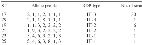
RDP typing results were known for 40 of the isolates within
the data set (Table 6). RDP type correlated closely with ST;
isolates of the same RDP type were identical by MLST or
differed at only a single locus.
Relationship between lineage, host, and country of origin.
The data set presented in this study was not specifically
de-signed to investigate the relationship between isolates and
country or host of origin. However, the following observations
can be made. ST-1 and ST-19 were significantly associated with
carriage (chi-squared test, Yates corrected,
P
⫽
0.004 and
P
⫽
0.008, respectively), and several different capsular serotypes
were represented in these STs. The 44 strains within ST-17
were all serotype III. ST-17 was significantly associated with
invasive neonatal disease (chi-squared test, Yates corrected,
P
⫽
0.0000001). ST-23 contained serotype Ia strains (11 of 16,
68.6%) from carriage and invasive disease. ST-1, ST-19, ST-17,
and ST-23 each contained strains isolated from Australasia,
Europe, Asia, and North America, suggesting global dispersal.
DISCUSSION
We describe an MLST scheme for GBS based on seven
housekeeping genes which was validated with a worldwide
collection of capsule-typed strains that included a subgroup of
strains previously characterized by RDP.
The percentage of variable sites (1.2 to 2.5%) in the seven
selected GBS genes was comparable to that seen by Tettelin et
al. (25) in their analysis of sequence variation in 19 genes from
11 GBS strains. The percentage of variable sites was less than
that seen in the related species, group A streptococcus (10)
(5.1 to 7.6%), and considerably less than that of
Campylobacter
jejuni
(7) (9.2 to 21.7%), a gram-negative organism. The
dn
/
ds
ratios for the seven GBS genes were all less than 1, which
suggests that there is selection against amino acid change and
is consistent with most of the variation being selectively
neu-tral. The genes chosen were distributed around the
chromo-some and were located in the same approximate locations in
both of the published GBS genome sequences, NEM316 (11)
and 2603V/R (25). MLST results for the two published GBS
genome sequences showed that NEM316 was ST-23 and that
2603V/R was a single-locus variant of ST-19.
The DNA sequence data obtained with MLST are amenable
to storage on Internet-based web sites (16), where the STs of
strains from geographically distinct laboratories can be
ob-tained and compared with those on a web-based MLST
data-base. A web site for GBS MLST has been set up and can be
accessed at http://sagalactiae.mlst.net. This offers investigators
a valuable typing tool that will promote further
epidemiologi-cal investigations of this organism. Studies investigating the
differences between strains sampled in well-defined frames
from neonatal invasive disease, adult disease, and carriage in
humans or bovines with MLST will offer the opportunity of
determining whether specific clones are associated with
dis-ease. Furthermore, analysis of the sequence data may give
information on the evolutionary origins and transmission
pat-terns of this organism.
[image:4.603.46.279.85.511.2]The aim of this work was to establish an MLST typing
system, but there were sufficient numbers of strains to make
early observations about the population structure of GBS. The
most common STs in the data set were ST-1, ST-17, ST-19, and
ST-23. These four STs represented two-thirds of the strain
collection. ST-19 and ST-1 contained several different capsular
serotypes and were significantly associated with the carrier
state. ST-17 was more homogeneous and consisted of serotype
III strains predominantly associated with neonatal invasive
dis-ease. The STs were grouped together in similar fashion by
UPGMA and Burst. A better understanding of the relationship
between clones and clonal complexes and disease and the full
extent of diversity in GBS will await the examination of much
larger collections of diverse isolates that will now be possible.
FIG. 1. Polymorphic nucleotide sites in GBS MLST genes. Only
the variable sites are shown. The nucleotide at each site is shown for
allele 1; only those that differ from the nucleotide in allele 1 are shown
for the other alleles. Nucleotide sites are numbered in vertical format.
on May 15, 2020 by guest
http://jcm.asm.org/
The MLST findings are in accord with the results of Musser
et al. (17). These authors used multilocus enzyme
electro-phoresis to study the population structure of GBS. They found
that two distantly related evolutionary lineages of GBS could
be distinguished. The first lineage contained a single
electro-phoretic type (ET-1) and consisted of serotype III isolates
which had been isolated from neonatal disease. This
presum-ably corresponds to ST-17 of MLST. The second lineage of
multilocus enzyme electrophoresis was more diverse and
con-tained several subdivisions and numerous electrophoretic types
which may correspond to the ST-19 complex or ST-1 complex,
which are more diverse, with several STs and different capsular
serotypes. Similar relationships between GBS isolates have
also been found by RDP typing (3) and pulsed-field gel
elec-trophoresis (20).
MLST shows that isolates with the same ST can have
differ-ent capsular serotypes. This could imply that the MLST
scheme has insufficient discriminatory power and groups
iso-lates that are not closely related in genotype. However, a
sim-ilar variation in the serotype of isolates within a single
geno-type was also shown by multilocus enzyme electrophoresis
(17). The variation in serotype within a single ST and the
presence of genetically diverse isolates with the same serotype
suggest that the capsular biosynthesis genes of GBS are subject
to relatively frequent horizontal gene transfer, as is seen in
[image:5.603.57.530.107.485.2]Streptococcus pneumoniae
(6). It has been demonstrated that a
single gene confers serotype specificity in GBS of capsular
types III and Ia (5), and recombinational replacement of this
gene with that from an isolate of a different serotype would
result in a change of capsular type. However, thus far,
hori-zontal transfer of capsular genes has not been shown for GBS
other than in the laboratory. An alternative, perhaps less likely
explanation is that capsular serotyping may be prone to
mis-takes and is difficult to interpret. Confirmation of serotypic
FIG. 2. UPGMA dendrogram showing genetic relationships between the 29 STs. The allelic profile of each ST is shown in parentheses. The four
most common STs are indicated by an asterisk.
on May 15, 2020 by guest
http://jcm.asm.org/
TABLE 5. The collection of 152 isolates of GBS described according to ST, country of isolation, host type, epidemiology, and capsule type
aIsolate no. ST Country Host Epidemiology Capsule Isolate no. ST Country Host Epidemiology Capsule
IS11 1 Is A C V
IS19 1 Is A I V
IS2 1 Is A I V
U64 1 J A C III
U65 1 J A C III
U88 1 J A C VI
U89 1 J A C VI
U90 1 J A C VI
U92 1 J A C VI
U93 1 J A C VIII
U94 1 J A C VIII
U95 1 J A C VIII
U96 1 J A C VIII
NZ14 1 NZ N I V
NZ18 1 NZ A C V
NZ23 1 NZ A I V
T3 1 T A C NT
Z12 1 UK A C V
Z84 1 UK A C V
Z95 1 UK A C V
UK22 1 UK N I Ib
IS28 2 Is A I NT
NZ7 2 NZ A I II
U63 4 J A C Ia
UK6 4 UK N I Ia
U62 5 J A C Ia
Z78 6 UK A C Ib
U72 7 J A C Ia
U75 7 J N I Ia
U71 7 J N I Ia
U79 8 J A C Ib
NZ19 8 NZ A I Ib
NZ4 8 NZ A I Ib
A8 8 S A I NT
Z111 8 UK A C Ib
Z72 8 UK A C Ib
UK13 8 UK N I Ib
IS13 9 Is A C Ib
U78 10 J N I Ib
NZ17 10 NZ A I Ib
NZ20 10 NZ A I II
Z73 10 UK A C Ib
Z41 10 UK A C NT
A2 11 S A I III
A3 11 S A I III
A4 11 S A I III
A5 11 S A I III
A6 11 S A I III
U80 12 J N I Ib
U81 12 J N I Ib
Z69 12 UK A C Ib
T5 13 T A C VI
T1 14 T A C VI
NZ15 15 NZ A I Ib
NZ16 15 NZ N I Ib
IS56 16 Is A I Ia
U11 17 J A C III
U23 17 J A C III
U25 17 J A C III
U3 17 J A C III
U4 17 J A C III
U5 17 J A C III
U1 17 J N I III
U12 17 J N I III
U13 17 J N I III
U14 17 J N I III
U15 17 J N I III
U2 17 J N I III
U26 17 J N I III
U24 17 J N I III
NZ10 17 NZ N I III
Z34 17 UK A C III
Z37 17 UK A C III
UK3 17 UK N I III
UK4 17 UK N I III
UK5 17 UK N I III
aAbbreviations: ST, sequence type; NT, nontypeable; NK, not known; A, adult; N, neonatal; I, invasive strain; C, carried strain; J, Japan; UK, United Kingdom; Is, Israel; NZ, New Zealand; T, Thailand; S, Singapore.
UK8 17 UK N I III
UK10 17 UK N I III
UK12 17 UK N I III
UK15 17 UK N I III
UK17 17 UK N I III
UK20 17 UK N I III
UK21 17 UK N I III
UK18 17 UK N I III
U21 17 USA A I III
U22 17 USA A I III
U29 17 USA A C III
U10 17 USA N I III
U17 17 USA N I III
U19 17 USA N I III
U20 17 USA N I III
U27 17 USA N I III
U28 17 USA N I III
U31 17 USA N I III
U7 17 USA N I III
U8 17 USA N I III
U9 17 USA N I III
U18 17 USA N I III
U30 17 USA N I III
U32 17 USA N I III
UK11 18 UK N I III
U54 19 J A C III
U84 19 J A C V
NZ1 19 NZ A I III
NZ11 19 NZ A C III
NZ2 19 NZ N I III
NZ21 19 NZ A I III
NZ3 19 NZ N C III
UK16 19 UK A C III
Z101 19 UK A C III
Z117 19 UK A C III
Z77 19 UK A C II
8541 19 UK A C NT
Z50 19 UK A C III
UK7 19 UK N I III
UK19 19 UK N I III
U55 19 USA A C III
U56 19 USA A C III
U57 19 USA A C III
U58 19 USA A C III
U59 19 USA A C III
UK1 20 UK N I III
U53 21 J N I III
IS1 22 Is A I II
IS12 22 Is A C II
IS9 23 Is A I III
U69 23 J A C Ia
U70 23 J A C Ia
U60 23 J A C III
NZ12 23 NZ N I Ia
NZ13 23 NZ N C Ia
NZ22 23 NZ A I Ia
NZ5 23 NZ N I III
NZ6 23 NZ A C Ia
NZ8 23 NZ A C Ia
NZ9 23 NZ A I Ia
A7 23 S N I Ia
Z81 23 UK A C Ia
Z87 23 UK A C V
UK14 23 UK N I Ia
NEM316 23 NK N I III
Z18 24 UK A C Ia
U61 25 USA N I III
U86 26 J A C V
U87 26 J A C V
UK2 26 UK N I V
IS31 27 Is A I III
U82 28 J A C II
U83 28 J A C II
UK9 28 UK N I II
U16 29 J N I III
U85 30 J A C V
on May 15, 2020 by guest
http://jcm.asm.org/
identity may be possible when a DNA sequence-based
sero-typing method is more readily available. Preliminary findings
of one such method have recently been described (15).
In conclusion, the GBS MLST system appears to be
suffi-ciently discriminatory for epidemiological studies and provides
a precise and unambiguous way of characterizing isolates of
GBS. The results have confirmed previous findings that a
sin-gle clone of GBS (ST-17) seems to be frequently represented
in neonatal invasive disease. ST-17 is a natural choice for
future study of the virulence of GBS, and it is unfortunate
perhaps that neither of the recently published genome
se-quences for GBS represent this important clone.
ACKNOWLEDGMENTS
This work was supported by the Medical Research Council and
Action Research.
We acknowledge the following for provision of strains: N. Tee,
Clinical Microbiology Laboratory, KK Women’s and Children’s
Hos-pital, Singapore; D. Martin and J. Morgan, Communicable Disease,
ESR, Porirua, New Zealand; N. White and J. Short, Faculty of
Trop-ical Medicine, Mahidol University, Bangkok, Thailand; and Centre for
Tropical Medicine, Nuffield Department of Clinical Medicine, John
Radcliffe Hospital, Oxford, United Kingdom. We also acknowledge
A. A. Whiting (Department of Pediatrics, University of Utah Health
Sciences Center, Salt Lake City, Utah) for capsular serotyping of
strains.
REFERENCES
1. Bliss, S. J., S. D. Manning, P. Tallman, C. J. Baker, M. D. Pearlman, C. F. Marrs, and B. Foxman.2002. Group B streptococcus colonization in male and nonpregnant female university students: a cross-sectional prevalence study. Clin. Infect. Dis.34:184–190.
2. Bohnsack, J. F., S. Takahashi, S. R. Detrick, L. R. Pelinka, L. L. Hammitt, A. A. Aly, A. A. Whiting, and E. E. Adderson.2001. Phylogenetic classifica-tion of serotype III group B streptococci on the basis of hylB gene analysis and DNA sequences specific to restriction digest pattern type III-3. J. Infect. Dis.183:1694–1697.
3. Bohnsack, J. F., S. Takahashi, L. Hammitt, D. V. Miller, A. A. Aly, and E. E. Adderson.2000. Genetic polymorphisms of group B streptococcusscpBalter functional activity of a cell-associated peptidase that inactivates C5a. Infect. Immun.68:5018–5025.
4. Bohnsack, J. F., A. A. Whiting, R. D. Bradford, B. K. Van Frank, S. Taka-hashi, and E. E. Adderson.2002. Long-range mapping of theStreptococcus agalactiaephylogenetic lineage restriction digest pattern type III-3 reveals clustering of virulence genes. Infect. Immun.70:134–139.
5. Chaffin, D. O., S. B. Beres, H. H. Yim, and C. E. Rubens.2000. The serotype of type Ia and III group B streptococci is determined by the polymerase gene within the polycistronic capsule operon. J. Bacteriol.182:4466–4477. 6. Coffey, T. J., M. C. Enright, M. Daniels, J. K. Morona, R. Morona, W.
Hryniewicz, J. C. Paton, and B. G. Spratt.1998. Recombinational exchanges at the capsular polysaccharide biosynthetic locus lead to frequent serotype changes among natural isolates ofStreptococcus pneumoniae. Mol. Micro-biol.27:73–83.
7. Dingle, K. E., F. M. Colles, D. R. Wareing, R. Ure, A. J. Fox, F. E. Bolton, H. J. Bootsma, R. J. Willems, R. Urwin, and M. C. Maiden.2001. Multilocus sequence typing system forCampylobacter jejuni. J. Clin. Microbiol.39:14– 23.
8. Embley, T. M.1991. The linear PCR reaction: a simple and robust method for sequencing amplified rRNA genes. Lett. Appl. Microbiol.13:171–174. 9. Enright, M. C., and B. G. Spratt.1998. A multilocus sequence typing scheme
forStreptococcus pneumoniae:identification of clones associated with serious invasive disease. Microbiology144:3049–3060.
10. Enright, M. C., B. G. Spratt, A. Kalia, J. H. Cross, and D. E. Bessen.2001. Multilocus sequence typing ofStreptococcus pyogenesand the relationships betweenemmtype and clone. Infect. Immun.69:2416–2427.
11. Glaser, P., C. Rusniok, C. Buchrieser, F. Chevalier, L. Frangeul, T. Msadek, M. Zouine, E. Couve, L. Lalioui, C. Poyart, P. Trieu-Cuot, and F. Kunst.
2002. Genome sequence ofStreptococcus agalactiae, a pathogen causing invasive neonatal disease. Mol. Microbiol.45:1499–1513.
12. Hauge, M., C. Jespersgaard, K. Poulsen, and M. Kilian.1996. Population structure ofStreptococcus agalactiaereveals an association between specific evolutionary lineages and putative virulence factors but not disease. Infect. Immun.64:919–925.
13. Henning, K. J., E. L. Hall, D. M. Dwyer, L. Billmann, A. Schuchat, J. A. Johnson, and L. H. Harrison.2001. Invasive group B streptococcal disease in Maryland nursing home residents. J. Infect. Dis.183:1138–1142. 14. Jolley, K. A., E. J. Feil, M. S. Chan, and M. C. Maiden.2001. Sequence type
analysis and recombinational tests (START). Bioinformatics17:1230–1231. 15. Kong, F., S. Gowan, D. Martin, G. James, and G. L. Gilbert.2002. Serotype identification of group B streptococci by PCR and sequencing. J. Clin. Microbiol.40:216–226.
16. Maiden, M. C., J. A. Bygraves, E. Feil, G. Morelli, J. E. Russell, R. Urwin, Q. Zhang, J. Zhou, K. Zurth, D. A. Caugant, I. M. Feavers, M. Achtman, and B. G. Spratt.1998. Multilocus sequence typing: a portable approach to the identification of clones within populations of pathogenic microorganisms. Proc. Natl. Acad. Sci. USA95:3140–3145.
17. Musser, J. M., S. J. Mattingly, R. Quentin, A. Goudeau, and R. K. Selander.
1989. Identification of a high-virulence clone of type IIIStreptococcus aga-lactiae(group B streptococcus) causing invasive neonatal disease. Proc. Natl. Acad. Sci. USA86:4731–4735.
18. Public Health Laboratory Services.2002. Incidence of group B streptococcal disease in infants aged less than 90 days. CDR Wkly.12:3.
19. Quentin, R., H. Huet, F. S. Wang, P. Geslin, A. Goudeau, and R. K. Selander.
1995. Characterization ofStreptococcus agalactiaestrains by multilocus en-zyme genotype and serotype: identification of multiple virulent clone families that cause invasive neonatal disease. J. Clin. Microbiol.33:2576–2581. 20. Rolland, K., C. Marois, V. Siquier, B. Cattier, and R. Quentin.1999. Genetic
features ofStreptococcus agalactiaestrains causing severe neonatal infec-tions, as revealed by pulsed-field gel electrophoresis andhylBgene analysis. J. Clin. Microbiol.37:1892–1898.
21. Schrag, S., R. Gorwitz, K. Fultz-Butts, and A. Schuchat.2002. Prevention of perinatal group B streptococcal disease. Revised guidelines from CDC. Morb. Mortal. Wkly. Rep. Recommun. Rep.51:1–22.
22. Schuchat, A.1998. Epidemiology of group B streptococcal disease in the United States: shifting paradigms. Clin. Microbiol. Rev.11:497–513. 23. Schuchat, A.1999. Group B streptococcus. Lancet353:51–56.
24. Staden, R.1996. The Staden sequence analysis package. Mol. Biotechnol.
5:233–241.
25. Tettelin, H., V. Masignani, M. J. Cieslewicz, J. A. Eisen, S. Peterson, M. R. Wessels, I. T. Paulsen, K. E. Nelson, I. Margarit, T. D. Read, L. C. Madoff, A. M. Wolf, M. J. Beanan, L. M. Brinkac, S. C. Daugherty, R. T. DeBoy, A. S. Durkin, J. F. Kolonay, R. Madupu, M. R. Lewis, D. Radune, N. B. Fedorova, D. Scanlan, H. Khouri, S. Mulligan, H. A. Carty, R. T. Cline, S. E. Van Aken, J. Gill, M. Scarselli, M. Mora, E. T. Iacobini, C. Brettoni, G. Galli, M. Mariani, F. Vegni, D. Maione, D. Rinaudo, R. Rappuoli, J. L. Telford, D. L. Kasper, G. Grandi, and C. M. Fraser.2002. Complete genome sequence and comparative genomic analysis of an emerging human pathogen, serotype V
Streptococcus agalactiae. Proc. Natl. Acad. Sci. USA99:12391–12396. 26. Wilder-Smith, E., K. M. Chow, R. Kay, M. Ip, and N. Tee.2000. Group B
[image:7.603.42.280.89.166.2]streptococcal meningitis in adults: recent increase in Southeast Asia. Aust. N. Z. J. Med.30:462–465.
TABLE 6. Restriction digest patterns compared with
ST for 40 GBS isolates
ST Allelic profile RDP type No. of strains