Copyright © 2004, American Society for Microbiology. All Rights Reserved.
Comparison of Results of Fluconazole Disk Diffusion Testing for
Candida
Species with Results from a Central Reference Laboratory in the
ARTEMIS Global Antifungal Surveillance Program
M. A. Pfaller,
1,2* K. C. Hazen,
3S. A. Messer,
1L. Boyken,
1S. Tendolkar,
1R. J. Hollis,
1and D. J. Diekema
1,4Departments of Pathology,
1Epidemiology,
2and Medicine,
4Roy J. and Lucille A. Carver College of Medicine and
College of Public Health, University of Iowa, Iowa City, Iowa 52242, and Department of Pathology,
University of Virginia, Charlottesville, Virginia
3Received 10 February 2004/Returned for modification 4 April 2004/Accepted 24 April 2004
The accuracy of antifungal susceptibility tests is important for accurate resistance surveillance and for the
clinical management of patients with serious infections. Our main objective was to compare the results of
fluconazole disk diffusion testing of
Candida
spp. performed by ARTEMIS participating centers with disk
diffusion and MIC results obtained by the central reference laboratory. A total of 2,949 isolates of
Candida
spp.
were tested by NCCLS disk diffusion and reference broth microdilution methods in the central reference
laboratory. These results were compared to the results of disk diffusion testing performed in the 54
partici-pating centers. All tests were performed and interpreted following NCCLS recommendations. Overall
cate-gorical agreement between participant disk diffusion test results and reference laboratory MIC results was
87.4%, with 0.2% very major errors (VME) and 3.3% major errors (ME). The categorical agreement between
the disk diffusion test results obtained in the reference laboratory with the MIC test results was similar: 92.8%.
Likewise, good agreement was observed between participant disk diffusion test results and reference laboratory
disk diffusion test results: 90.4%, 0.4% VME, and 3.4% ME. The disk diffusion test was especially reliable in
detecting those isolates of
Candida
spp. that were characterized as resistant by reference MIC testing. External
quality assurance data obtained by surveillance programs such as the ARTEMIS Global Antifungal
Surveil-lance Program ensure the generation of useful surveilSurveil-lance data and result in the continued improvement of
antifungal susceptibility testing practices.
Disk diffusion testing of fluconazole against
Candida
spp.
was developed in order to provide a simple inexpensive
meth-od for monitoring fluconazole susceptibility in a variety of
laboratory settings (2, 4, 6–8, 12, 13). Barry and colleagues (2)
demonstrated that the accuracy and precision of fluconazole
disk diffusion testing, using a 25-
g disk and Mueller-Hinton
agar supplemented with 2% glucose and 0.5
g of methylene
blue per ml (MH-MB), were comparable to those of reference
broth microdilution (BMD) MIC testing. Recently, the
Na-tional Committee for Clinical Laboratory Standards (NCCLS)
Subcommittee on Antifungal Testing has approved this agar
disk diffusion method, M44-A, for testing fluconazole against
yeasts (11).
The M44 fluconazole disk diffusion test with MH-MB has
also been used for more than 4 years as part of the ARTEMIS
Global Antifungal Surveillance Program (4, 8, 12). In this
program, fluconazole disk diffusion testing is performed in
more than 80 different laboratories in 35 countries, and the
data are used to follow trends in fluconazole susceptibility
patterns worldwide (4, 8). Isolates of
Candida
spp. collected by
ARTEMIS participants are also sent to a central reference
laboratory at the University of Iowa for additional testing using
NCCLS disk diffusion and BMD methods (12–14). Data
gen-erated from the ARTEMIS Program have been useful in
doc-umenting the sustained activity of fluconazole versus clinical
isolates of
Candida
(4, 8, 12, 14).
In addition to assessing the scale of the resistance problem
at the local, national, or international level, antimicrobial
re-sistance surveillance may also provide an opportunity for
im-proving the quality of susceptibility testing among those taking
part in the surveillance (5). The ARTEMIS Program
demon-strates two different, and complementary, approaches to the
performance of antifungal resistance surveillance. The central
reference laboratory provides a means to overcome differences
between methods, or differences in the same method
per-formed in different laboratories. The centralized approach
en-sures that standardized, internationally recognized
quantita-tive methods are used and that isolates are available for further
studies of resistance mechanisms and for epidemiological
typ-ing (5, 15, 16). Alternatively, fluconazole disk diffusion testtyp-ing
performed on-site in the participant laboratories results in the
generation of large amounts of data in a short period of time,
using a standard protocol and with results submitted to a
cen-tral database (4, 8). Despite the use of a standard protocol, it
is recognized that any surveillance system based on
suscepti-bility tests performed by the participating laboratories needs to
include some measure of quality assurance, beyond simple
quality control (QC) testing, in order to provide an
indepen-dent assessment of laboratory performance and validation of
results generated from the various laboratories (5, 18). The
ARTEMIS DISK Surveillance Study (4) generated massive
* Corresponding author. Mailing address: Medical Microbiology
Di-vision, C606 GH, Department of Pathology, University of Iowa
Col-lege of Medicine, Iowa City, IA 52242. Phone: (319) 384-9566. Fax:
(319) 356-4916. E-mail: [email protected].
3607
on May 15, 2020 by guest
http://jcm.asm.org/
amounts of data but was limited by a lack of validation and
comparison with reference MIC results. In the present study,
we assess the accuracy of the ARTEMIS participant disk
dif-fusion test results by comparison to those obtained for the
same isolates tested in the central reference laboratory by disk
diffusion and BMD methods recommended by the NCCLS.
MATERIALS AND METHODS
Study design.The ARTEMIS Program was established to monitor the species and antifungal susceptibility patterns ofCandidaspp. isolated from clinically significant sites of infection (e.g., blood and normally sterile sites [NSSI]) via a broad network of sentinel hospitals in North America, Latin America, Europe, Africa, and Asia.Candidaspp. causing invasive disease (in blood and NSSI) and the accompanying fluconazole disk zone diameters were reported from 54 dif-ferent medical centers in 2001 and 2002.
Each participant medical center contributed results (organism identification and fluconazole disk zone diameter) for consecutive blood and NSSI culture isolates (one isolate per patient) ofCandidaspp. judged to be clinically signifi-cant by local criteria and detected in each calendar month during the study. All isolates were saved on agar slants and were sent to the University of Iowa College of Medicine (Iowa City) for storage and further characterization by reference identification methods and susceptibility testing (3, 10, 11). In the central refer-ence laboratory, isolates were tested by both NCCLS disk diffusion (11) and BMD (10) methods.
Organism identification.AllCandidaspp. isolates were identified at partici-pating institutions by the routine method used in each laboratory. Upon receipt at the University of Iowa, the isolates were subcultured onto potato dextrose agar (Remel, Lenexa, Kans.) and CHROMagarCandidamedium (Hardy Laborato-ries, Santa Maria, Calif.) to ensure viability and purity. Confirmation of species identification was performed with Vitek and API products (bioMerieux, St. Louis, Mo.), as recommended by the manufacturer, or by conventional methods, as required (3). Isolates were stored as suspensions in water or on agar slants at ambient temperature until needed.
Susceptibility testing.Disk diffusion testing of fluconazole was performed in both participant laboratories and the central reference laboratory according to the methods described in NCCLS document M44-A (11). MIC testing was performed by the NCCLS BMD reference method M27-A2 (10). Standard flu-conazole reference powder was obtained from Pfizer Pharmaceuticals (Groton, Conn.), and 25-g fluconazole disks were obtained from Becton Dickinson (Sparks, Md.).
Disk diffusion testing of fluconazole was performed as described by Hazen et al. (4) and in NCCLS document M44-A (11). Agar plates (150-mm diameter) containing MH-MB at a depth of 4.0 mm were used. The agar surface was inoculated by using a swab dipped in a cell suspension adjusted to the turbidity of a 0.5 McFarland standard. The plates were incubated in air at 35 to 37°C and read at 18 to 24 h. Zone diameter endpoints were read at 80% growth inhibition by using the BIOMIC image analysis plate reader system (version 5.9; Giles Scientific, Santa Barbara, Calif.) (4, 12).
MIC interpretive criteria for fluconazole were those published by Rex et al. (17) and the NCCLS (10) and were as follows: susceptible, MIC ofⱕ8g/ml; susceptible dose dependent, MIC of 16 to 32g/ml; resistant, MIC ofⱖ64g/ml. The interpretive criteria for the fluconazole disk diffusion test were those pub-lished by Barry et al. (2) and the NCCLS (11): susceptible, zone diameter ofⱖ19 mm; susceptible dose dependent, zone diameter of 15 to 18 mm; resistant, zone diameter ofⱕ14 mm.
QC.QC was performed for BMD in accordance with NCCLS document M27-A2 (10) by using Candida kruseiATCC 6258 andCandida parapsilosis ATCC 22019 (1, 10). QC for disk diffusion was performed usingCandida albicans ATCC 90028 andC. parapsilosisATCC 22019 (2, 4).
Analysis of results.The diameters of the zones of inhibition (in millimeters) surrounding the fluconazole disks at 24 h of incubation obtained in the partici-pant laboratories and in the reference laboratory were plotted against their respective BMD MICs read at 48 h (see Fig. 1 and 2, below) (2, 12, 13). Similarly, the diameters of the zones of inhibition surrounding the fluconazole disks at 24 h of incubation obtained in the participant laboratories were plotted against their respective zones obtained in the reference laboratory read at 24 h. The inter-pretive breakpoints described by the NCCLS (10, 11) were used to determine the categorical agreement between disk diffusion and BMD results and between participant and reference disk diffusion results. Major errors (ME) were classi-fied as resistant by disk diffusion (participant or central laboratory) and suscep-tible by BMD or resistant by the participant disk diffusion test and suscepsuscep-tible by
the central laboratory disk diffusion test. Very major errors (VME) were classi-fied as susceptible by the participant or central laboratory disk diffusion test and resistant by BMD or susceptible by the participant disk diffusion test and resis-tant by the central laboratory disk diffusion test. Minor errors (M) occurred when the result of one of the tests was susceptible or resistant and that of the other test was susceptible dose dependent.
RESULTS AND DISCUSSION
During the 2001-2002 study period, a total of 2,949 isolates
of
Candida
from blood and other NSSI and their
accompany-ing fluconazole disk diffusion zone diameters were submitted
from 54 participating centers in North America (8 centers),
Latin America (12 centers), Europe (20 centers), and the
Asia-Pacific region (14 centers). The frequencies of infections due
to the various species of
Candida
identified in the central
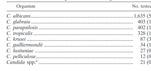
reference laboratory are presented in Table 1 and represent a
total of 14 different species.
[image:2.603.300.541.97.211.2]In vitro susceptibility testing performed in both participant
laboratories (disk diffusion) (Fig. 1) and the reference
labora-tory (disk diffusion and MIC test) (Fig. 2) indicated that
resis-tance to fluconazole was uncommon among isolates of
Can-dida
from blood and NSSI (Table 2). Notably, more isolates of
all species appeared resistant to fluconazole by disk diffusion
testing in both participant and reference laboratories, with the
greatest resistance observed with disk diffusion testing
per-formed in the participant laboratories. Overall, the level of
categorical agreement between the disk diffusion test results
and the reference MIC results was quite good in both
partic-ipant (87.4%) and reference (92.8%) laboratories (Table 2).
Importantly, there were very few VME, indicating that the disk
diffusion test was reliable in detecting isolates resistant to
flu-conazole by MIC testing. The level of agreement between disk
diffusion and MIC results was highest with
C. albicans
and
lowest with
Candida glabrata
. The low level of agreement
be-tween disk diffusion and MIC results with
C. glabrata
was
similar to that observed previously (9, 12) and was due almost
entirely to M-type errors. Interestingly, the M-type errors
ob-served with
C. glabrata
involved shifts between the susceptible
and susceptible–dose-dependent categories in the reference
laboratory and between the susceptible–dose-dependent and
resistant categories in the participant laboratories. This was
likely the result of the distribution of both zones and MICs
around the respective breakpoints and a more conservative
TABLE 1. Isolates of
Candida
spp. tested by participant and
reference laboratories in the ARTEMIS Global
Antifungal Surveillance Program
Organism No. tested (%)
C. albicans
... 1,635 (55.4)
C. glabrata
... 403 (13.7)
C. parapsilosis
... 402 (13.6)
C. tropicalis
... 328 (11.2)
C. krusei
...
87 (3.0)
C. guilliermondii
...
34 (1.1)
C. lusitaniae
...
27 (0.9)
C. pelliculosa
...
12 (0.4)
Candida
spp.
a...
21 (0.7)
Total ... 2,949 (100)
aIncludesC. kefyr(n⫽9), C. rugosa(n⫽5), C. dubliniensis(n⫽2), C. lipolytica(n⫽2),C. zeylanoides(n⫽2), andC. famata(n⫽1).on May 15, 2020 by guest
http://jcm.asm.org/
FIG.
1.
Zones
of
inhibition
around
25-
g
fluconazole
disks
tested
in
ARTEMIS
participant
laboratories
plotted
against
the
MICs
determined
at
48
h
by
using
the
reference
BMD
method
for
2,949
isolates
of
Candida
spp.
The
numbers
represent
the
number
of
results
(isolates)
for
each
data
point.
on May 15, 2020 by guest
http://jcm.asm.org/
FIG.
2.
Zones
of
inhibition
around
25-
g
fluconazole
disks
tested
on
MH
agar
supplemented
with
2%
glucose
and
methylene
blue
(0.5
g/ml)
in
the
central
reference
laboratory
plotted
against
the
MICs
at
48
h
determined
by
using
the
reference
BMD
method
for
2,949
isolates
of
Candida
spp.
The
numbers
represent
the
number
of
results
(isolates)
for
each
data
point.
on May 15, 2020 by guest
http://jcm.asm.org/
reading of zone diameters in the participant laboratories.
Im-portantly, the number of VME remained low for
C. glabrata
disk diffusion tests in both participant and reference laboratory
settings.
A similar level of agreement was seen when disk diffusion
results obtained in the participant laboratories were compared
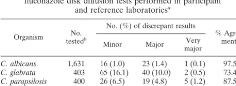
with those obtained in the reference laboratory (Table 3). The
overall categorical agreement was excellent at 90.4%, and the
number of VME, ME, and M errors was quite low. As with the
comparison to BMD testing, agreement was highest with
C.
albicans
and lowest with
C. glabrata
, with most of the errors in
the M category.
This study is the largest one of its kind validating the
per-formance of disk diffusion testing in surveillance program
par-ticipant laboratories by testing the same isolates in a central
reference laboratory. We demonstrate that fluconazole disk
diffusion testing using the NCCLS M44-A method can be
per-formed with a high level of accuracy in more than 50 different
laboratories worldwide. The participant results were not only
validated by fluconazole disk diffusion testing performed in the
central reference laboratory, but also by reference BMD MIC
testing. Importantly, the participant laboratories tended to err
on the side of calling isolates more resistant than the reference
laboratory. This was most pronounced with
C. glabrata
and
other non-
C. albicans
species. False susceptible results were
uncommon. Similar results have also been reported by Morace
et al. (9).
Previously, Barry et al. (2) demonstrated that when replicate
fluconazole disk diffusion tests were performed in three
differ-ent laboratories, 94 to 97% of zone diameters fell within a
range of the median zone
⫾
6 to 8 mm. Although a formal
precision analysis was not conducted in the present study, we
found that among replicate zone diameters generated in both
reference and participant laboratories, 80% fell within
⫾
8 mm
(data not shown).
In summary, we have shown that fluconazole disk diffusion
testing performed in accordance with the NCCLS M44-A
method can be performed with an acceptable degree of
accu-racy in a wide range of laboratory settings throughout the world.
The test was especially reliable in identifying those strains of
Candida
characterized as resistant by reference BMD MIC
testing. False resistant results may be an issue with disk
diffu-sion, especially with
C. glabrata
and other non-
C. albicans
spe-cies. Further assessment of such isolates by BMD should be
performed if clinically indicated. Continued collaboration and
external validation of antifungal susceptibility testing results
such as that presented here for the ARTEMIS Global
Anti-fungal Surveillance Program will ensure the generation of
use-ful antifungal surveillance data and result in continued
im-provement of antifungal susceptibility testing practices.
ACKNOWLEDGMENTS
This study was supported in part by research and educational grants
from Pfizer Inc., Pfizer Global Pharmaceuticals, New York, N.Y.
[image:5.603.44.543.79.251.2]We thank Linda Elliott and Shanna Duffy for secretarial assistance
in the preparation of the manuscript. We appreciate the participation
TABLE 3. Overall interpretive agreement between results of
fluconazole disk diffusion tests performed in participant
and reference laboratories
aOrganism testedNo.b
No. (%) of discrepant results
% Agree-mentc Minor Major majorVery
C. albicans 1,631 16 (1.0) 23 (1.4) 1 (0.1) 97.5
C. glabrata 403 65 (16.1) 40 (10.0) 2 (0.5) 73.4
C. parapsilosis 400 26 (6.5) 19 (4.8) 5 (1.2) 87.5
C. tropicalis 327 21 (6.4) 11 (3.4) 1 (0.3) 89.9
AllCandidaspp. 2,949 170 (5.8) 100 (3.4) 13 (0.4) 90.4
aThe fluconazole disk diffusion method was performed according to NCCLS method M44-A.
bTotal number of isolates tested in 54 laboratories participating in the ARTEMIS program and also in the ARTEMIS reference laboratory.
[image:5.603.44.283.588.675.2]cPercent total agreement, the categorical agreement between participant and reference laboratory results.
TABLE 2. Interpretive agreement between results of fluconazole disk diffusion tests and standard 48-h BMD
aOrganism
(no. tested) Test methodd
% of results in each categoryb
% Agreementc
% errors
S SDD R VME ME M
C. albicans
(1,631)
Ref.-MIC
99.8
0.2
0.0
Ref.-disk
99.5
0.4
0.1
99.6
0.0
0.1
0.3
Part.-disk
97.6
0.7
1.7
97.7
0.0
1.4
0.9
C. glabrata
(403)
Ref.-MIC
69.5
26.3
4.2
Ref.-disk
93.6
2.2
4.2
71.7
0.8
0.5
27.0
Part.-disk
71.5
14.4
14.1
60.6
0.7
7.4
31.3
C. parapsilosis
(400)
Ref.-MIC
93.3
6.2
0.5
Ref.-disk
91.0
3.0
6.0
93.3
0.0
1.0
5.7
Part.-disk
85.5
4.0
10.5
85.5
0.0
4.8
9.7
C. tropicalis
(327)
Ref.-MIC
99.1
0.3
0.6
Ref.-disk
98.8
0.6
0.6
97.9
0.6
0.6
0.9
Part.-disk
89.6
7.0
3.4
88.7
0.6
3.4
7.3
All
Candida
spp. (2,949)
Ref.-MIC
91.6
6.7
1.7
Ref.-disk
94.1
2.2
3.7
92.8
0.2
0.4
6.6
Part.-disk
87.9
4.5
7.6
87.4
0.2
3.3
9.1
aFluconazole disk diffusion testing was performed according to NCCLS method M44-A, and fluconazole BMD MIC testing was performed according to NCCLS method M27-A2.
bFluconazole susceptibility categories: S, susceptible, MIC ofⱕ8g/ml (ⱖ19 mm); SDD, susceptible dose dependent, MIC of 16 to 32g/ml (15 to 18 mm); R, resistant, MIC ofⱖ64g/ml (ⱕ14 mm).
cPercent categorical agreement between disk diffusion and MIC test results.
dRef.-MIC, MIC testing performed by ARTEMIS reference laboratory; Ref.-disk, disk testing performed by ARTEMIS reference laboratory; Part.-disk, disk testing performed by ARTEMIS participants.
on May 15, 2020 by guest
http://jcm.asm.org/
of all ARTEMIS site participants. A list of ARTEMIS participants
can be found on the following website: http://www.medicine.uiowa
.edu/pathology/path_folder/research/acknowledgments/artemis
_participants.pdf.
REFERENCES
1. Barry, A. L., M. A. Pfaller, S. D. Brown, A. Espinel-Ingroff, M. A. Ghan-noum, C. Knapp, R. P. Rennie, J. H. Rex, and M. G. Rinaldi.2000. Quality control limits for broth microdilution susceptibility tests of ten antifungal agents. J. Clin. Microbiol.38:3457–3459.
2. Barry, A. L., M. A. Pfaller, R. P. Rennie, P. C. Fuchs, and S. D. Brown.2002. Precision and accuracy of fluconazole susceptibility tests by broth microdi-lution, Etest, and disk diffusion methods. Antimicrob. Agents Chemother. 46:1781–1784.
3. Hazen, K. C., and S. A. Howell.2003.Candida,Cryptococcus, and other yeasts of medical importance, p. 1693–1711.InP. R. Murray, E. J. Baron, J. H. Jorgensen, M. A. Pfaller, and R. H. Yolken (ed.), Manual of clinical microbiology, 8th ed. ASM Press, Washington, D.C.
4. Hazen, K. C., E. J. Baron, A. L. Colombo, C. Girmenia, A. Sanchez-Sousa, A. del Palacio, C. de Bedout, D. L. Gibbs, and The Global Antifungal Surveillance Group.2003. Comparison of the susceptibilities ofCandidaspp. to fluconazole and voriconazole in a 4-year global evaluation using disk diffusion. J. Clin. Microbiol.41:5623–5632.
5. Kahlmeter, G., and D. F. J. Brown.2002. Resistance surveillance studies— comparability of results and quality assurance of methods. J. Antimicrob. Chemother.50:775–777.
6. Kirkpatrick, W. R., T. M. Turner, A. W. Fothergill, D. I. McCarthy, S. W. Redding, M. G. Rinaldi, and T. F. Patterson.1998. Fluconazole disk diffu-sion susceptibility testing ofCandidaspecies. J. Clin. Microbiol.36:3429– 3432.
7. Lee, S.-C., C.-P. Fung, N. Lee, L.-C. See, J.-S. Huang, C.-J. Tsai, K.-S. Chen, and W.-B. Shieh.2001. Fluconazole disk diffusion test with methylene blue-and glucose-enriched Mueller-Hinton agar for determining susceptibility of Candidaspecies. J. Clin. Microbiol.39:1615–1617.
8. Meis, J., M. Petrou, J. Bille, D. Ellis, D. Gibbs, and The Global Antifungal Surveillance Group.2000. A global evaluation of the susceptibility of Can-didaspecies to fluconazole by disk diffusion. Diagn. Microbiol. Infect. Dis. 36:215–223.
9. Morace, G., G. Amato, F. Bistoni, G. Fadda, P. Marone, M. T. Montagna, S. Oliveri, L. Polonelli, R. Rigoli, I. Mancuson, S. La Face, L. Masucci, L. Romano, C. Napoli, D. Tato, M. G. Buscema, C. M. C. Belli, M. M. Piccirillo,
S. Conti, S. Covan, F. Fanti, C. Cavanna, F. D’Alo, and L. Pitzurra.2002. Multicenter comparative evaluation of six commercial systems and the Na-tional Committee for Clinical Laboratory Standards M27-A broth microdi-lution method for fluconazole susceptibility testing of Candidaspecies. J. Clin. Microbiol.40:2953–2958.
10. National Committee for Clinical Laboratory Standards.2002. Reference method for broth dilution testing of yeasts. Approved standard, 2nd ed., M27-A2. National Committee for Clinical Laboratory Standards, Wayne, Pa. 11. National Committee for Clinical Laboratory Standards.2004. Method for antifungal disk diffusion susceptibility testing of yeasts: approved guideline M44-A. National Committee for Clinical Laboratory Standards, Wayne, Pa. 12. Pfaller, M. A., D. J. Diekema, S. A. Messer, L. Boyken, and R. J. Hollis.2003. Activities of fluconazole and voriconazole against 1,586 recent clinical iso-lates ofCandidaspecies determined by broth microdilution, disk diffusion, and Etest methods: report from the ARTEMIS Global Antifungal Suscep-tibility Program, 2001. J. Clin. Microbiol.41:1440–1446.
13. Pfaller, M. A., D. J. Diekema, L. Boyken, S. A. Messer, S. Tendolkar, and R. J. Hollis.2003. Evaluation of the Etest and disk diffusion methods for determining susceptibilities of 235 bloodstream isolates ofCandida glabrata to fluconazole and voriconazole. J. Clin. Microbiol.41:1875–1880. 14. Pfaller, M. A., S. A. Messer, L. Boyken, R. J. Hollis, C. Rice, S. Tendolkar,
and D. J. Diekema.2004. In vitro activities of voriconazole, posaconazole, and fluconazole against 4,169 clinical isolates ofCandidaspp. and Crypto-coccus neoformanscollected during 2001 and 2002 in the ARTEMIS Global Antifungal Surveillance Program. Diagn. Microbiol. Infect. Dis.48:201–205. 15. Pujol, C., M. Pfaller, and D. R. Soll.2002. Ca3 fingerprinting ofCandida albicansbloodstream isolates from the United States, Canada, South Amer-ica, and Europe reveals a European clade. J. Clin. Microbiol.40:2729–2740. 16. Pujol, C., M. A. Pfaller, and D. R. Soll.2004. Flucytosine resistance is restricted to a single genetic clade ofCandida albicans. Antimicrob. Agents Chemother.48:262–266.
17. Rex, J. H., M. A. Pfaller, J. N. Galgiani, M. S. Bartlett, A. Espinel-Ingroff, M. A. Ghannoum, M. Lancaster, M. G. Rinaldi, T. J. Walsh, and A. L. Barry. 1997. Development of interpretive breakpoints for antifungal susceptibility testing: conceptual framework and analysis of in vitro-in vivo correlation data for fluconazole, itraconazole, andCandidainfections. Clin. Infect. Dis. 24:235–247.
18. Tenover, F. C., M. J. Mohammed, J. Stelling, T. O’Brien, and R. Williams. 2001. Ability of laboratories to detect emerging antimicrobial resistance: proficiency testing and quality control results from the World Health Orga-nizations’ external quality assurance system for antimicrobial susceptibility testing. J. Clin. Microbiol.39:241–250.