JOURNAL OFCLINICALMICROBIOLOGY,
0095-1137/00/$04.00⫹0 Nov. 2000, p. 4201–4207 Vol. 38, No. 11
Copyright © 2000, American Society for Microbiology. All Rights Reserved.
Application of tRNA Intergenic Spacer PCR for Identification
of
Enterococcus
Species
MARGO BAELE,1* PAUL BAELE,1MARIO VANEECHOUTTE,2VIRGINIE STORMS,3 PATRICK BUTAYE,1LUC A. DEVRIESE,1GERDA VERSCHRAEGEN,2
MONIQUE GILLIS,3ANDFREDDY HAESEBROUCK1
Department of Pathology, Bacteriology and Poultry Diseases, Faculty of Veterinary Medicine, University of Ghent, B-9820 Merelbeke,1Department of Clinical Chemistry, Microbiology and Immunology, Faculty of Medicine,
University of Ghent,2and Laboratorium voor Microbiologie, Faculteit Wetenschappen,
Universiteit Gent,3B-9000 Ghent, Belgium
Received 13 June 2000/Returned for modification 25 July 2000/Accepted 31 August 2000
tRNA intergenic spacer PCR (tDNA-PCR) was evaluated for its usefulness in the differentiation of entero-coccal species of human and animal origin. This technique was carried out for 124 strains belonging to 17 enterococcal species and generated DNA fragments, which were separated by capillary electrophoresis.
tDNA-PCR enabled us to discriminate for all species tested.Enterococcus faeciumshowed minor but reproducible
differences withEnterococcus durans, whileEnterococcus hiraewas easily distinguishable.Enterococcus avium,
Enterococcus malodoratus, andEnterococcus raffinosusgenerated highly similar though distinctive patterns.
Enterococci are regarded as one of the leading causes of nosocomial infections (11), and cases of endocarditis, bactere-mia, urinary tract infection, and neonatal sepsis have fre-quently been reported. Several authors have highlighted the need for rapid and accurate identification of enterococcal strains (3, 6, 7, 21). AlthoughEnterococcus faecalisand Entero-coccus faeciumare responsible for about 95% of all nosoco-mial infections caused by enterococci, most of the described species have been encountered in human infections (20).
The increasing occurrence of antibiotic resistance, for in-stance to-lactam antibiotics and more recently to glycopep-tides, has caused great concern (11). Some species, such asE. faecium, are likelier to be more resistant to antimicrobial agents than are others, andEnterococcus gallinarum, Entero-coccus casseliflavus, andEnterococcus flavescensshow intrinsic low-level resistance to glycopeptide antibiotics. Rapid species identification therefore can be of substantial help in the choice of antibiotic therapy (15).
In the study of outbreaks it is helpful to know that the isolates involved are all the same species of enterococci (1, 5, 23). Fingerprinting techniques acting at infraspecies level, such as restriction analysis in combination with pulsed-field gel elec-trophoresis, can then be applied (3).
Sequencing of conserved regions, such as the 16S rRNA gene (16) and thesodAgene encoding superoxide dismutase (18), is useful in the identification of enterococci. Several other molecular identification methods have recently been evaluated (3, 6, 14, 16, 19, 24, 29). PCR assays using genes involved in peptidoglycan synthesis (D-alanine:D-alanine ligase genes) and
in vancomycin resistance (vanC-1, vanC-2/3) have also been used for identification, classification, or detection of entero-cocci (7, 9, 17).
tRNA intergenic spacer PCR (tDNA-PCR) (26) has been applied for the species differentiation of streptococci (27),
Acinetobacterspp. (8, 28), staphylococci (12), andListeriaspp.
(25) and consists of amplification of the tDNAs by use of consensus primers, which are complementary to the highly conserved edges of the flanking tRNA genes and are directed outwardly. The resulting PCR fragments can be separated by capillary electrophoresis. tDNA-PCR makes use of primers complementary to regions conserved throughout the bacteria and should therefore be applicable to a wide range of genera.
MATERIALS AND METHODS
Bacterial strains.Seventy-one well-characterized enterococcal strains from different origins, identified by whole-cell protein analysis using sodium dodecyl sulfate-polyacrylamide gel electrophoresis, were obtained from the Belgian Co-ordinated Collection of Microorganisms culture collection (University of Ghent, Ghent, Belgium). This series was extended to include 19 strains isolated from different animal species, which were identified with a biochemical test scheme as described by Devriese et al. (4, 5).E. faecium,E. faecalis,E. gallinarum, andE. casseliflavusstrains were confirmed by a specific multiplex PCR using thevanand
ddlprimers as described by Dutka-Malen et al. (7). All reference strains are listed in Table 1.
Thirty-four strains originating from the intestines of different animal species and from humans were identified by biochemical tests, multiplex PCR according to the method of Dutka-Malen et al. (7), and tDNA-PCR and are shown in Table 2.
DNA preparation.Bacterial cells were grown overnight on Columbia agar (Gibco Technologies, Paisley, Scotland) with 5% sheep blood for 24 h at 37°C in a 5% CO2-enriched environment and were checked for purity. A 1-l loopful of
cells was suspended in 20l of lysis buffer (0.25% sodium dodecyl sulfate, 0.05 N NaOH) and heated at 95°C for 5 min. The cell lysate was spun down by brief centrifugation at 16,000⫻gand diluted by adding 180l of distilled water. The cell debris was removed by centrifugation at 16,000⫻gfor 5 min. Supernatants were directly used as the template for PCR or were frozen at⫺20°C until further use.
tDNA-PCR.PCR was carried out using the outwardly directed tRNA gene consensus primers T5A (5⬘AGTCCGGTGCTCTAACCAACTGAG) and T3B (5⬘AGGTCGCGGGTTCGAATCC), as described by Welsh and McClelland (26). Reactions were carried out in a 10-l volume containing 9.1l of High Fidelity Mix 1.1⫻(Gibco Life Technologies). Primers were added at a final concentration of 0.1M. Primer T3B consisted of a mixture of 1/5 fluorescent TET-labeled oligonucleotides and 4/5 nonlabeled oligonucleotides (Perkin-Elmer Applied Biosystems, Foster City, Calif.). A volume of 0.7l of sample DNA was added (the template was diluted 15 times). After 2 min at 94°C, reaction mixtures were cycled 30 times in a Perkin-Elmer Cetus 9600 thermo-cycler under the following conditions: 30 s at 94°C, 1 min at 50°C, and 1 min at 72°C. The final extension was 30 min at 72°C. Reaction vials were then cooled to 10°C and kept on ice until used in electrophoresis.
Capillary electrophoresis.Twelve microliters of deionized formamide was mixed with 0.5l of an internal size standard mixture, containing 0.3l of the GS-400 High Density size standard and 0.2l of the GS-500 size standard, which both have ROX-labeled fragments in the range of 50 to 500 bp (Perkin-Elmer
* Corresponding author. Mailing address: Department of Pathology, Bacteriology and Poultry Diseases, Faculty of Veterinary Medicine, University of Ghent, Salisburylaan 133, B-9820 Merelbeke, Belgium. Phone: 32 9 264 74 34. Fax: 32 9 264 74 94. E-mail: Margo.Baele@rug .ac.be.
4201
on May 15, 2020 by guest
http://jcm.asm.org/
Applied Biosystems). One microliter of tDNA-PCR product was added. The mixtures were denatured by heating at 95°C for 3 min and placed directly on ice for at least 15 min.
Capillary electrophoresis was carried out using an ABI-Prism 310 Genetic Analyzer (Perkin-Elmer Applied Biosystems) at 60°C, a constant voltage of 1.5 kV, and a more or less constant current of approximately 10 mA. Capillaries with a length of 47 cm and a diameter of 50m were filled with Performance Optimized Polymer 4. Electropherograms were normalized using Genescan Analysis software, version 2.1 (Perkin-Elmer Applied Biosystems). The fragment lengths were derived from the peak positions after interpolation with the peak positions of the size standard fragments.
Data analysis.Electropherograms were interpreted visually and with a soft-ware program developed at our laboratory (available upon request from the authors).
The software compares samples which are derived from the ABI310 Genescan Analysis program as molecular weight tables. A library, containing one entry per species, was constructed manually as a text file (see Table 3 for an example). Each entry is a list of numerical values which represent those fragment lengths (i.e., peaks) differentiating the species from each other, as established by visual interpretation of superimposed fingerprints obtained with the Genescan soft-ware. A positive value indicates that a peak is present in the fingerprint of a particular species, while a negative value penalizes the presence of a peak with this value. After elimination of peaks lower than 50% of the average height of all peaks, the fingerprint of an unknown strain was compared with all entries in the library. The number of fragments in common between the unknown fingerprint and the species entry, divided by the total number of fragments of the species entry in the library, was taken as a measure of similarity. The library does not contain irreproducible peaks or peaks considered irrelevant after visual compar-ison, which therefore are not taken into consideration by the program when comparing unknown fingerprints with entries in the library. The program enables one to enlarge the peak position tolerance, which corrects for small base pair shifts.
For clustering analysis, the distance matrix was calculated with the in-house software. The similarity between two samples was calculated as described above in a pairwise manner (first considering one sample as the library entry), whereby peaks below a user-defined background threshold were not taken into consider-ation and the second sample was taken as the library entry. The similarity between the two samples was the average of the two calculated similarity values. Clustering was done with Neighbor software (Phylip) (http://evolution.genetics .washington.edu/phylip.html), employing the algorithm for the unweighted-pair group method using average linkages (UPGMA).
RESULTS
tDNA-PCR.Capillary electrophoresis of tDNA-PCR
ampli-fication products of enterococcal strains generated fingerprint patterns with three to five large, reproducible peaks and sev-eral smaller, irreproducible ones. The reproducibility of tDNA-PCR was evaluated by carrying it out for strains LMG 11423 (E. faecium), LMG 13129 (E. gallinarum), and LMG 13595 (E. faecalis) four times, each time using different PCR mixtures, thermal cycling runs, and electrophoresis runs. For each strain, one of these four tDNA-PCR products was run three times in capillary electrophoresis. Similarity was calculated with the in-house software and a background noise level of 50%. Clus-tering was done using the UPGMA algorithm. The minimal similarity level for the six tDNA-PCR fingerprints obtained for each strain named above was 88.6, 89.3, and 90%, respectively. For all six samples, the standard deviation of the peaks, which are used in the library of the in-house software, ranged from 0.045 bp (for a fragment length of 266.5 bp) to 0.364 bp (for a fragment length of 110.8 bp). Repeated electrophoresis runs of the same PCR product gave a minimum standard deviation of 0.007 bp (for a fragment length of 266.5 bp) and a maximum of 0.418 bp (for a fragment length of 110.8 bp). Repeats of the entire PCR assay (different PCR mixtures, PCR runs, and electrophoresis runs) gave a minimum standard deviation of 0.035 bp (for a fragment length of 269.3 bp) and a maximum of 0.371 bp (for a fragment length of 65.8 bp). The highest re-producibility and the most reliable identifications were ob-tained by not taking into account those peaks lower than 50% of the average peak height within the range of 60 to 400 bp.
Visual interpretation showed that tDNA-PCR fingerprints of strains belonging to the same species were similar, while strains belonging to different species exhibited different pat-TABLE 1. Enterococcal strains used in this study
Enterococcusspecies LMG-BCCM collection type strain no.
E. faecalis...266/5371,acc193613,aCDC 149-77,aLMG 7937T, LMG 11395, LMG 11636, LMG 11637, LMG 11734, LMG 12161, LMG 13595
E. faeciumspecies group
E. faecium...LMG 11423T, LMG 11397, LMG 11635, LMG 8148, LMG 16198, LMG 16200, LMG 9431, LMG 9448
E. durans...Lab 848, LMG 10741, LMG 12691, LMG 12903, LMG 13604, LMG 14197, LMG 16887, LMG 16888, LMG 16889
E. hirae...LD 13216,accm 2423T,aLab 1342,aLMG 6399T, LMG 11746, LMG 14198, LMG 14200, LMG 14489, LMG 17189, LMG 17190, LMG 17191, LMG 17192, LMG 17285
E. mundtii...LMG 12308, LMG 13044, LMG 10748
E. gallinarumspecies group
E. gallinarum...LMG 12313, LMG 12904, LMG 13129T, LMG 14166, LMG 14405, LMG 16201, LMG 16202, LMG 16204
E. casseliflavus...LMG 12306, LMG 12307, LMG 12311, LMG 12314, LMG 14406
E. flavescens...LMG 13518T, LMG 13597, LMG 16313, LMG 16314
E. aviumspecies group
E. avium...LMG 10744T, LMG 11394 LMG 12171, LMG 12304, LMG 12305
E. pseudoavium...266/7909,a266/7910a
E. malodoratus...LMG 12300, LMG 12301, LMG 12905, LMG 15718
E. raffinosus...LMG 12172, LMG 12888T
E. cecorumspecies group
E. cecorum...2508/122a, 2508/331a, 2508/334a, 98/2539a, LMG 11743, LMG 11744, LMG 12902T, RS15Aa, S566a
E. columbae...LMG 11740T, LMG 12295, LMG 12296, LMG 14175, LMG 14595
E. dispar...SS 1295Tb, 266/8667a
E. saccharolyticus...CCUG 27643
E. asini...SS 1501Tb
aProvided by L. A. Devriese, University of Ghent, Ghent, Belgium.
bProvided by R. R. Facklam, Centers for Disease Control and Prevention, Atlanta, Ga.
4202 BAELE ET AL. J. CLIN. MICROBIOL.
on May 15, 2020 by guest
http://jcm.asm.org/
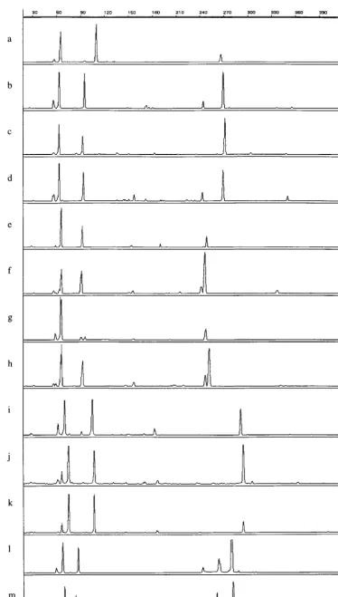
terns, except for those belonging to the speciesE. casseliflavus
andE. flavescens(Fig. 1). Some species, for instanceE. faecium
and Enterococcus durans, could be differentiated by the 1-bp length difference of a single tDNA spacer fragment. Entero-coccus avium and Enterococcus malodoratus differed in the lengths of two fragments: E. avium strains showed peaks at 65.8, 92.9, and 253.8 bp, whereas the patterns of isolates be-longing to E. malodoratuswere composed of peaks of 65.8, 91.6, and 251.5 bp.
For use with the in-house software, the tDNA-PCR finger-print library (Table 3) was constructed using all reference strains (Table 1) from our collection. Each entry contains for each species all reproducible fragment length values that are present in the fingerprints of its different isolates, and some also include negative values to distinguish between highly sim-ilar patterns. Using this library, all strains were identified cor-rectly, including those belonging to the speciesE. duransand
E. faeciumand those belonging to the speciesE. avium, En-terococcus raffinosus, andE. malodoratus.
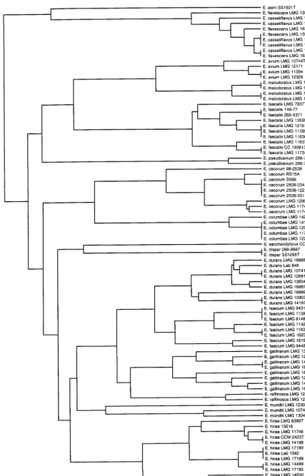
A dendrogram was constructed and is shown in Fig. 2. All species clustered separately exceptE. casseliflavusand E. fla-vescens, which clustered together.
Identification of unknown strains with tDNA-PCR.
[image:3.612.54.551.92.427.2]Thirty-four strains isolated from humans and animals were identified in a polyphasic approach using biochemical tests (4, 5) and TABLE 2. Strains isolated from intestines of various animals and from humans and used for blind testing
in order to evaluate the ability of tDNA-PCR to identify enterococci
Strain no. Origin biochemical methodsSpecies according to Species according tovan-ddlPCR Species according totDNA-PCR
PAT1084 Pigeon E. avium NDa E. avium
PAT1123 Horse E. avium ND E. avium
KOMA042 Pig E. avium ND E. avium
UHG 98 11 2049 Human E. faecium E. faecium E. faecium
PAT843 Finch E. gallinarumgroup E. casseliflavus E. casseliflavus
PAT755 Goat E. gallinarumgroup E. casseliflavus E. casseliflavus
UHG 98 08 5234 Human ND E. casseliflavus E. casseliflavus
PAT999 Parakeet E. gallinarumgroup E. casseliflavus E. casseliflavus
PAT233 Pigeon E. cecorumorE. columbae ND E. cecorum
PAT664 Pigeon E. cecorumorE. columbae ND E. cecorum
PAT047 Pigeon E. cecorumorE. columbae ND E. columbae
PAT495 Pigeon E. cecorumorE. columbae ND E. columbae
PAT499 Pigeon E. cecorumorE. columbae ND E. columbae
PAT914 Chicken E. faecalis E. faecalis E. faecalis
UHG 98 11 0487 Human ND E. faecalis E. faecalis
PAT397 Squirrel E. faecalis E. faecalis E. faecalis
PAT052 Turkey E. faecalis E. faecalis E. faecalis
UHG 98 06 1902 Human ND E. faecium E. faecium
UHG 98 07 5596 Human ND E. faecium E. faecium
UHG 98 12 2683 Human ND E. faecium E. faecium
UHG 98 12 3779 Human ND E. faecium E. faecium
UHG 99 01 0517 Human ND E. faecium E. faecium
PAT612 Pig E. faecium E. faecium E. faecium
PAT236 Horse E. faecium E. gallinarum E. gallinarum
UHG 98 07 4441 Human ND E. gallinarum E. gallinarum
UHG 98 08 0962 Human ND E. gallinarum E. gallinarum
UHG 98 08 1527 Human ND E. gallinarum E. gallinarum
UHG 98 12 4026 Human ND E. gallinarum E. gallinarum
PAT426 Rabbit E. gallinarumgroup E. gallinarum E. gallinarum
PAT1100 Pigeon E. hirae/durans ND E. hirae
PAT1105 Pigeon E. hirae/durans ND E. hirae
PAT1236 Parakeet E. hirae/durans ND E. hirae
PAT1238 Pigeon E. hirae/durans ND E. hirae
PAT882 Rabbit E. faecium Negative2 E. hirae
aND, not done.
bNegative, no amplification product obtained. Implies that the strain does not belong to the speciesE. faecalis,E. faecium,E. gallinarum, orE. casseliflavusorE.
flavescens.
TABLE 3. Manually constructed tDNA-PCR library, composed of entries that each consists of a list of
tDNA spacer fragment lengths (in base pairs)
Species entry Lengths of fragments taken intoconsiderationa
E. faecalis ...65.8,110.8,266.6
E. faecium ...63.8,95.5,244,269.2
E. hirae ...63.9,93,271.5,303.5
E. durans ...63.8,94.3,⫺95.5,243.3,269.2
E. mundtii ...63.5,102.5,269.6
E. avium ...65.8,92.9,253.8
E. pseudoavium ...66.2,86.6,220
E. malodoratus...65.8,91.6,251.5,⫺256.5
E. raffinosus...65.8,96,252.5
E. gallinarum ...64.9,95.3,261.5
E. casseliflavusorE. flavescens...61.3,69.1,97.5,264.6
E. cecorum ...63.8,81.2,⫺154,⫺155,240.9,254.7
E. columbae ...66,78.4,237.5,241,255.6
E. dispar ...64,79.2,231,266,284.1
E. saccharolyticus ...64,107.8,231,266
E. asini...73,98,261.6
aValues represent peaks that ought to be present (x) or absent (⫺y) in the
fingerprint of an unknown strain in order to be identified as a certain species.
VOL. 38, 2000 PCR IDENTIFICATION OF ENTEROCOCCI 4203
on May 15, 2020 by guest
http://jcm.asm.org/
[image:3.612.55.294.537.710.2]FIG. 1. tDNA-PCR fingerprint patterns of enterococcal strains. a,E. faecalis266/5371; b,E. faeciumLMG 11423; c,E. hiraeLMG 6399T; d,E. duransLMG 13604; e,E. aviumLMG 107444T; f,E. malodoratusLMG 12300; g,E. raffinosusLMG 12888T; h,E. raffinosusLMG 14595; i,E. gallinarumLMG 16204; j,E. casseliflavusLMG 10745; k,E. flavescensLMG 13518; l,E. cecorumLMG 11744; m,E. columbaeLMG 11740T. Thexaxis represents the fragment length in base pairs; theyaxis represents the peak intensity.
4204
on May 15, 2020 by guest
http://jcm.asm.org/
FIG. 2. Dendrogram obtained from tDNA-PCR fingerprints after similarity calculation with the in-house software and clustering with UPGMA using Neighbor software. Bar, distance of 10%.
4205
on May 15, 2020 by guest
http://jcm.asm.org/
van-ddl PCR (7) to verify identification results from tDNA-PCR. The results are summarized in Table 2. tDNA finger-prints were analyzed with the in-house software. All 34 strains were identified correctly, regardless of whether peaks lower than 50% of the average peak height were eliminated.
The fingerprints of the enterococcal species were compared with fingerprints obtained from about 400 species belonging to over 40 different genera. All enterococcal strains could be correctly identified.
DISCUSSION
For the identification of the most important enterococci, a simple, conventional biochemical test scheme exists (10) and a more complex, phylogenetically based differential identifica-tion scheme has been described (4). However, the results of these tests are sometimes unreliable or ambiguous, and some species are too closely related to show biochemical differences. This is especially the case forEnterococcus hiraeandE. durans,
E. gallinarumand E. casseliflavus, and Enterococcus cecorum
and Enterococcus columbae(4). In this study, we evaluated a universally applicable genotypic method for the identification of enterococci.
tDNA-PCR enabled us to differentiate between all species tested, exceptE. casseliflavusandE. flavescens. However, these species are most probably synonymous, as is also apparent from several other studies (3, 7, 19, 22). Closely related organ-isms showed peak shifts of no more than one or a few base pairs. Therefore, the high resolution of capillary electrophore-sis was needed to separate fragments differing by 1 bp in length.
Values for similarity between fingerprints of the same strain obtained in different PCR and electrophoresis runs were very high, around 90%. In the reproducibility test, standard devia-tions of the peak posidevia-tions in base pairs were not higher than 0.364 bp, which indicates that the peak positions in the finger-prints are highly reproducible.
To be able to compare large numbers of unidentified strains to a database of well-characterized strains, a suitable software package was necessary. When comparing complete finger-prints, as is done when using the Dice coefficient, calculation of the similarity between visually highly identical fingerprints can still give low values (as a consequence of small peak shifts and variable presence of minor peaks). Software which enabled a different approach was developed. A library in which only reproducible peaks were included was manually constructed. Peaks in new fingerprints were regarded to be identical to a peak of a reference pattern when their positions lay within a range of⫺0.7 bp to⫹0.7 bp of the reference peak. This is twice the maximum standard deviation obtained in the reproducibil-ity tests. Because the peak position of each fragment is Gauss distributed, 95% of the electrophoretic profiles of the same sample should have a peak within this range. The use of a manually constructed library also prevents nonreproducible peaks from influencing the calculation of similarity values, which in turn leads to a higher discriminatory power.
Analysis of the molecular weight tables with the in-house software permitted discrimination of the highly similar tDNA patterns ofE. faeciumand E. duransstrains. Blind testing of enterococcal strains which were previously identified with bio-chemical tests or multiplex PCR showed that this software enabled us to process tDNA-PCR fingerprints originating from an ABI Prism 310 Genetic Analyzer. One of the most impor-tant advantages of tDNA-PCR is the use of universal primers. In theory, this technique can be used for species identification for a wide range of genera. It requires little time and manual
labor. tDNA-PCR takes about 3 h; the GeneAmp 9600 PCR cycler permits testing of 96 strains at once. Capillary electro-phoresis requires half an hour per run; one run can include up to three samples if primers are labeled with different fluores-cent dyes. Its high reproducibility and satisfactory discrimina-tory power make it possible to develop an identification tool which can be used by different laboratories. Normalization of the fingerprints is done automatically by the Genescan Anal-ysis program, and quality control of the different steps in the protocol takes between 2 and 10 min for approximately 50 strains. Using our software, a list of identifications for up to 50 strains at once is available within 5 min of exportation in table format of the normalized fingerprints as obtained on ABI310. Taken together, the whole procedure as described above, starting from a pure culture to a final identification, can be completed in 24 h for 45 strains, requiring only 4 to 5 h hands-on time. The cost per strain, comprising DNA prepara-tion, tDNA-PCR, and capillary electrophoresis reagents (cap-illary, buffer, size marker, POP4 gel, and laser wear, excluding PCR and electrophoresis equipment), was calculated to add up to $2.50 (U.S.) (labor not included). tDNA-PCR fingerprints can be obtained within 8 h after colony picking for the first five electrophoresis samples, which can contain PCR products of up to three strains.
From these results, we conclude that tDNA-PCR is very suitable for rapid, discriminatory, and reliable identification of all currently described enterococcal species and can be ex-tended to include newly described ones.
In addition to the high discriminatory power of tDNA-PCR for other groups, likeAcinetobacter(8, 28),Listeria(25), staph-ylococci (12), and streptococci (2, 13), one can start to consider the applicability of this technique for the species identification of cultured organisms in the average laboratory.
ACKNOWLEDGMENTS
M.V. is indebted to FWO Flanders for an appointment as research associate.
We are grateful to A. Vandekerckhove, L. Van Simaey, and F. Grillaert for excellent technical assistance.
REFERENCES
1.Alonso-Echanove, J., B. Robles, and W. R. Jarvis.1999. Proficiency of clin-ical laboratories in Spain in detecting vancomycin-resistantEnterococcus
spp. J. Clin. Microbiol.37:2148–2152.
2.Degheldre, Y., P. Vandamme, H. Goossens, and M. Struelens.1999. Identi-fication of clinically relevant viridans streptococci by analysis of transfer DNA intergenic spacer length polymorphism. Int. J. Syst. Bacteriol.49:1591– 1598.
3.Descheemaeker, P., C. Lammens, B. Pot, P. Vandamme, and H. Goossens.
1997. Evaluation of arbitrarily primed PCR analysis and pulsed-field gel electrophoresis of large genomic DNA fragments for identification of en-terococci important in human medicine. Int. J. Syst. Bacteriol.47:555–561. 4.Devriese, L. A., B. Pot, and M. D. Collins.1993. Phenotypic identification of the genusEnterococcusand differentiation of phylogenetically distinct en-terococcal species and species groups. J. Appl. Bacteriol.75:399–408. 5.Devriese, L. A., B. Pot, K. Kersters, S. Lauwers, and F. Haesebrouck.1996.
Acidification of methyl-␣-D-glucopyranoside: a useful test to differentiate
Enterococcus casseliflavusandEnterococcus gallinarumfromEnterococcus faeciumspecies group and fromEnterococcus faecalis. J. Clin. Microbiol.
34:2607–2608.
6.Donabedian, S., J. W. Chow, D. M. Shlaes, M. Green, and M. J. Zervos.1995. DNA hybridization and contour-clamped homogeneous electric field elec-trophoresis for identification of enterococci to the species level. J. Clin. Microbiol.33:141–145.
7.Dutka-Malen, S., S. Evers, and P. Courvalin.1995. Detection of glycopep-tide resistance genotypes and identification to the species level of clinically relevant enterococci by PCR. J. Clin. Microbiol.33:24–27.
8.Ehrenstein, B., A. T. Bernards, L. Dijkshoorn, P. Gerner-Smidt, K. J. Towner, P. J. M. Bouvet, F. D. Daschner, and H. Grundmann.1996. Acin-etobacterspecies identification by using tRNA spacer fingerprinting. J. Clin. Microbiol.34:2414–2420.
9.Evers, S., B. Casadewall, M. Charles, S. Dutka-Malen, M. Galimand, and P.
4206 BAELE ET AL. J. CLIN. MICROBIOL.
on May 15, 2020 by guest
http://jcm.asm.org/
Courvalin.1996. Evolution of structure and substrate specificity inD-alanine: D-alanine ligases and related enzymes. J. Mol. Evol.42:706–712.
10. Facklam, R., and M. D. Collins.1989. Identification ofEnterococcusspecies isolated from human infections by a conventional test scheme. J. Clin. Mi-crobiol.27:731–734.
11. Huycke, M. M., D. F. Sahm, and M. S. Gilmore.1998. Multiple-drug resis-tant enterococci: the nature of the problem and an agenda for the future. Emerg. Infect. Dis.4:239–249.
12. Maes, N., Y. De Gheldre, R. De Ryck, M. Vaneechoutte, H. Meugnier, J. Etienne, and M. J. Struelens.1997. Rapid and accurate identification of
Staphylococcus species by tRNA intergenic spacer length polymorphism analysis. J. Clin. Microbiol.35:2477–2481. (Erratum,36:1468, 1998.) 13. McClelland, M., and J. Welsh.1992. Length polymorphisms in tRNA
inter-genic spacers detected by using the polymerase chain reaction can distin-guish streptococcal strains and species. J. Clin. Microbiol.30:1499–1504. 14. Monstein, H. J., M. Quednau, A. Samuelsson, S. Ahrne, B. Isaksson, and J.
Jonasson.1998. Division of the genus Enterococcus into species groups using PCR-based molecular typing methods. Microbiology144:1171–1179. 15. Murray, B. E.1990. The life and times of theEnterococcus. Clin. Microbiol.
Rev.3:46–65.
16. Patel, R., K. E. Piper, M. S. Rouse, J. M. Steckelberg, J. R. Uhl, P. Kohner, M. K. Hopkins, F. R. Cockerill, and B. C. Kline.1998. Determination of 16S rRNA sequences of enterococci and application to species identification of nonmotile Enterococcus gallinarum isolates. J. Clin. Microbiol.
36:3399–3407.
17. Patel, R., J. R. Uhl, P. Kohner, M. K. Hopkins, and F. R. Cockerill III.1997. Multiplex PCR detection ofvanA,vanB,vanC-1, andvanC-2/3genes in enterococci. J. Clin. Microbiol.35:703–707.
18. Poyart, C., G. Quesnes, and P. Trieu-Cuot.2000. Sequencing the gene encoding manganese-dependent superoxide dismutase for rapid species identification of enterococci. J. Clin. Microbiol.38:415–418.
19. Quednau, M., S. Ahrne, A. C. Petersson, and G. Molin.1998. Identification of clinically important species of Enterococcus within 1 day with randomly
amplified polymorphic DNA (RAPD). Curr. Microbiol.36:332–336. 20. Ruoff, K. L., L. de la Maza, M. J. Murtagh, J. D. Spargo, and M. J. Ferraro.
1990. Species identities of enterococci isolated from clinical specimens. J. Clin. Microbiol.28:435–437.
21. Schaberg, D. R., D. H. Culver, and R. P. Gaynes.1991. Major trends in the microbial etiology of nosocomial infection. Am. J. Med.91:72S–75S. 22. Teixeira, L. M., M. G. Carvalho, V. L. Merquior, A. G. Steigerwalt, M. G.
Teixeira, D. J. Brenner, and R. R. Facklam.1997. Recent approaches on the taxonomy of the enterococci and some related microorganisms. Adv. Exp. Med. Biol.418:397–400.
23. Toye, B., J. Shymanski, M. Bobrowska, W. Woods, and K. Ramotar.1997. Clinical and epidemiologic significance of enterococci intrinsically resistant to vancomycin (possessing thevanCgenotype). J. Clin. Microbiol.35:3166– 3170. (Erratum,36:1469, 1998.)
24. Tyrrell, G. J., R. N. Bethune, B. Willey, and D. E. Low.1997. Species identification of enterococci via intergenic ribosomal PCR. J. Clin. Micro-biol.35:1054–1060. (Erratum,35:2434.)
25. Vaneechoutte, M., P. Boerlin, H. V. Tichy, E. Bannerman, B. Ja¨ger, and J. Bille.1998. Comparison of PCR-based DNA fingerprinting techniques for the identification ofListeriaspecies and their use for atypicalListeriaisolates. Int. J. Syst. Bacteriol.48:127–139.
26. Welsh, J., and M. McClelland.1991. Genomic fingerprints produced by PCR with consensus tRNA gene primers. Nucleic Acids Res.19:861–866. 27. Welsh, J., and M. McClelland.1992. PCR-amplified length polymorphisms
in tRNA intergenic spacers for categorizing staphylococci. Mol. Microbiol.
6:1673–1680.
28. Wiedmann-Al-Ahmad, M., H. V. Tichy, and G. Scho¨n.1994. Characterization ofAcinetobactertype strains and isolates obtained from wastewater treat-ment plants by PCR fingerprinting. Appl. Environ. Microbiol.60:4066–4071. 29. Williams, A. M., U. M. Rodrigues, and M. D. Collins.1991. Intrageneric relationships of enterococci as determined by reverse transcriptase sequenc-ing of small-subunit rRNA. Res. Microbiol.142:67–74.
VOL. 38, 2000 PCR IDENTIFICATION OF ENTEROCOCCI 4207