ORIGINAL RESEARCH ARTICLE
EFFECT OF SURFACE PROTECTION ASSOCIATED TO DIFFERENT BONDING PROTOCOLS
ON THE BOND STRENGTH TO ERODED DENTIN
*1
Vivian Leite Martins,
2Rodrigo Ramos,
3Max José Pimenta Lima,
4Roberto Paulo Correia
Araújo
and
5Andrea Nóbrega Cavalcanti
1
MSc, School of Medicine and Public Health of Bahia (BAHIANA), Salvador, BA, Brazil
2
Private Practice, Federal University of Bahia. Salvador, Ba, Brazil
3
Assistant Professor, Federal University of Bahia, Salvador, BA, Brazil
4
Full Professor, Federal University of Bahia, Salvador, BA, Brazil
5
Adjunct Professor, Dentistry Course, School of Medicine and Public Health of Bahia (BAHIANA) and School of
Dentistry, Federal University of Bahia (FOUFBA), Salvador, BA, Brazil
ARTICLE INFO ABSTRACT
Erosion, also known as biocorrosion, is a type of dental wear, promoted by chemical process in absence of bacteria. The objective of the present study was to evaluate the effect of superficial protection and bonding protocols on bond strength to eroded dentin. Sound human molars had occlusal dentin exposed and were allocated into 16 groups (n = 10) according to the association between three main factors: simulation of endogenous erosion (absent or 18 DES-RE cycles); previous surface protection (absent or glass-ionomeric sealant), and protocol for dentin bonding with an universal adhesive system (with or without phosphoric acid etching; and exposed or not to chlorhexidine). Composite resin buildups were constructed on the dentin surfaces, the specimens were sectioned and submitted to the microtensile test. The sticks obtained from each tooth were divided in two groups. The first one was tested after 24 hours and the second was stored in water for seven months. Results for each period were analyzed by means of 3-way ANOVA and Tukey test. The comparison between the two periods was done by Student's t-test, for paired data. According to the statistical analysis, bonding procedures didn’t interfere on immediate bond strength values. The erosive challenge reduced the immediate bond strength in the absence of surface protection, but not in the presence of the glass-ionomeric sealant. After storage, the effect of the erosive challenge couldn’t be noted; and the use of chlorhexidine resulted in decreased bond strength in groups previously coated with glass-ionomer sealant. It could be concluded that eroded dentin surfaces previously coated with glass-ionomer sealant might have impaired bonding; and that chlorhexidine was not able to increase bonding stability after storage. Finally, both self-etch and etch-and-rinse protocols seems to be feasible for the use of the universal adhesive system on eroded dentin surfaces.
Copyright © 2019,Vivian Leite Martins et al. This is an open access article distributed under the Creative Commons Attribution License, which permits unrestricted use, distribution, and reproduction in any medium, provided the original work is properly cited.
INTRODUCTION
Erosion, also known as biocorrosion, is a type of dental wear, promoted by chemical process in absence of bacteria. Its etiology is complex, being the consumption of acidic foods and beverages and gastroesophageal disorders mainly responsible for its high incidence and prevalence (Rajitkar, 2012).
*Corresponding author: Vivian Leite Martins
MSc, School of Medicine and Public Health of Bahia (BAHIANA), Salvador, BA, Brazil
The endogenous hydrochloric acid, present in the gastric juice, seems to have high corrosive potential due to the low pH and titratable acidity (Bartlet, 2001). Contact with this acid is frequent in patients with eating disorders and gastroesophageal reflux disease (GERD), leading to dental lesions, mainly in enamel (Rajitkar, 2012; Bartlet, 2001). However, when etiological factors aren’t controlled, the erosive wear may result in dentin exposure over time (Cruz, 2015). To prevent dissolution of dental surfaces by acids, surface protection techniques may be employed from the early diagnosis of
ISSN: 2230-9926
International Journal of Development Research
Vol. 09, Issue, 02, pp.25986-25992, February, 2019
Article History:
Received 03rd November, 2018
Received in revised form 26th December, 2018
Accepted 16th January, 2019 Published online 28th February, 2019
Key Words:
Tooth Erosion. Dentin. Matrix Metalloproteinases. Chlorhexidine.
Citation: Vivian Leite Martins, Rodrigo Ramos, Max José Pimenta Lima, Roberto Paulo Correia Araújo and Andrea Nóbrega Cavalcanti, 2019.“Effect of surface protection associated to different bonding protocols on the bond strength to eroded dentin”, International Journal of Development Research, 9, (02), 25986-25992.
erosion, such as the use of the glass-ionomer sealant (Austin, 2011; Zhou, 2012). Studies have shown that glass-ionomer-based materials have good remineralizing ability due to the action of calcium and great potential for fluoride release (Zhou, 2012; Elkassas, 2014). However, there is a question to be answered which concerns the interference of the residual sealant on the bonding capacity to dentin if the preventive method was not sufficient to stop the loss of substance and a subsequent adhesive restoration may be necessary. Dentin erosion presents specific histology, since the progression of the demineralization is mediated by the presence of the demineralized organic matrix (DOM). If this organic matrix is affected by enzymes and by chemical degradation, the progression of the loss of structure increases over time, so its preservation is extremely important (Zarela, 2015; Comar, 2015). However, the demineralized organic matrix may generate a substrate of difficult bonding, since the exposed collagen may be inadequately infiltrated by the resinous monomers. This fact may affect the durability of adhesive restorations performed to rehabilitate sequelae left by corrosion (Hebling, 2005). The selection of materials that promote efficient adhesion to different substrates, such as enamel and dentin, is an important challenge for restorative dentistry. Traditionally, enamel adhesion is considered safe and reliable, however, bonding in dentin is less predictable and more complex due to its histological differences (Manfroi, 2016). Considering the individuality of each substrate, a new adhesive system, classified as "multi-mode" or "universal", has been recently launched. It promises superior results due to its versatility in relation to the adhesive technique (self-etching or etch-and-rinse). However, there is still no consensus about the best technique for the use of this adhesive on the dentin substrate and not much is known about its performance on the
eroded substrate. Finally, it is not clear whether the use of
protease inhibitors to maintain the demineralized organic matrix would help increasing the bonding stability when using such systems. Therefore, the aim of the present study was to evaluate the effect of previous dentin protection with glass-ionomer sealant associated with different adhesive protocols in the bond strength to eroded dentin. The null hypotheses tested are that there is no influence of the surface protection method, and the way of application of the adhesive system in the immediate and long-term bond strength to eroded dentine.
MATERIALS AND METHODS
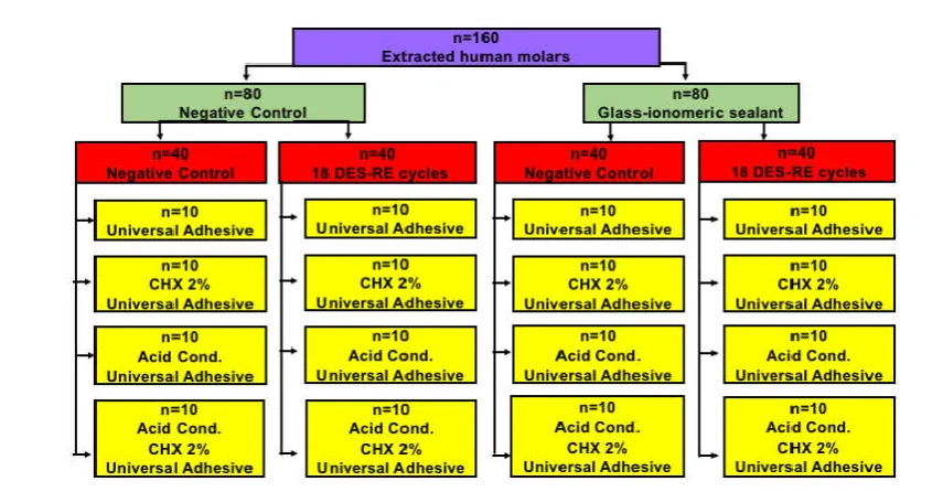
Sample preparation: The materials used in the present study, their respective manufacturers and compositions are described in Table 1. In the present study, 160 healthy human molars were extracted less than 6 months before the use. After approval by the Ethics and Research Committee of the Federal University of Bahia (CAAE: 63689716.6.0000.5024/1895081), the teeth were cleaned and then stored in 0.1% thymol solution (Cromato Produtos Químicos LTDA, Diadema - SP), under refrigeration at 4oC. Each tooth was individually fixed with hot glue on an acrylic plate, positioned in a high precision cutter
(Extec Corp.®, Enfield CT - USA) and sectioned mesio-distally to total exposure of coronal dentin surface, with
diamond-cut disc (Extec Corp.®, Enfield CT-USA) under
constant refrigeration. Afterward a second cut was made in the same direction, with a distance of 6mm from the first one, for high standardization. After, the enamel surface was protected with nail polish, keeping exposed only the surface of coronary dentin. Finally, the 160 specimens were divided in 16 groups (n = 10) according to the surface protection method, the degree
of corrosion and the way how the adhesive restoration was performed (Figure 1).
Methods of surface protection
The methods of surface protection were performed as follows:
Control: the samples were not subjected to any form of surface protection, only kept in relative humidity at 37°C.
Glass-ionomer sealant:the product (Clinpro XT Varnish, 3M-ESPE, Sumaré, SP, Brazil) was applied according to the recommendations from the manufacturer. Initially, equal portions of the two pastes were placed and manipulated for 15 seconds. Straight after, a thin layer was applied on the dentine surfaces with a disposable applicator followed by photo-activation for 20 seconds (Radii Plus, SDI Brazil, São Paulo, SP, Brazil). Finally, they were kept in relative humidity at 37°C.
Simulation of erosion by gastric acid
After the use of the methods for surface protection, the specimens were divided into two groups according to the simulation of erosion by gastric acid.
Control: The samples of this subgroup were kept in relative humidity of 37°C as the cycles were carried out in the remaining subgroups.
18 cycles of DES-RE: Each completed cycle consisted of immersing the sample in 10 ml solution of hydrochloric acid (5% HCl, pH = 2.2) for two minutes in room temperature. After this, the specimens were washed with the help of disposable syringe containing 20 ml distilled water and
immersed in remineralizing solution for 60 minutes(Pashley,
2004). Its composition included 1,5 mmol/L Ca, 0,9 mmol/L
PO4, 0.075 mol/L of acetate with pH 7,0(Tjäderhane, 2013).
The cycles were performed in 2 consecutive days, among them, the units were stored in relative humidity of 37°C (Martins, 2018).
Bonding procedures
The four experimental conditions of the adhesive restorations follow the description below:
Universal Adhesive System in self-etching form and without
exposure to chlorhexidine: In these specimens, only the
application of adhesive system was performed actively for 20 seconds, in two consecutive layers, light cured for 20 seconds with light intensity of 1500 mW / cm² (Radii Plus, SDI Brasil Indústria e Comércio LTDA, São Paulo - SP).
Universal Adhesive System in the self-etching form and with exposure to chlorhexidine: the specimens were submitted to the application of 1ml of 2% Chlorhexidine Digluconate solution (Chlorhexidine S, FGM Dental Products - Joinville SC) for 60 seconds. Afterwards, the excess of solution was gently removed, keeping the surface moist, and the adhesive system was applied as previously reported.
(Biodynamic Chemistry and Farm, LTDA, Ibiporã - PR); washed abundantly for 30 seconds and dried with absorbent paper. After the adhesive system was applied as described above.
Universal Adhesive System in the etch-and-rinsing form and
with exposure to chlorhexidine: the specimens were
submitted to phosphoric acid etching as previously described. After, 1 ml of 2% chlorhexidine Digluconate solution (Chlorhexidine S, FGM Dental Products - Joinville S.C) was applied in the dentin surface for 60 seconds and then the solution was dried gently. Then, the adhesive was applied as described above. The restorations in composite resin (Filtek Z350, color A3B, 3M-ESPE, Sumaré - SP) were performed after application of the adhesive system, in 3 increments of 2 mm each, totaling 6 mm. Each increment was light cured for 20 seconds; and after the final increment, the whole restoration was light cured for 40 seconds.
Microtensile bond strength test: Each specimen was fixed to an acrylic plate, which was attached to the high precision cutter arm. The cuts were executed serially, in the direction of the "x" and "y" axis. Sticks having an approximate dimension of 0.81 mm² of cross-section were obtained in the same way.
The sticks for each unit were divided into 2 groups. One half was tested after 24 hours of cutting and the other half was
stored in water at 37oC for 7 months. At the time of the test,
each specimen was fixed with cyanocrylate glue
(SuperBonder, Henkel Loctite Adhesives Ltda., Itapevi, SP, Brazil, Lot EA) to a microtensile test device, which was coupled to the universal test machine (EMIC, São José dos Pinhais, PR, Brazil). Tensile stress occurred perpendicular to the adhesive interface at a speed of 0.5 mm / min. For the calculation of the tensile strength of each specimen, in MPa, the cross-sectional area of the specimens was measured with the aid of a digital caliper and converted to mm². After the test, the fractured specimens were examined in an optical microscope with a 10x magnification, by the same evaluator, and classificated as (1) Adhesive: when the adhesive was removed from the dental surface without fracture; (2) Cohesive: when failure was observed only in dentin or composite resin and; (3) Mixed: when failure was identified simultaneously on dentin surface and restorative material.
Statistical analysis
[image:3.595.40.550.78.248.2]Initially the exploratory analysis of the data was performed to verify the homogeneity of the variances and to determine if the experimental errors had normal distribution (Variance Table 1. Composition of the materials used in the study, according to the manufacturer
Material Commercial Name Composition
Glass-ionomer sealant Clinpro XT Varnish (3M-ESPE, Sumaré - SP)
Part A: silane treated glass, HEMA, water, silane treated silica, EDMAB, Bis-GMA.
Part B: Copolymer of acrylic acid and itaconic acid, water, HEMA, calcium glycerophosphate, diphenyliodonium hexafluorophosphate.
Chlorhexidine Digluconate at 2%
Clorexidina S (FGM Odontologic Products - Joinville S.C)
2% Digluconate, Water, Glycerin, Ethanol, Polysorbate 20, Sodium saccharate, FD & C Blue No. 1.
Adhesive system Single Bond Universal (3M-ESPE, Sumaré – SP) – SB without acid and SB with fosfóric acid at 35-37%.
BIS-GMA, HEMA, silica treated silica, ethyl alcohol, decamethylene
dimethacrylate, water, 10-decanediol phosphate methacrylate, acrylic copolymer and itaconic acid, camphorquinone, N, N-dimethylbenzocaine,
2-dimethylamonoethyl methacrylate, methyl ethyl ketone. Phosphoric acid Phosphoric acid 37% (Biodinâmica Quím.
e Farm. LTDA, Ibiporã – PR)
Orthophosphoric acid, water, thickener and pigments
Composite Resin Filtek Z350, color A3B, 3M-ESPE, Sumaré – SP)
Treated silanized ceramics, treated silane silica, UDMA, bisphenol A polyethylene glycol diether dimethacrylate, BisGMA, treated silanized zirconia, polyethylene glycol dimethacrylate, TEGDMA, 2,6-Di-tert-butyl-p-cresol.
EDMAB: Ethyl 4-dimethylaminobenzoate; BIS-GMA: Bisphenol A diglycidyl ether dimethacrylate; HEMA: 2-hydroxyethyl methacrylate; UDMA: Dimethacrylate diurethane; TEGDMA: Triethyleneglycol dimethacrylate.
Figure 1. Distribution of experimental groups. (CHX 2%: application of 2% Chlorhexidine Digluconate solution;
[image:3.595.85.513.259.482.2]Analysis parameters). The inferential statistical analysis at each study time was made by means of the 3-criteria Analysis of Variance (protection x erosive challenge x bonding procedure), with all possible interactions included in the model. Multiple comparisons between the means were performed with the Tukey post-hoc test. The comparison between the results obtained in the two studied times was done by Student's t-test, for paired data. These analyses were done in the statistical program SAS, version 9.1, and Bioestat, version 5.0; with significance level of 5%.
RESULTS
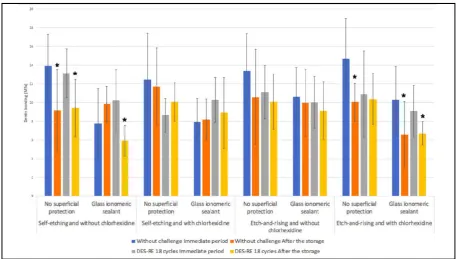
The Figure 2 shows the average and standard deviation of the bond strength found in the immediate period and after-storage and the statistical differences found between each group in both times. There are differences between the two evaluation periods only in experimental conditions associated with the
self-etching adhesive technique without chlorhexidine
application and etch-and-rinsing with chlorhexidine
application. Under such situations, the bond strength significantly decreased after storage.
[image:4.595.44.560.337.381.2]The statistical analysis also demonstrated that no significant triple interaction between the factors "surface protection", "erosive challenge" and "bonding procedure" was observed neither in the immediate period (p = 0.26) nor after storage (p = 0.17). Among the double interactions, there was statistical significance between the factors "surface protection" vs. "erosive challenge" (p = 0.002) in the immediate period and between "surface protection" and "bonding procedure" (p = 0.05) after storage. These statistical interactions were assessed by the Tukey test (Tables 2 and 3). In the initial period, a reduction in bond strength was observed after 18 cycles of DES-RE in the absence of surface protection. However, when the ionomer sealant was used, no differences were observed between the bond strength values, in the presence and absence of the erosive challenge (Table 2). In relation to the effect of surface protection, it can be seen that in the absence of erosive challenge, the use of ionomeric sealant significantly reduced the average of bond strength. On the other hand, when 18 cycles of DES-RE were performed, groups with and without glass-ionomeric sealant maintained similar means. After storage, it was noted that the presence of surface protection with sealant reduced the bond strength in the groups where the
Table 2. Average (standard deviation) of the bond strength in the experimental groups, grouping the data of the bonding agent factor
Surface Protection Erosive challenge
Absent (control) 18 DES-RE cycles
Absent (control) 13.66 (4.13) Aa 10.95 (3.44) Ab
Glass-ionomeric sealant 9.17 (3.38) Ba 9.96 (2.74) Aa
[image:4.595.46.558.429.479.2]Averages followed by distinct letters represent statistical significance (3-way ANOVA / Tukey, alpha = 5%). Upper case letters compare levels of the surface protection within each level of the erosive challenge. Lowercase letters compare levels of the erosive challenge within each level of the surface protection.
Table 3. Average (standard deviation) of the bond strength in the experimental groups, grouping the erosive challenge factor data
Surface protection Bonding Procedure Self-etching and without chlorhexidine
Self-etching and with chlorhexidine
Etch-and-rinsing and without chlorhexidine
Etch-and-rinsing and with chlorhexidine
Absent control 9.29 (3.74) Aa 11.01 (3.4) Aa 10.32 (3.87) Aa 10.46 (3.20) Aa Glass- ionomeric sealant 8.23 (2.64) Aa 8.51 (2.95) Ba 9.45 (3.15) Aa 6.66 (2.61) Ba
Means followed by distinct letters represent statistical significance (3-way ANOVA / Tukey, alpha = 5%). Upper case letters compare levels of the surface protection within each level of the bonding procedure. Lowercase letters compare levels of the bonding procedure within each level of the surface protection.
[image:4.595.71.529.506.765.2]application of the bonding agent included chlorhexidine (Table 3). However, within each protection method, no differences were observed between the bond averages according to the application technique.
DISCUSSION
The exposure of dental surfaces to acids can be simulated in different forms, among them, the use of demineralization and remineralization cycles (DES-RE) (Ranjitkar, 2012; Cruz, 2015; Austin, 2011; Zarela, 2015). The use of DES-RE cycles has been chosen for this work, because it simulates the endogenous erosion promoted by hydrochloric acid (gastric juice) in patients with gastroesophageal reflux (Ranjitkar, 2012; Bartlet, 2001; Cruz, 2015; Austin, 2011). Usually each acid episode lasts 2 minutes, being always associated to the remineralizing action of saliva, raising the pH of the oral cavity (Ranjitkar, 2012; Martins, 2018). In this study, a negative effect of the erosive challenge was noticed in the groups that didn’t receive surface protection, considering that after 18 DES-RE cycles the immediate values of bond strength
were significantly lower. That result can be justified by the
dentin erosion pattern, from the exposure and degradation of the collagen organic matrix by acids (Cruz, 2011; Hebling,
2005; Carrilho, 2007; Tjäderhane, 2015; Flurry, 2013).On the
other hand, after the 7-months storage, the effect of the erosive challenge could not be noticed. Probably because the hydrolytic degradation due to the storage in water in the form of sticks was even more severe for dentin bonding than the modification of the substrate by DES-RE cycles. During the resin restorations, the mechanisms of bonding to dentin, whether they are etch-and-rinse or self-etching, are based on the formation of a hybrid layer that covers the underlying dentin. Except for resin tags that extend a few micrometers to the dentin tubules, the only physical continuity between the hybrid layer and the underlying dentin are the collagen fibrils
(Tjäderhane, 2013; Tjäderhane, 2015).Thus, since the eroded
dentin substrate has degradation of the organic matrix, it may present faults in the formation of the hybrid layer and,
consequently, worse bonding strength.
Various materials have been used to protect the dentin surface from the effects of the erosive challenge, among them is the light-curing glass-ionomeric sealant. Studies have shown that this material has the capacity to release a large quantity of F and Sr ions. These ions can penetrate the dentin and can react with the hydroxyapatite, making fluorapatite and strontium-apatite, thus generating resistance to demineralization (Zhou,
2012; Arita, 2017).In addition, the glass-ionomeric sealant has
more fluoride content and shows a controlled pattern of release of these minerals, having greater remineralization capacity of the tooth structure (Zhou, 2012). However, in the present study, in the immediate period, after 18 DES-RE cycles, the groups with and without glass-ionomeric sealant showed similar means. It is not possible to say whether this result is because of the intensity of the challenge or to the interference that the physical barrier created by the sealant promotes in the
ability to bond to the dentine.After all, the groups exposed to
the glass-ionomeric sealant showed similar means regardless of the DES-RE challenge. In the absence of the erosive challenge, the use of the glass-ionomeric sealant promoted inferior average of immediate bond strength compared to its absence. These results can be justified by the findings of Arita et al. (2017), which evaluated the bond strength between the glass-ionomer sealant and dentin and observed that the bond
strength to dentin with the glass-ionomeric sealant was inferior in comparison with the bonding produced by the resin-based materials. This can be justified by the fact that the bonding of the glass-ionomeric cement to the dentin is carried out chemically, from the incorporation of polyelectrolyte chains in hydroxyapatite, which is produced by a reaction of replacement between the phosphate ion and such chains. In contrast, the bonding of the adhesive to dentin occurs mechanically through the penetration of adhesive monomers in the superficial dentin matrix and the dentin tubules, forming the hybrid layer and resin tags after light-curing (Arita, 2017). No statistically significant differences were found between the protocol of adhesive application in the initial period. Previous studies that evaluated the immediate bonding strength to dentin of universal adhesive systems, compared the etch-and-rise and self-etching techniques. Results showed no statistically significant differences between the average of bond strength of the universal adhesive when both ways of application were compared, corroborating with the results of the present investigation (Manfroi, 2016; Muñoz, 2013; Vermelho, 2016; Tsujimoto, 2017). This can be partially justified by the
presence of the 10-Methacryloyloxydecyl dihydrogen
phosphate) (10-MDP) in the composition of this universal adhesive system. This phosphate monomer can provide acidity to the adhesive and, consequently, the capacity of dentin surface etching, promoting formation of hybrid layer and monomer infiltration, as well as present some chemical affinity the residual hydroxyapatite (Cruz, 2015; Manfroi, 2016; Muñoz, 2013). On the other hand, other authors reported that universal adhesives applied on the etch-and-rinse mode show higher bond strength than in the self-etching mode, due to the better hybridization or resin infiltration within the exposed collagen fibrils of the dentin surface (Morabak, 2010). However, some other research show that the results of bond strength of these systems are substrate and product dependent, and it is necessary to perform more studies, especially in long-term to improve the knowledge about such systems (Muñoz, 2013; Vermelho, 2016; Tsujimoto, 2017).
Another contemporary concern regarding dentin bonding is the degradation of the collagen and the resin hydrophilic over time (Tay, 2004; Tjäderhane, 2013). The demineralized collagen matrix of eroded dentin might become more susceptible to the action of host-derived matrix metalloproteinases (MMPs), a family of zinc and calcium dependent endopeptidases present in dentin and saliva that are capable of degrading the components of the extracellular matrix, including the collagen (Cruz, 2015; Hebling, 2005; Francisconi-dos-Rios, 2015; Pashley, 2004). Some substances such as the chlorhexidine
digluconate are able to inhibit the endogenous
metalloproteinases present in human dentin, mainly after being submitted to erosive challenge (Zarella, 2015; Comar, 2015; Francisconi-dos-Rios, 2015; Carrilho, 2007; Moraback, 2010; De- Melo, 2013; Dursun, 2013; Montagner 2014). The chlorhexidine digluconate can be incorporated into the acid, the adhesive system or applied as solution directly onto the dentine surface. This latter form is the most tested in the
studies and shows some promising in-vitro results in the
reduction of the degradation of the hybrid layer and in the stability of resin-dentin bonding in long term (Hebling 2005;
Carrilho, 2007). Among several concentrations of
to the bond strength of adhesive systems (Francisconi-dos-Rios, 2015; Carrilho, 2007). The present study was not able to demonstrate a significative effect of chlorhexidine digluconate, both in immediate and in stored bond strength values. These results are corroborated by the study of Moraback et al. (2010), that reported that the application of 2% and 5% chlorhexidine before the use of a self-etching adhesive system in healthy dentin or artificially affected by caries did not change the bond strength. In addition, de-Melo et al (2013) evaluated the short-term effect of the application of 2% chlorhexidine prior to a self-etching adhesive system on a bond strength of the composite resin to the healthy and demineralized dentin, showing that the chlorhexidine did not affect the immediate bonding of self-etching adhesive systems with both dentin substrates.
After 7-months storage, it was observed that the groups that received protection with glass-ionomeric sealant and the adhesive protocol with the application of 2% chlorhexidine showed inferior bond strength when compared to the other
groups. This can be explained by the fact that chlorhexidine
digluconate has strong cationic properties, which may have
reacted with the anionic carboxyl groups of the glass ionomer sealant, preventing the formation of calcium-carboxyl bonds, reducing the adhesion capacity to dentin (Dursun, 2013). The null hypothesis tested under the present investigation were all rejected, considering that: the eroded dentin surfaces coated with glass-ionomer sealant presented impaired bonding under some circumstances and the protocol for adhesive bonding resulted in some negative long-term bonding when the chlorhexidine digluconate was used on previously sealed surfaces. Such findings demonstrate that there is still much to learn about bonding to eroded substrates, once several material and clinical variables may interfere with long-term outcomes. Finally, the clinical follow-up of patients with erosive lesions cannot be discarded as well as the correct diagnosis, early treatment of the etiological factor and conservative management of sequelae. It is clear that anticipation might help minimizing the damages caused by this condition. Within the limitations of the present study, it is possible to conclude that the erosive challenge compromised the bonding strength in the absence of surface protection in the immediate period, but the degradation in water seems to have masked this effect in the long term. Chlorhexidine was not able to increase bonding stability after storage; and both self-etch and etch-and-rinse protocols seems to be feasible for the use of the universal adhesive system on eroded dentin surfaces. The physical presence of the glass-ionomeric sealant appeared to disrupt the bond strength in some experimental conditions.
Acknowledgments
This work should be attributed to Dentistry Course, School of Medicine and Public Health of Bahia (BAHIANA). This investigation has the material donation by 3M-ESPE.
REFERENCES
Arita S, Suzuki M, Kazama-Koide M, Shinkai K. 2017. Shear bond strengths of tooth coating materials including the experimental materials contained various amounts of multi-ion releasing fillers and their effects for preventing dentin demineralization. Odontology.
Austin RS, Stenhagen KS, Hove LH, Dunne S, Moazzez R,
Bartelett DW, et al. 2011. A qualitative and quantitative
investigation into the effect of fluoride formulations on enamel erosion and erosion–abrasion in vitro. J Dent. 39; pp 648-655.
Bartlett DW and Coward PY. 2001. Comparison of the erosive
potential of gastric juice and a carbonated drink in vitro. J
Oral Rehab. 28, pp 1045-1047.
Carrilho MRO, Carvalho RM, de Goes MF, di Hipólito V, Geraldeli S, Tay FR et al. 2007. Chlorhexidine Preserves
Dentin Bond in vitro. J Dent Res. 86(1); pp 90–94.
Comar LP, Cardoso CAB, Charone S, Grizzo LT, Buzalaf MAR, Magalhães AC. 2015. TiF4 and NaF varnishes as anti-erosive agents on enamel and dentin erosion
progression in vitro. J Appl Oral Sci. 23(1); pp 14-18.
Cruz JA, Bonini G, Lenzi TL, Imoarato JCP, Raggio DP. 2015. Bonding stability of adhesive systems to eroded dentin. Braz Oral Res. 29(1); pp 1-6.
De-Melo MA, Goes DC, de-Moraes MD, Santiago SL, Rodrigues LK. 2013. Effect of chlorhexidine on the bond strength of a self-etch adhesive system to sound and demineralized dentin. Braz Oral Res. 27; pp 218-224. Dursun E, Le Goff S, Ruse DN. 2013. Effect of Chlorhexidine
Application on the Longterm Shear Bond Strength to
Dentin of a Resin modified Glass Ionomer. Oper Dent.
38(3); pp 275-281.
Elkassas D and Arafa A. 2014. Remineralizing efficacy of different calcium-phosphate and fluoride-based delivery
vehicles on artificial caries like enamel lesions. J Dent. 42;
pp 466-474.
Flury S, Koch T, Peutzfeldt A, Lussi A, Ganss C. 2013. The effect of a tin-containing fluoride mouth rinse on the bond between resin composite and erosively demineralised
dentin. Clin Oral Invest. 17; pp 217–222.
Francisconi-dos-Rios LF, Casas-Apayco LC, Calabria MP, Francisconi PAS, Borges AFS and Wang L. 2015. Role of chlorhexidine in bond strength to artificially eroded dentin
over time. J Adhes Dent. 17; pp 133–139.
Hebling J, Pashley DH, Tjäderhane L, Tay FR. 2005. Chlorhexidine arrests subclinical breakdown of dentin hybrid layers in vivo. J Dent Res. 84; pp 741-746.
Kato MT, Leite AL, Hannas AR, Calabria MP, Magalhães AC,
Pereira JC et al. 2012. Impact of protease inhibitors on
dentin matrix degradation by collagenase. J Dent Res.
91(12); pp 1119-1123.
Manfroi FB, Marcondes ML, Somacal DC, Borges GA, Burnett Júnior LH, Spohr AM. 2016. Bond Strength of a Novel One Bottle Multi-Mode Adhesive to Human Dentin
After Six Months of Storage. The Open Dentistry Journal.
10; pp 268-277.
Martins VL, Ramos, RVC, Lima, MJP, Araújo RPC, Cavalcanti AN. 2018. Effect of surface protection on the
permeability of eroded dentin. J Conserv Dent; 21; pp
16-20.
Mobarak EH, El-Korashy DI, Pashley DH. 2010. Effect of chlorhexidine concentrations on micro shear bond strength of self-etch adhesive to normal and caries-affected dentin.
Am J Dent. 23; pp 217-222.
Montagner AF, Sarkis-Onofre R, Pereira-Cenci T, Cenci TMS. 2014. MMP Inhibitors on Dentin Stability: A Systematic
Review and Meta-analysis. J Dent Res. 93(8); pp 733-743.
Muñoz MA, Luque I, Hass V, Reis A, Loguercio AD, et al. 2013. Immediate bonding properties of universal adhesives
to dentine. J Dent Res.41; pp 404-411.
Ranjitkar S, Kaidonis JA, Smales RJ. 2012 Gastroesophageal
reflux disease and tooth erosion. Int J Dent. pp 1-10.
Sano H, Shono T, Sonoda H, Takatsu T, Ciucchi B, et al. 1994. Relationship between surface area for adhesion and tensile bond strength- Evaluation of a micro-tensile bond test. Dent Mater.10; pp 236-240.
Tay FR, Pashley DH, Suh B, Carvalho R, Miller M. 2004. Single-step, self-etch adhesives behave as permeable membranes after polymerization. Part I. Bond strength and
morphologic evidence. Am j Dent. 17; pp 000-000.
Tjaderhane L, Buzalaf MAR, Carrilho M, Chaussain C. 2015. Matrix Metalloproteinases and Other Matrix Proteinases in Relation to Cariology: The Era of ‘Dentin Degradomics’.
Caries Res. 49; pp 193-208.
Tjäderhane L, Nascimento FD, Breschi L, Mazzoni A,
Tersariol ILS, Geraldeli S et al. 2013 Strategies to prevent
hydrolytic degradation of the hybrid layer - A review. Dent. Mat. 29; pp 999-1011.
Toda S and Featherstone JD. 2008. Effects of fluoride
dentifrices on enamel lesion formation. J Dent Res. 87(3);
pp 224-227.
Tsujimoto A, Barkmeier WW, Takamizawa T, Wilwerding TM, MA Latta MA, Miyazaki M. 2017. Interfacial Characteristics and Bond Durability of Universal Adhesive
to Various Substrates. Oper Dent.42 (2); pp e59-e70.
Vermelho PM, Reis AF, Ambrosano GMA, Giannini M. 2016. Adhesion of multimode adhesives to enamel and dentin after one year of water storage. Clin Oral Invest
Zarella BL, Cardoso CAB, Pela VT, Kato MT, Tjaderhane L, Buzalaf MAR. 2015. The role of matrix metalloproteinases and cysteine-cathepsins on the progression of dentine
erosion. J Adhes Dent. 17; pp 133–139.
Zhou SL, Zhou J, Watanabe S, Watanabe K, Wen LY, Xuan K. 2012. In vitro study of the effects of fluoride-releasing dental materials on remineralization in an enamel erosion
model. J Dent. 40; pp 255-263.