AND VARIATIONS WITH ITS CLINICAL
APPLICATIONS
Dissertation submitted for
M.S. ANATOMY BRANCH - V
DEGREE EXAMINATION
THE TAMIL NADU DR.M.G.R.MEDICAL UNIVERSITY
CHENNAI, TAMIL NADU
This is to certify that the dissertation on "BRACHIAL ARTERY,
ITS BRANCHING PATTERN AND VARIATIONS WITH ITS
CLINICAL APPLICATIONS" is the bonafide work done by
Dr.V.SATHIA LAKSHMI, in the Institute of Anatomy, Madras Medical
College, Chennai - 600 003, during 2003 - 2006 under my supervision
and guidance in partial fulfilment of the regulation laid down by Tamil
Nadu Dr.M.G.R. Medical University, for the M.S. Anatomy Branch V
examination to be held in September 2006.
Dr.KALAVATHY PONNIRAIVAN, B.Sc., M.D., Dr.CHRISTILDA FELICIA
Dean JEBAKANI M.S.,
Madras Medical College, Director
Chennai - 600 003. Madras Medical College, Chennai - 600 003.
Date : Date :
I wish to acknowledge my indebtedness to my teacher and guide
Dr.CHRISTILDA FELICIA, JEBAKANI, Director and Professor, Institute of Anatomy, Madras Medical College, who led me like a light throughout my study. Without her valuable guidance, kind concern and support, this work would not have been possible. Working under her has given me the great experience of working with a person who is a blend of a teacher.
I wish to acknowledge my thankfulness to Dr.KALAVATHY
PONNIRAIVAN, B.Sc., M.D., Dean, Madras Medical College, Chennai, for permitting me to avail the facilities in the college during the course of my study.
I wish to acknowledge my gratefulness to Dr.I.JAYARAJ, Dr.B.CHEZIAN and Dr.N.M.PICKTHAL, for their encouragement to my study.
I wish to acknowledge my sincere thanks to Mrs.M.S.THENMOZHI
for her appreciation and encouragement throughout my dissections.
I wish to acknowledge my sincere thanksfulness to Dr.S.A.HUSSAIN, M.S., M.Ch., F.R.C.S., Professor and Head of Department of Vascular Surgery, Madras Medical College, Chennai - 600 003, for guiding me through the clinical study and helping me to collect material for the study.
I wish to acknowledge my sincere thankfulness to
and encouraging throughout my study.
I wish to acknowledge my thankfulness to Dr.A.SHARMILA, Dr.M.KAVIMANI, Dr.IVAN JAMES and Dr.S.SUMATHI LATHA, for being supportive and encouraging throughout my study.
I sincerely thank Mr.M.ABDUL JABBAR, Mr.P.DEVARAJAN, Mr.G.ANKIAH and Mr.SUGENDRAN, who helped with making arrangements for dissections.
I thank my daughter K.ANITHA my nieces K.NANDITA, G.UMA my nephew ASHWIN RAVI, for providing tactful, constructive criticism and patience during the computer preparation of the manuscript.
CONTENTS
Chapter Title Page No.
I AIM OF THE STUDY 1
II. REVIEW OF LITERATURE 3
III. DEVELOPMENTAL ANATOMY 19
IV. MATERIALS AND METHODS 23
V. OBSERVATION 27
VI DISCUSSION 40
VII. CONCLUSION 58
"Any thing out of sight is out of
mind"
A basic law of anatomy is that the only thing which remain
constant is its variability. Striking variations in origin and course of the
principal arteries of the upper extremities have long received the attention
of anatomists and surgeons. Now a days cardiologists and radiologists are
utilizing the brachial artery with increasing frequency for catheter based
diagnostic and therapeutic intervention procedures. It is gaining
importance because brachial approach allows early ambulation and
discharge.
Brachial artery is used in diagnostic angiography, cardiac
catheterisation for angioplasty, carotid stenting, transbrachial access for
endovascular renal artery intervension, embolectomy through arteriotomy
on branchial artery is done. Apart from the above mentioned procedures,
accidental intra arterial injections, ligation of artery instead of vein have
been reported. In order to avoid all these catastrophes accurate knowledge
of this major arterial conduit in relation with its course and particularly of
Thus an interest in the anatomy of this clinically so important
brachial artery was a stimulus for me to pursue this study in a detailed
manner. Isolation of the brachial artery and tracing of the branches was
done to know more about it than already documented and thereby hoping
to add more information to guide the operating surgeons, cardiologists,
vascular surgeons and anaesthetists.
The present study of brachial artery was undertaken by me to study
the variation in the branching pattern. It was also studied under the
following parameters.
1. The length of the brachial artery from the teres major tendon
to the inter condylar line,
2. Point of bifurcation in relation to intercondylar line,
3. Variation in branches :
a. profunda brachii
b. superior ulnar collateral artery c. nutrient artery of the humerus d. inferior ulnar collateral
e. terminal branches
f. superficial brachial artery
REVIEW OF LITERATURE
Quains (1844) was believed to be the first person to provide data
sufficient for useful statistical evaluation regarding brachial artery. In his
series of dissection with 481 extremities he encountered 19.5% of
anomalies of radial artery. He reported 0.2% prevalence superficial
brachial in 506 extremities he dissected, presence of an anastomotic
vessel in the antecubital fossa in 2.5% and presence of superficial ulnar
artery 1.7%.
Gruber (1848) came across 8.6% of variations of the brachial
artery in 1200 extremities. He reported 5 cases (0.4%) of superficial
brachial artery with an incidence of 0.5% of the anastomotic vessel in the
antecubital fossa.
Gray (1858) in his "Text Book of Grays Anatomy" He describes
that the brachial artery is the continuation of the axillary artery, which
begins at the inferior border of Teres Major Muscle and ends about 1 cm
Poirier (1886) has discussed in his text book of anatomy regarding
the superficial brachial artery, in hundred dissection he found superficial
brachial in 6% cases i.e. brachial artery crossed superficial to the median
nerve in 6 limbs. High origin of the ulnar artery was found 20 times in
440 dissection. High origin of the radial artery according to Poirier was
rare.
Muller (1903) appreciated 14% of variation in the brachial artery
in his dissection with 100 limbs, and came across 1% superficial brachial
artery, 2% superficial ulnar artery and pointed out 6% presence of an
anastomotic vessel in the antecubital fossa.
Hofer & Hofer (1910) described a case in which brachial artery
passed between the two heads of pronator teres instead of dividing above
it, but they were unable to find a similar case in literature.
Linell (1921) among the 34 limbs dissected he demonstrated two
instances in the same body in which the median nerve crossed deep to the
brachial artery.
Beuntaro Adachi (1928), a Japanese anatomist described that
arterial trunk runs superficial to the nerve, this is the "arteria brachialis
superficialis". It may replace the main trunk, or it may be accompanied
by an equally important, less important, or more important trunk running
parallel and deep to the median nerve in the normal position. In these
cases the superficially placed vessel may continue as the radial or more
rarely as the ulnar artery. He further subdivided the "arteria brachial
superficialis into superior, medial and inferior according to its point of
origin for the main arterial trunk. The point of origin may be from the
axillary, most frequently it is from the upper part of the brachial, but a
"superficial brachial artery" may also arise from the lower part of the
brachial artery nearer the elbow.
Degaris & Swartley (1928), the authors recognised 23 different
patterns of axillary artery and its braches in their study based on 512
dissection and about 0.8% of high origin of ulnar artery and 7.7% of high
origin of radial artery. He also pointed out 9% of prevalence of
superficial brachial artery.
Huber (1930), describes that inferior ulnar collateral artery a
branch of brachial artery begins 4 cm above the termination of the
behind the supracondylar process of humerus from which a fibrous arch
is most often thrown over the artery. This condition resembles the normal
condition in some carnivores.
Piersol (1930) in his text of human anatomy had written that
variations which the brachial artery presents are numerous and important,
in that they affect materially the origin of the two terminal branches, the
radial and ulnar. In cases in which there is a well-developed
supracondylar process on the humerus the brachial artery accompanies
the median nerve behind it, and only passes upon the anterior surface of
the arm after it has passed it. In such cases there generally arises from the
upper part of the brachial or even from the axillary, a vessel which
descends upon the anterior surface of the arm, lying superficially and
sending branches to the biceps and brachialis muscles. This has been
variously termed the vas aberrans, the a. brachialis superficialis, or the a.
radialis superficialis, and it appears to be normally present, but much
reduced in size and included among the muscular branches. The majority
of the modifications of the brachial artery are due to an extraordinary
development of the superficial brachial. Thus it may enlarge and become
termed a "high" origin of the radial; more rarely it may unite with the
ulnar artery, producing a "high" origin of ulnar vessel.
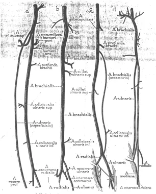
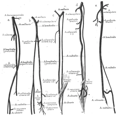
Charles et al., (1931) made a study of the types of origin of the
profunda brachii artery in 300 dissections and specified 7 types of origin
for profunda brachii artery.
Type I : Branch of brachial artery in 54.7% cases.
Type Ia : Origin of arteria profunda brachii by 2 separate
branches seen in 0.7% dissections.
Type Ib : Origin of arteria profunda brachii by 3 separate
branches seen in 0.3% dissections.
Type II : Arising as a common trunk with superior ulnar
collateral in 22.3% cases.
Type III : Arising at lower border of teres major so can be
considered to be arising from axillary or brachial in
8% cases.
Type IV : Branch of 3rd part of axillary artery in 8.7% cases.
Type V : Arising as a common trunk with posterior circumflex
Type VI : Arising as a common trunk with subscapular and both
circumflex humerals from axiallary artery in 0.7%
cases.
Type VII : Absent arteria profunda brachii in 0.7% cases.
Polanskaja (1932) pointed out that the smaller branches of
brachial artery, especially those which anastomose around the elbow to
form the collateral circulation, have no constant pattern. He further added
that he was never able to find the same pattern even on the 2 sides of one
body.
Singer (1933) Considered the high origin of radial artery as a kind
of persistent superficial brachial artery.
Schwyzer & DeGaris (1935) reported two cases of superficial
brachial artery dividing into radial and ulnar arteries in the cubital fossa
and then the ulnar artery going superficial to all flexor muscles of forearm
in one case and all but palmaris longus in the other; the brachial artery
continued in the arm as interosseous complex. They called this ulnar
J.E. Frazer, Professor, University of London (1937). In his text
book of 'A manual of anatomy". Had written that the brachial artery may
divide at a higher level than usual. In most cases the abnormally early
branch is the radial; more rarely it is the ulnar, and in these cases the
interosseous trunk arises from the radial; still more rarely the premature
branch is the interosseous trunk, or a large vas aberrans. The level at
which a high division takes place is most frequently in the upper third of
the arm, less frequently in the lower third, and rarely in the middle third.
When two arteries are present, they usually lie one in front of the median
nerve and the other behind it. When a vas aberrans is present, it usually
arises from the upper part of the brachial artery, lies in front of the
median nerve, and terminates below by joining, most commonly, the
radial artery. In rare cases the brachial artery divides high up into two
vessels of equal size, which become reunited into one trunk a little above
the elbow.
Miller (1939) in his dissection with 480 bodies came across about
3% superficial brachial artery. And he believed that superficial brachial
artery is an atavistic condition, since a main brachial artery crossing
superficial to median nerve is said to be the usual arrangement in the
Massie (1944) said arteria profunda brachii is also known as
superior profunda, superior ulnar collateral as inferior profunda and
inferior ulnar collateral as arteria anastomotica.
Treves & Rogers (1947) described the presence of 2 arteries
instead of one brachial artery. These 2 arteries may be (a) radial & ulnar,
(b) 2nd branch may be interosseous which has originated high up from
arteries at normal position, (c) the 2 vessels may be normal brachial and a
vas aberrans.
Thorek (1951) says that brachial artery gives 3 main branches
(profunda brachii, superior ulnar collateral and inferior ulnar collateral).
He called superior ulnar collateral as inferior profunda.
Lawrence J. McCormack, et al.,(1953) in their total series which
comprised of 750 consecutive upper extremities. 139 (18.53%) presented
major variations in respect to the origin and course of brachial or
antebrachial arteries. Instances of origin of the radial artery proximal to
the inter condylar line form the largest group of the gross variation which
represented 14.27%. He pointed out the presence of an anastomatic vessel
artery he demonstrated the presence of a large anastomotic connection
between the radial and ulnar arteries in cubital fossa (Fig.1 & 2)
J.C.Boileau Grant (1958) in his book "A Method of anatomy
Descriptive and Deductive" had written that the brachial artery is the
largest artery whose walls can be felt satisfactorily in the living subject. It
may be palpated along the medial bicipital furrow throughout the length
of the arm to the point where it disappears behind the bicipital
aponeurosis. At the level of the neck of the radius, 1" below the
transverse crease of the elbow, it divides into its two terminal branches,
larger ulnar artery and smaller radial artery.
W.Henry Hollinshead (1958) in his book "Anatomy for surgeons :
Volume III" has described brachial artery as the continuation of axillary
artery, the change in name occurring at the lower border of the teres
major muscle.
Skopakoff (1959) gives the percentage frequency of all types of
"superficial brachial artery" as 19.7% (610 dissections). The author
includes several instances of comparatively small branches of the brachial
which ran superficial to the median nerve and resolved themselves into
J.A.Keen (1961) in his series of 284 dissections found that
profunda brachii was often represented by more than one trunk which
follow the radial nerve. In 26% (284 dissections) the profunda brachii
arose from the terminal part of the axillary artery, i.e., the origin was
from the main arterial trunk at a level above the lower border of the teres
major tendon. In 6% the profunda brachii arose as a branch of the
posterior circumflex humeral, or of the subscapular in the case of a
common trunk for these two vessels. A third abnormality of the profunda
brachii is its origin from the posterior circumflex humeral artery. He also
encountered about 12.3% of superficial brachial artery. He sub divides
superficial brachial artery into 3 types.
a. Those superficial brachial artery which continue in the
cubital fossa and bifurcate as usual into radial and ulnar
arteries (3.6%).
b. Superficial brachial continues as radial artery and known as
high origin radial artery (5.9%).
c. Superficial brachial artery continue as ulnar artery and
Romanes (1964) has described that brachial artery some times
accompanies median nerve behind the supra condylar process of humerus
from which a fibrous arch is most often thrown over the artery. This
condition resembles normal condition in carnivores. He also pointed out
high origin of ulnar artery but with out statistics.
Anson (1966) in his dissection came across, 15% of high origin of
radial artery. In the arm, it lies anterior to median nerve and in the
forearm it takes normal course. He also demonstrated the profunda
brachii arising from brachial artery in 55%. 22% profunda brachii arising
as a common trunk with superior ulnar collateral. 16% as a branch of
third part of the axillary artery and 7% as a common trunk from posterior
circumflex humeral artery. According to him superficial brachial arises
from axillary artery or from proximal 1/3 of brachial artery usually
between the contributions of medial and lateral cords of brachial plexus
to the median nerve. It is superficial to muscles of the arm under brachial
fascia lying slightly more lateral than the brachial artery and in the elbow
Vare and Bansal (1969) reported a case with high division of
brachial artery - the superficial brachial artery giving rise to radial and
ulnar artery while deep division continuing in the forearm as interosseous
complex and giving a large median artery which in the palm formed
superficial palmar arch with ulnar artery.
C.J.Romanes (1972) in Cunningham's Textbook of Anatomy had
written that the brachial artery sometimes divides at a higher level than
usual. In such cases the ulnar artery may cross superficial to the flexor
muscles, and may even be subcutaneous; and the radial artery may
descend in the superficial facsia of the forearm. In performing
venesection at the elbow such variations have to be borne in mind.
Sometimes the brachial artery accompanies the median nerve behind a
supracondylar process, or ligament; and it may pass in front of the
median nerve instead of behind it.
Karisson and Niechajev (1982) in angiographic observations,
found high origin of radial artery in 10% patients, the parent trunk being
axillary artery in 12.5%, proximal 1/3 of brachial in 62.5% and middle
1/3 of brachial in 25%. They could find high origin of ulnar artery in 1%
Lippert and Pabst (1985) had reported 22% prevalence of
superficial brachial artery and also considered high origin of radial artery
as a kind of persistent superficial brachial artery.
Jurjus a, Sfeir R, Bezirdjian R. (1986). They had reported an
anomalous brachial artery, after giving off a profunda brachii artery with
no collaterals, divides in its upper one-third into two equal-sized arteries,
brachial arteries 1 and 2. These arteries lie next to each other in the
normal path of the brachial artery. Brachial artery 1 is possibly a
high-origin and persisting radial artery. It gives no collaterals in the arm. At
the cubital fossa, it becomes subcutaneous and divides into two equal -
sized radial and ulnar arteries. These arteries run completely superficial to
flexor muscles of the forearm and are terminated by branches running
above the thenar and hypothenar eminences, respectively. Brachial artery
2 is possibly a high origin artery of the common interosseous. The course
of this artery resembles the course of the brachial axial artery of the
embryo. It supplies the anterior compartment of brachial muscles and
continues as the common interosseous artery. This common interosseous
artery in turn branches into the superior and inferior ulnar collaterals, and
the anterior and posterior interossei. It does not regress, but has a major
Lengele B, Dhem A.(1989) in their routine dissection came across
three cases of multiple anomalies involving the vessels and nerves of the
axilla. The main common characteristic between them was the unilateral
existence of a superficial brachial artery.
Nakatani T, Tanaka S, Mizukami S. (1996) they described rare
anomalies of the bilateral superficial brachial arteries in a dissected
69-year-old Japanese man in the gross anatomical course. The right and left
superficial brachial arteries were observed to originate from the axillary
artery, pass over the lateral root of the median nerve, course laterally and
superficially to the median nerve, and split into the radial and ulnar
arteries in the cubital fossa. The right brachial artery ended in the
posterior aspect of the elbow. The left brachial artery ended in the
anastomosis with the ulnar artery at the site opposite to the origin of the
common interosseous artery. These arterial patterns can be explained by
the existence, during the developmental process of the arteries of the arm,
of a superficial brachial artery and an anastomotic branch between the
Yucel AH (1999) described unilateral variation in the origin and
distribution of the arterial pattern of the human upper extremity on the
right side. Apart from its usual branches, the third part of the right
axillary artery gave origin to a common branch, the profunda brachii
artery and the superior ulnar collateral artery. The right brachial artery, at
a point 5.0 cm distal to its origin, bifurcated into the radial and ulnar
arteries; their origin was in a position opposite the usual location. The
radial artery continued on the medial side of the arm for 2.5 cm and
crossed the ulnar artery anteriorly to gain a lateral position in the arm.
The inferior ulnar collateral artery arose not from the brachial artery, but
from the ulnar artery.
William et al., (1999) described the brachial artery, as the
continuation of the axillary artery from the distal (inferior) border of the
tendon of teres major and ends about a centimeter distal to the elbow joint
(at the level of the neck of the radius) by dividing into radial and ulnar
arteries. At first it is medial to the humerus, but gradually spirals anterior
to it until it lies midway between the humeral epicondyles. Its pulsation
can be felt throughout. They also said the nutrient artery of the humerus
arises near the mid-level of the upper arm, and enters the nutrient canal
Rodriguez-Baeza A, Nebot J, Ferreira B, Reina F, Perez J,
Sanudo JR, Roig M. (2000) they reported 23 cases with variations in the
brachio-antebrachial arterial pattern of the human upper limb are
reported. According to them the brachial artery showed, 4 groups of
variation;
1. Isolated persistence of the median artery;
2. High origin of the ulnar artery;
3. High origin of the radial artery; and
4. Duplication of the brachial artery, either with or without
anastomosis at the cubital fossa.
Kumar MR. (2004) in a routine dissection of a female cadaver,
came across a variation in the course of the radial artery in the cubital
fossa and a communication between the brachial artery and radial artery
were observed. A rare origin and course of the median artery was also
DEVELOPMENTAL ANATOMY OF
UPPER LIMB VASCULATURE
NORMAL DEVELOPMENT
When the upper limb buds are formed in the 4th week of intra
uterine life, a number of small arteries arise from the dorsal aorta and
pass into the limb bud to form capillary network which drain into the
anterior cardinal veins. Out of these small arteries only one persists as the
axis artery of the upper limb. And it shifts its origin to the lateral branch
of the 7th cervical into segmental artery. This axis artery grows deep to
the muscle mass in the forearm and terminates in superficial and deep
capillary plexuses of the developing hand. Digital arteries develop from
the capillary plexus. (Fig.3)
Proximal part of the main trunk forms the axillary artery and
brachial arteries, and its distal part persists as the anterior interosseous
artery and the capillary plexus in which it ends forms the deep palmar
arch.
High in the forearm a branch from the axis artery passes dorsally
A little below that point, another branch of the axis artery accompanies
the median nerve and called the median artery reaches the superficial
capillary plexuses of the hand. Thereafter the distal part of the axis artery
loses its connection with the plexus.
A little below the elbow, the axis artery gives a branch which
becomes the ulnar artery. It grows distally to unite with the superficial
capillary plexus of the palm which later becomes the superficial palmar
arch. Now the median artery regresses from about the middle of the arm,
another branch is given off from the axis artery, and is called the primary
radial artery. The proximal part of the radial artery above this level
degenerates and disappears. The radial artery grows down to join the
deep capillary plexus of the hand which forms the deep palmar arch. Thus
the following structures are derived from the axis artery of the upper
limb. (a) axillary artery (2) brachial artery (c) proximal part of ulnar
EMBRYOLOGICAL JUSTIFICATION OF THE EXISTENCE OF ARTERIAL VARIATIONS IN THE ADULT UPPER LIMB
Based on the results of (Rodriguez - Niedenfuhr et al., 2001b)
injection studies on animal embryos and experimental data which showed
that when an endothelial tube gets a muscular coating it looses its
remodeling ability. They proposed that the sprouting theory described in
many of the embryological and anatomical textbooks and reproduced in
(Fig.4A) was absolute. The new findings suggest that the arterial pattern
of the upper limb develops from an initial capillary plexus by a proximal
to distal differentiation of certain capillary vessels, and the regression of
others (Fig.4B). It is suggested that the persistant, enlargement and
differentiation of capillaries forming the initial capillary plexus, which
would normally remain in a capillary state or even regress, gives rise to
arterial variations of the definitive arterial pattern, rather than the
sprouting of aberrant vessels.
The establishment of the superficial brachial, accessory brachial
and the brachial part of those variations affecting the arm and forearm
must be determined before stage 17 as then, the arteries until the elbow
would have already got a definitive morphology of its wall and no further
arteries as well as the ante-brachial part of those variations affecting the
arm and forearm, have to be established before stage 18 as then the
forearm arteries have got their definitive wall morphology except the
distal part of the radial. The superficial radial and the distal part of the
superficial brachioradial arteries would be determined before stage 21
MATERIAL AND METHODS
STUDY MATERIAL
The study material consists of
a) 40 upper limb specimens from 20 adult cadavers (14 males,
6 females)
b) 10 upper limb specimens from 5 full term foetuses
c) 2 clinical cases
METHODS OF STUDY
I. Conventional dissection method a) In adult cadavers
b) Foetal cadavers
II. Clinical Study
I. Conventional dissection method
a) Adult cadaveric study
Twenty adult human cadavers were selected from the cadavers
allotted to the I MBBS students at the Institute of Anatomy, Madras
Medical College. The age and sex of the human cadavers were noted
The skin and superficial fascia over the arm up to lower 1/3 of
forearm were carefully reflected along the conventional anatomical lines
from anterior aspect. Then the deep fascia was removed, flaps reflected to
uncover biceps brachii muscle, the muscle was displaced laterally.
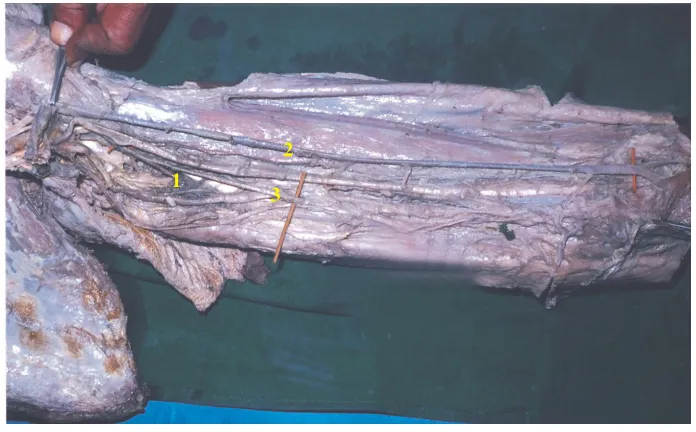
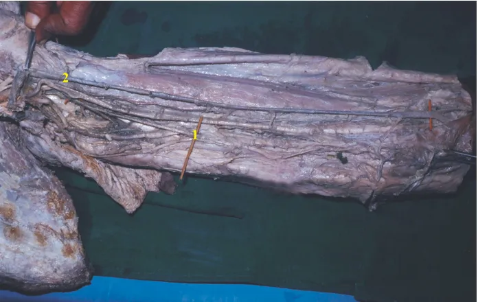
The principal neurovascular bundle of the arm medial to biceps
was identified. Then the structures in the arm were traced proximally upto
the insertion of the corocobrachialis. The neurovascular bundle consist of
the brachial artery and the two venae comittants. The brachial artery was
dissected from the distal border of the teres major muscle and was found
medial to the humerus but gradually spirals anterior to it and descended
midway between the humeral condyles.
During the above procedure the profunda brachii artery was
identified. It arose from the posteromedial aspect of the brachial artery
distal to the teres major muscle. It was traced along with the radial nerve
running between the long and medial heads of the triceps up to the radial
groove.
Superior ulnar collateral was dissected a little distal to the upper
the medial inter muscular septum and descended between the medial
epicondyle and olecranon.
The nutrient artery was traced from its origin near the mid level of
the upper arm, and found to enter the nutrient canal near the attachment
of coracobrachialis.
Inferior ulnar collateral was dissected about 5 cms proximal to the
elbow joint. It was traced between the median nerve and brachialis
muscle upto the point where it pierced the medial intermuscular septum.
The branchial artery was dissected upto the point of bifurcation and the
origin of ulnar artery and radial identified.
b) Foetal cadaveric study
Full term foetuses 5 in number were obtained from the Institute of
Obstetrics and Gynaecology, Egmore, and was embalmed in formalin
solution and studied.
In full term foetuses, the skin was reflected from the upper arm
upto the elbow joint. The neurovascular bundle was identified. The
muscles mass was pushed laterally, the brachial artery with the
branching pattern of the brachial artery was noted. The profunda brachii
artery, superior ulnar collateral artery and inferior ulnar collateral artery
were identified.
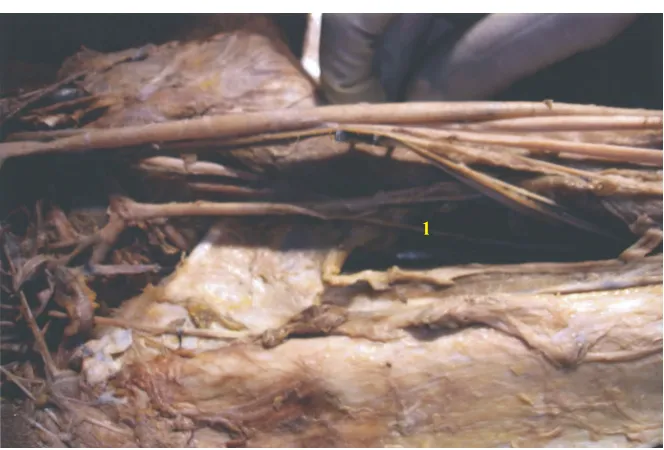
During the above cadaveric dissection in adult as well as in foetus
the variations of the brachial artery and its branches were photographed
for documentation.
The findings of the observation were noted down as per the
parameter taken for this study.
II. Clinical Study
Two cases from the Vascular Surgery Department, Government
General Hospital, Chennai - 3 have been selected for the study (2 male
patients of age group 20 and 25) with the following diagnosis.
Sl.No. Name Age Diagnosis
1. Navamani 20 Aneurysm of subclavian artery
2. Kannan 25 Ulnar artery occlusion
In the above two cases the branching pattern of the brachial artery
OBSERVATION
The brachial artery and its branching pattern was studied by the
following methods.
I. Conventional Methods
a. In adult cadavers
b. Foetal cadavers
II. Clinical Study
Conventional dissection method
Ia. Adult cadaveric study :
40 upper limbs were taken for study. After the conventional
dissection was carried out the findings were noted.
1. The length of the brachial artery from the teresmajor tendon
to the intercondylar line.
2. Point of the bifurcation inrelation to the intercondylar line.
3. Branches :
a. Profunda brachii artery also known as superior
b. Superior ulnar collateral artery also known as inferior
profunda.
c. Nutrient artery of the humerous
d. Inferior ulnar collateral or supra trochlear artery.
e. Two terminal branches - Ulnar and Radial
f. Superficial brachial artery
4. Brachial artery's relation to Median nerve
Ia. 1. Length of the brachial artery
In all 40 specimens dissected, the length of the brachial
artery was calculated as mentioned by Patnaik et al., 2002. For
measuring its length the following 2 points were taken.
a. The mid point of the width of the artery where it begins i.e.
at the lower border of teresmajor.
b. The point of termination of the artery.
First the distance between the lower border of the teresmajor
tendon and intercondylar line was measured along the artery (x)
termination of brachial artery where it divides into ulnar and radial
arteries was measured (y). Then the length of the brachial artery
was calculated by adding these two values (x + y) if the brachial
artery divided below the intercondylar line, or by substracting 2nd
distance from the first (x-y) if the brachial artery divided above the
intercondylar line. Measurement was taken with the help of inch
tape.
In one specimen the length of the brachial artery was 4cms
and in another it was 2cms. The Table-1 shows the complete length
of all the 40 specimens. The average length of the brachial artery
was 21.5 cms. Out of 40 specimens, 11 specimens were less than
the average length 21.5cms (28%) and 29 specimens were more
than the average length (72%).
Ia. 2. Point of bifurcation in relation to the intercondylar line
In 40 specimens dissected 36/40 (90%) (Table 2) specimens
bifurcated below the intercondylar line (Fig.5). In other 2/40 (5%)
specimens the bifurcation was above the intercondylar line (Fig.6).
In the remaining 2/40 (5%) the bifurcation of the brachial artery
Ia. 3. Branches
In 40 specimens dissected the profunda brachii artery,
superior ulnar collateral artery, nutrient artery collateral artery and
inferior ulnar and the terminal branches ulnar and radial were
studied.
3a. Profunda brachii artery
In 40 specimens dissected, profunda brachii artery arose
from the posteromedial side of the brachial artery in 35/40 (87.5%)
specimen (Fig.8) distal to the teresmajor tendon and followed the
radial nerve closely and it passed downward and outward between
the medial and long head of the triceps and reached posterior
surface of the humerus. (Tab.3).
In 2/40 specimens (5%) the profunda brachii artery and
superior ulnar collateral artery arose from a common trunk (Fig.9).
In 1/40 specimen (2.5%) the profunda brachii artery arose
from the posterior circumflex humeral artery a branch of the III
tendon. At the inferior border of teresmajor tendon it descended
posteriorly along with radial nerve (Fig.10).
In 1/40 specimen (2.5%) profunda brachii artery arose from
the axillary artery (Fig.11).
In 1/40 the profunda brachii arose as 2 separate branches and
both followed the course of radial nerve closely (Fig.12).
3b. Superior ulnar collateral artery
In 36/40 (90%) specimens (Fig.13) the superior ulnar
collateral arose from the medial side of the brachial artery a little
distal to the upper arm's mid level. It accompanied the ulnar nerve
and pierced the medial intermuscular septum and descended
between the medial epicondyle and the olecranon and terminated
deep to the flexor carpi ulnaris by anastomoting with posterior
ulnar recurrent and inferior ulnar collateral arteries. (Tab.4)
In 2/40 (5%) specimens the superior ulnar artery arose from
In 2/40 (5%) specimen superior ulnar collateral arose as a
branch of profunda brachii artery and descended between the
medial epicondyle and olecranon.
3c. Nutrient artery of the humerus
In all the 40 specimens the nutrient artery arose from the
brachial artery near the mid level of the upper arm, an entered the
nutrient canal on its medial surface near the attachment of the
corocobrachialis muscle.
3d. Inferior ulnar collateral artery
In all the 40 specimens dissected inferior ulnar collateral
artery arose about 5 cms proximal to the elbow no variation were
noted (Fig.15).
3e. Two terminal branches - Ulnar and Radial
In 40 specimens dissected 36/40 (90%) ulnar and radial
In 2/40 (5%) specimens the bifurcation of the brachial artery
was above the intercondylar line. So the radial and ulnar artery
originated from the mid level of the arm. (Fig.6)
In 1/40 (2.5%) ulnar artery originated from the third part of
the axillary artery and descended down to the palm and took part in
the superficial palmar arch (Fig.17).
In 1/40 (2.5%) the radial artery arose from the brachial artery
just below that teresmajor tendon and ran down the forearm up to
the wrist joint and turned to the dorsal surface (Fig.16).
3f. Superficial brachial artery
Superficial brachial artery has been already described
1. Brachial artery running superficial to median nerve.
2. The axillary artery at the inferior border of teresmajor tendon
gives off a superficial brachial artery branch and it continues
as a deep branch of the brachial artery (Fig.18).
3. The high origin of radial artery is also regarded as the
4. The high origin of ulnar artery is also termed as superficial
brachial artery (Fig.17).
In 38/40 (95%) specimens dissected the median nerve
formation was anterior to the brachial artery.
In 2/40 (5%) specimen the formation of median nerve was
posterior to the brachial artery, and the brachial artery and it ran
superficial to the median nerve (Fig.9).
In 1/40 (2.5%) specimen just below the inferior border of
teresmajor tendon a superficial vessel arose from the brachial
artery and it descended on the radial side superficial to all the
muscles upto the wrist joint and then turned to the dorsal aspect of
the hand. This vessel is termed as high origin of radial artery -a
type of superficial brachial artery (Fig.16).
In 1/40 (2.5%) specimen a vessel arose from the third part of
the axillary artery and descended superficially upto the palm and
took part in the superficial palmar arch. This is called the high
origin of ulnar artery. The brachial artery from the lower border of
intercondylar line bifurcated into radial and common interosseous
artery (Fig.17).
In one specimen 1/40 (2.5%) the brachial artery just below
its origin gave off a superficial branch which ran superficial to
median nerve and bifurcated into radial and ulnar artery below the
intercondylar line. From the deep vessel the profunda brachii, the
superior ulnar collateral nutrient and the inferior ulnar collateral
arteries arose (Fig.18).
4. Brachial artery's relation to median nerve
In 38/40 (95%) specimens the median nerve was formed
anterior to the brachial artery. In 2 specimens the median nerve
descended down on the medial side of the brachial artery
throughout. No crossing was seen (Fig.9).
In 2/40 (5%) specimens the median nerve was formed
Ib. Foetal cadaveric study
Length of the brachial artery
Ten arms of full term foetuses were dissected and the length
of brachial artery measured.
The Table - 6 shows the length of the brachial artery from
the lower border of the teresmajor tendon to the point bifurcation
below the intercondylar line. The average length of the artery was
8.35cms.
In 5/10 (50%) specimens the length of the brachial artery
was less than the average length (8.35cms).
In 5/10 (50%) specimens the length of the brachial artery
was more than the average length (8.35cms).
Point of bifurcation of the brachial artery
In 9/10 (90%) specimens the brachial artery bifurcating
below the intercondylar line.
In 1/10 (10%) specimens the brachial artery bifurcating
In all the 10 specimens dissected no gross branching
variations was noted.
Relation of the brachial artery to the median nerve
In 8/10 (80%) specimens the median nerve formed anterior
to the brachial artery and descended along the lateral side and
crossed to the medial side near the corocobrachialis muscle
incertion.
In 2/10 (20%) specimens the median nerve formed running
posterior to the brachial artery and descended medial to the
brachial artery throughout its course (Fig.20).
II. Clinical Study
Two clinical cases were selected from Vascular Surgery
Department, Government General Hospital, Chennai - 3 between the age
of 20 & 25 years (Fig.21).
Sl.No. Name Age Diagnosis
1. Navamani 20 Anneursym of subclavian
artery
For the above cases brachial arteriogram was done as an
investigation procedure. In case No.1 Anneursym of subclavian artery
was diagnosed and case No.2, Ulnar artery occlusion was made out.
Angiographic procedure
Retrograde axillary artery catheterization
First the patient was put in supine position. Then the left arm was
abducted to the extreme and the hand was placed under the patient's head.
The puncture site of the axillary artery located along the lateral axillary
fold over the proximal part of the humerus so that the underlying bone
provides support during compression.
The axillary artery was palpated and fixed by the left index and
middle finger, a small superficial skin nick was made with a No.11 blade
directly over the arterial pulse. The course of the artery was palpated
while a 18 guage needle Pott's Cournaud Needle having a sharp stylet
with a perforated hub was rapidly thrust down the artery. The needle was
angled at 45o with respect to the skin and gently advanced, when arterial
catheter (Seldinger) with two way tap attached to its hind end was passed
along the guide wire into the artery. The wire should not be forced.
Now, the urograffin solution (contrast) was injected to identify the
course of the vessel and to find out the clinical problems like thrombosis,
embolism, atheromatous plaque, stenosis or abnormal dilatations namely
anneurysm.
The study was done by visualising the pictures taken serially
starting from 5 minutes after injecting the contrast. Then the branches of
DISCUSSION
The present study of brachial artery was undertaken to study the
variations in the branching pattern. It was studied under the following
parameters.
1. Length of the brachial artery from the teres major tendon to
the point of bifurcation
2. Point by bifurcation in relation to intercondylar line.
3. Branches and variations:
a. Profunda brachii artery
b. Superior ulnar collateral artery
c. Nutrient artery
d. Inferior ulnar collateral artery
e. Terminal branches - radial and ulnar arteries
f. Superficial brachial artery
I.
Adult Cadaveric study
1. Length of the brachial artery from the teres major tendon to
the point of bifurcation
Quain (1844), Henry Gray (1858), Poirier (1886), Adachi
(1928), Piersol (1930), J.E.Frazer (1937), J.C.Boileau Grant (1957),
W.Henry Hollinshed (1958), Williams (1999) all the above scientists
had described the artery as the continuation of the axillary artery
commencing from the lower border of teres major to the point of
bifurcation of the artery below the inter condylar line. They had not
given any statistical data regarding the length of the brachial artery.
Patnaik et al. (2002), had said that brachial artery was the
continuation of the axillary artery from the lower border of teres major
tendon upto its bifurcation as radial and ulnar artery. According to his
study the total length of the brachial artery on an average was 26.29 cms
(ranging from 20.5 to 29.0 cms).
In the present study the average length of the brachial artery was
21.5 cms (ranging from 2 cms to 26 cms). So regarding the length of the
2. Point of bifurcation of brachial artery in relation to the
intercondylar line
Bifurcation of the brachial artery proximal to the inter condylar line
is considered as a variation.
Quains (1844), quoted that nearly 1.7% of brachial artery
bifurcated above the intercondylar line.
Gruber (1848), said that about 2% of the brachial artery in his
dissection bifurcated above the intercondylar line.
Muller (1903), described about 1% of brachial artery bifurcating
above the intercondylar line.
Buntaro Adachi (1928), reported 0.7% of the brachial artery
bifurcating above the intercondylar line.
Degaris and Swartley (1928), said that about 0.8% of the brachial
artery bifurcated above the intercondylar line.
Charles et al. (1931), reported that 10% of the brachial artery
Miller (1939), came across about 3% of the brachial artery
bifurcating above the intercondylar line.
J.A.Keen (1961), reported about 5-9% of the brachial artery
bifurcating above the intercondylar line.
Anson (1966), encountered about 15% of high bifurcation of
brachial artery.
Karisson and Niechajev (1982), in angiographic observation
found 10% of brachial artery bifurcating above the intercondylar line.
According to the above anatomists, bifurcation of the brachial
artery above the intercondylar line varies from 0.7% to 15.0%. All the
above anatomists have not given statistical data regarding the bifurcation
of the brachial artery below the intercondylar line and also bifurcation at
the intercondylar.
In the present study, in 5% cases brachial artery bifurcated above
the intercondylar line. In 90% cases brachial artery bifurcated below the
intercondylar line. And in 5% brachial artery bifurcated at intercondylar
intercondylar line, my study coincides with the anatomists Miller (1939)
and J.A.Keen (1961). (Table.8).
3. Branches and variation
a. Profunda brachii artery
The origin of arteria profunda brachii is quite variable. Charles et
al. (1931) specify 7 types of origins for this artery.
Type I : Branch of brachial artery in 54.7%
Type Ia : Origin of arteria profunda brachii by 2 separate
branches seen in 0.7% dissections).
Type Ib : Origin of arteria profunda brachii by 3 separate
branches seen in 0.3%.
Type II : Arising as a common trunk with superior ulnar
collateral in 22.3% cases
Type III : Arising at lower border of teres major so can be
considered to be arising from axillary or brachial in
8% cases.
Type V : Arising as a common trunk with posterior circumflex
humeral in 4% cases
Type VI : Arising as a common trunk with subscapular and both
circumflex humeral from axillary artery in 0.7% cases.
Type VII : Absent arteria profunda brachii in 0.7% cases.
J.A.Keen (1961) reported 61% of profunda brachii arising from as
a branch of brachial artery. In 13% cases profunda brachii artery arose as
a common trunk with superior ulnar collateral. In 26% of his dissection
the profunda brachii artery arose from axillary artery.
Anson (1966) said 55% of profunda brachii artery arose from the
brachial artery. 22% arose from a common trunk with superior ulnar
collateral artery. 7% of profunda brachii artery arose from posterior
circumflex humeral artery. He also said 16% of profunda brachii artery
arose from axillary artery.
Patnaik et al. (2002) said 94% of profunda brachii artery arose
from the postero medial aspect of brachial artery. 2% of profunda brachii
collateral artery and 2% from the axillary artery. 2% of profunda brachii
as 2 separate branches from brachial artery.
In the present study 87.5% of profunda brachii artery arose from
the brachial artery. 5% arose from the common trunk, with the superior
ulnar collateral artery, 2.5% from the posterior circumflex humeral artery
and 2.5% from the axillary artery. 2.5% of the profunda brachii artery
arose as to seperate branches. Regarding the origin of profunda brachii
from the brachial artery my study coincides with Patnaik et al. (2002).
(Table.9,10,11 & 12).
Regarding the origin of the profunda brachii from the common
trunk with superior ulnar collateral, the present study shows 4% which
coincides with the scientist Patnaik et al. (2002). Profunda brachii
originating from the posterior circumflex humeral was found to be 2.5%
which almost coincides with the scientist Charies et al. (1931) and
differs with the scientists J.A.Keen (1961) and Anson (1966). Profunda
brachii originating from the axillary artery was found to be 2.5% which
b. Superior ulnar collateral artery
Charles et al. (1931) said that the origin of superior ulnar collateral
artery in 22.3% cases was in common with the profunda brachii artery
and 77.7 from brachii artery.
Anson (1966) reported 22% of superior ulnar collateral artery
arising as a common trunk with profunda brachii artery and 78% from
brachial artery.
Patnaik et al. (2002) said 2% of superior ulnar artery arising as a
common trunk with profunda brachii artery and 2% as a branch of
prfounda brachial artery and 96% from brachical artery.
In the present study regarding the origin of superior ulnar
collateral artery as common trunk with profounda brachial artery 5%, as a
branch profunda brachial 5% from the brachial artery 90%.
c. Nutrient artery
Quains (1844) described that the nutrient artery arose from the
Gray (1858) described the nutrient artery as a branch of brachial
artery which entered the nutrient canal at the level of the attachment of
corocobrachialis muscle.
Poirier (1886) described that nutrient artery entered the nutrient
canal at the level of insertion of corocobranchialis muscle.
Piersol (1930) described that nutrient artery arises from brachial
artery and enters the nutrient foramen on its medial surface.
Frazer (1937) said that nutrient artery entered the nutrient foramen
opposite the lower border of the insertion of the cococobranchialis
muscle.
J.C.B.Grant (1957) said that nutrient artery arises from middle of
brachial artery and entered the nutrient foramen on the antero-medial
surface of the humerus.
C.J.Romanes (1971) said the nutrient artery a branch of the
brachial artery entered that nutrient canal on its medial surface.
In the present study, in all the 40 specimens the nutrient artery was
the branch of the brachial artery and it arose also out 5 cms proximal to
insertion of the corocobrachialis muscles. So my study coincides with the
all the above mentioned anatomists.
d. Inferior ulnar collateral artery
Gray (1858) said inferior ulnar collateral artery begins 5 cms
proximal to the elbow and passes behind the median nerve and brachialis
muscle.
Huber (1930) said that inferior ulnar collateral artery arises 4 cms
above the termination of the brachial artery.
Piersol (1930) said that inferior ulnar collateral artery a branch of
the brachial artery arose on its medial side just before its bifurcation.
Frazer (1930) described that the inferior ulnar collateral artery
arose 2 cms above the elbow and passes inward on the brachialis muscle.
Anson (1966) described that inferior ulnar collateral artery arose 5
cms proximal to the elbow.
C.J.Romanes (1971) said that inferior ulnar collateral artery arose
Williams et al. (1999) said that inferior ulnar collateral artery
begins above 5 cms proximal to the elbow.
Yucel A.H. et al. (1999) said that inferior ulnar collateral arose
from ulnar artery.
Patnaik et al. (2002) also discussed that inferior ulnar collateral
artery arose from the brachial artery above the elbow and in 4% of his
dissection inferior ulnar collateral artery was absent.
In my present study the origin and course of inferior ulnar
collateral artery in all the 40 specimen was similar to as described by the
Gray, Anson, C.J. Ramanes and Williams et al., But differed from Huber,
Persol, Frazwer, Putnaik and Yucel A.H. et al. in that I did not fine any
absence of inferior ulnar collateral artery.
e. Terminal branches
The brachial artery terminated as ulnar and radial artery below the
intercondylar line.
Quains (1844) reported 1.7% of high origin of ulnar artery.
Adachi (1928) quoted 0.7% of high bifurcation of brachial artery.
Degaris and Swartley (1928) reported, 0.8% of high origin of
radial artery.
McCormack et al. (1953) reported 2.26% of high origin of ulnar
artery.
J.A.Keen (1961) said 2.8% of ulnar artery arose above the
intercondylar line.
In my present study high origin of ulnar artery was observed in
5% of cases, which did not coincide with any of the above mentioned
scientists.
Degaris and Swartley (1928) reported 7.7% of high origin of
radial artery.
Miller (1939) said 3% of radial artery arose above the
intercondylar line.
McCormack et al. (1953) reported 14.27% of high origin of radial
artery.
Karisson & Niechajev (1982) reported 10% of high origin of
radial artery in their angiographic study.
J.A.Keen (1961) reported 5.9% of high origin of radial artery.
In my present study 5% specimen showed high origin of radial
artery, which all most coincide with J.A.Keen.
f. Superficial brachial artery
Quains (1844) reported 0.2% of superficial brachial artery.
Gruper (1848) quoted 0.4% prevalence of superficial brachial
artery.
Poirier (1886) said that he had encountered 6% of superficial
brachial artery.
Muller (1903) described 1.0% of superficial brachial artery.
Linell (1921) reported 6.0% of superficial brachial artery.
Degaris & Swartley (1928) came across 9% of superficial brachial
artery.
Treves and Rogers (1947) reported 15% of superficial brachial
artery.
McCormack et al. (1953) reported 5.75% of superficial brachial
artery.
Skopakoff (1959) presented 19.7% of superficial brachial artery.
Lanz & Wachsmith (1959) reported 25% of superficial brachial
artery.
J.A.Keen (1961) reported 12.3% of superficial brachial artery.
Fuss et al. (1985) reported 17.0% of superficial brachial artery.
Leppert and Pabst (1985) described 22% of superficial brachial
artery.
Baeza et al. (1995) described 11.9% of superficial brachial artery.
Kapur et al. (2000) described 5% of superficial brachial artery.
In my present study the prevalence of superficial brachial artery
12.5%. Which coincides with the study of J.A.Keen, Baeza et al. and
differ from the rest of the above mentioned anatomists. (Table.13).
4. Brachial artery's relation to median nerve
Gray (1848) described that median nerve was lateral to the
brachial artery proximally and crossed over to the medial side beyond the
insertions of coracobrachialis.
J.C.Boileau Grant (1957), said median nerve was lateral to the
branchial artery proximally and medial to it in the distal part of the arm.
Piorier (1886), said that 6% of brachial artery was found
superficial to the median nerve.
Linelle (1921), described 5.8% of brachial artery crossed
superficial to the median nerve.
Piersol (1930), said that the median nerve was anterolateral to the
brachial artery proximally and beyond the insertion of corocobrachialis
C.J.Romanes (1971), said that the median nerve crossed the
brachial artery at the middle of the arm from lateral to medial side.
Patnaik et al. (2002), reported 2% of brachial artery superficial to
the median nerve.
So in my present study 5% specimens of brachial artery was found
superficial to the median nerve. Hence my study coincides with Piorier
II. Foetal cadaveric study
Foetal cadaveric study of the brachial artery was not documented
by any author so far. My observatory finding of the fetal cadaveric study
in 10 specimens regarding the length of the brachial artery is given in the
Table - 6. The average length of the foetal brachial artery was 8.35.
It was noted that in 90% specimens the brachial artery bifurcated
below the intercondylar line and in 10% above the intercondylar line.
No variation in the branching pattern of the brachial artery in the
foetal study was noted.
The relation of brachial artery to the medial nerve was observed in
the foetal study. In 10% specimens the median nerve was found medial to
the brachial artery through out its course. In the remaining 90% it was
CLINICL STUDY
I have studied 2 clinical cases in the age group of 20-25 years; In
the above cases alongwith the general investigative procedures, specific
investigations like brachial arteriogram (Angiogram) was done.
With the help of the angiograms, I found that brachial artery was
the continuation of the the axillary artery in both the cases. Profunda
brachial artery arose from the postero medial aspect of the brachial artery
CONCLUSION
The present study included both (adult and foetal) cadaveric
dissection with clinical studies. The results of the study are based on the
routine dissection methods, radiological methods and clinical studies. The
branching pattern and the variation of the brachial artery in the present
study has contributed to our knowledge regarding the relationship, course
and the variation of the branching pattern which is of considerable
practical importance in the conduct of reparative surgery in arm, forearm
and hand.
Their importance lies in the fact that the large artery may occur
where the existence of capacious vessels would not ordinarily be
expected, because the greater number of aberrant radial and ulnar arteries
arise in the proximal half of the arm. The occurrence of accessory major
channels through the greater extent of arm is precluded. Consequently,
serious secondary haemorrhage might occur in the depth of the wound
when the operator has successfully ligated or identified only those vessels
Careful scrutiny of the anti cubital area, should be made
preceeding simple venipuncture, there is a possibility of entering an
aberrant ulnar artery.
Accidental intra arterial injections may lead to gangrene of fingers,
hand and fore arm. This accident is facilitated by the superficial course of
the ulnar artery of the high origin and by its consistent relationship to the
medial basilic and medial anti-brachial veins. The brachial artery itself
may be located superficially in the cubital fossa just medial to the biceps
tendon. The superficial position of the arteries make them vulnerable to
trauma and also make them more accessible to cannulation if needed.
The superficial radial artery and the superficial ulnar artery have
been encountered during elevation of the radial forearm flap. The
superficial ulnar artery has been suggested on the basis for skin flap.
Arteriographic misinterpretation results when the contrast dye is injected
distal to the origin of these variant arteries. The existence of the
superficial radial artery implies the absence of the normal radial pulse at
the wrist level. The recently reported clinical case says that the absence of
the ulnar artery was responsible for hand ischaemia after radial artery
In my extensive studies, I have found many variations regarding
the branches of the brachial artery namely profunda brachii, superior
ulnar collateral and the presence of superficial brachial artery high origin
of radial and ulnar arteries. All these variations pointed out in this study
will warn the medical people before they finalised the therapeutic use.
In this way my findings about the above said "brachial artery and
its branching pattern and variations will be definitely helpful and useful
1. Adachi, B : Das Arteriensystem des japaner, Kyoto Vol 1.p 205-10. (1928).
2. Anson, B.J : Morris ` Human Anatomy In : The Cardiovascular system - Arteries & Veins. Thomas, M.Edr. McGraw Hill Book C.New York : pp 708-724 (1966.)
3. Anson, B.J. & Maddock, W.G: Callander's Surgical Anatomy. In: Arm or Brachial region. 3rd Edn. W.B. Saunder's Co. Philadelphia: pp 762-64 (1952.)
4. Baeze, R.A; Nebot, J; Ferreira, B ; Reina, F; Perer, J; Saundo, J.R. & Roig, M. (1995) : An anatomical study & ontogenic explanation of 23 cases with variations in main pattern of brachio antebrachial arteries. Journal of Anatomy. 187(2) : 473-9.
5. Bradley M. Patten - Human Embrology 2nd Edn.p.632-637.
6. Charles, C.M; Pen, L; Holden, H.F; Miller, R.A. & Elvis, E.B. (1931) : The origin of the deep brachial artery in American White & American Negro males. Anatomical Record 50 : pp 299-302.
7. De Garis, C.F. & Swartley, W.B. (1928) : The axillary artery in white & negro stocks. American Journal of Anatomy 41 : pp 353 - 97.
8. Frazer (1937) : A manual of anatomy, pp.454 - 457.
9. Fuss, F.K; Matula, C.H.W. & Tschabhcher, M. (1985) : Die arteria brachialis superficialis Anatomical Anzeles. 160 : 285-94.
10. George A.Piersal, 1930 Human anatomy p:773-778.