Diagnostic accuracy of high-resolution
MRI as a method to predict potentially
safe endoscopic and surgical planes in
patients with early rectal cancer
Svetlana Balyasnikova,1,2,3James Read,1,2,4Andrew Wotherspoon,1S Rasheed,1 Paris Tekkis,1,2 Diana Tait,1David Cunningham,1G Brown,1,2
To cite:Balyasnikova S, Read J, Wotherspoon A,
et al. Diagnostic accuracy of
high-resolution MRI as a method to predict potentially safe endoscopic and surgical planes in patients with early rectal cancer.BMJ Open
Gastro2017;4:e000151.
doi:10.1136/bmjgast-2017-000151
Received 28 April 2017 Revised 8 June 2017 Accepted 19 June 2017
1
The Royal Marsden Hospital, NHS Foundation Trust, Sutton, Surrey, UK 2
Imperial College London, London, UK
3
The State Scientific Center of Coloproctology, Moscow, Russia
4
Croydon University Hospital, Croydon, Surrey, UK Correspondence to Professor Gina Brown; [email protected]
ABSTRACT
Introduction:Early rectal cancer (ERC) assessment should include prediction of the potential excision plane to safely remove lesions with clear deep margins and feasibility of organ preservation.
Method:MRI accuracy for differentiating≤T1sm2 ( partially preserved submucosa) or≤T2 ( partially preserved muscularis) versus >T2 tumours was compared with the gold standard of pT stage T1sm1/2 versus≤pT2 versus >pT2. N stage was also compared. The MRI protocol employed a standard surface phased array coil with a high resolution (0.6×0.6×3 mm resolution). The staging data were analysed from a prospectively recorded database of all ERC (≤mrT3b) treated by primary surgery.
Results:Of 65 <mrT3b tumours, 45 were≤pT2 and 14 were≤pT1sm2. MRI accuracy for≤T1sm2 was 89% (95% CI 63% to 87%), positive predictive value (PPV) 77% and negative predictive value (NPV) 92%, and for≤T2 89% (95% CI 79% to 95%), PPV 93% and NPV 81%. Interobserver agreement between two experienced radiologists was >0.7 suggesting good agreement. 44 out of 65 patients underwent radical surgery and 22 out of 44 were≤mrT2. MRI accuracy to predict lymph node status was 84% (95% CI 70% to 92%), PPV 71% and NPV 90%. Among the 21 out of 65 (32%) patients undergoing local excision or TEM, 20 out of 21 were staged as MR≤T2 and confirmed as such by pathology. On follow-up, none had relapse. If the decision had been made to offer local excision on MRI TN staging rather than clinical assessment, a significant increase in organ preservation surgery from 32% to 60% would have been observed (difference 23%, 95% CI 9% to 35%).
Conclusions:MRI is a useful tool for
multidisciplinary teams (MDTs) wishing to optimise treatment options for ERC; these study findings will be validated in a prospective multicentre trial.
INTRODUCTION
The slow evolution of cancer in polyps was
first reported in 1975 by Muto et al1 at St Marks Hospital in 1975 and led directly to the implementation of colorectal screening.
Through screening, an increase in detection of early rectal cancers (ERC) and significant polyps may improve survival outcomes by intercepting the polyp to cancer sequence. An unfortunate consequence of screening has been the high rate of patients with ERC
Summary box
What is already known about this subject? ▸ The proportion of detected T1 colorectal cancers
is highly increasing due to a number of ing programmes with up to 30% of screen-detected cancers being identified as Dukes’A. ▸ Endoluminal ultrasound has not proven to be
effective, as preoperative staging does not offer sufficient accuracy or influence the rate of R1 resection and outcomes.
▸ High-resolution MRI is widely accepted as a gold standard of advanced rectal tumours spread assessment; however, MRI is not always relied on as a primary tool for staging early rectal cancer (ERC) as a tendency to inaccurately stage lesions as T1/2 without precisely specify-ing the depth of invasion has previously been observed.
What are the new findings?
▸ High-resolution MRI enables differentiation of partial versus full submucosal invasion with 89% accuracy when tested prospectively in patients with ERC.
▸ Eighty-nine per cent accuracy was observed of detection of partial or full preservation of the muscularis propria.
▸ MRI specificity to predict node-negative disease in≤pT2 tumours was 84%.
How might it impact on clinical practice in the foreseeable future?
▸ This paper suggests that MRI could be used as a guiding tool to plan endoscopic and surgical planes in ERC and can be a useful tool for multi-disciplinary teams (MDTs) wishing to optimise treatment options for ERC.
copyright.
on May 30, 2020 by guest. Protected by
been found in the depth of TEM excision or R1 rate between the patients who underwent endoluminal ultra-sound (EUS) before TEM and those who did not ( p=0.73) with EUS understaging taking place in 32.7% of patients.3 Therefore, the current standards of endo-scopic and EUS assessment appear insufficient to iden-tify patients suitable for potentially less radical treatment and the lack of any current robust preoperative staging method means that decision and discussion of such
treatment options cannot currently be offered
consistently.
The national SPECC (Significant Polyp and Early Colorectal Cancer) initiative was a consequence of a major problem we are currently facing—an overtreat-ment and undertreatovertreat-ment of ERC/significant polyps. MRI has largely been disregarded as a method of staging ERC; however, the majority of publications have reported that MRI is not as useful compared with EUS for staging these lesions, although this has not been supported by significant evidence in either direction.4–7 In the late 1990s, a high-resolution technique for staging advanced rectal cancers and the principles for staging advanced rectal cancers were developed that enabled identifying the layers of the rectal wall with sufficient resolution to assess the submucosa and the MP.8In subsequent studies, when formally compared with pathology, the depth of spread on MRI has been found to agree with the corre-sponding histopathology measurements to within 1 mm.9 10The degree of agreement was the best for the earlier stage tumours; therefore, if the attempt is made to make these measurements within the rectal wall, it is logical to assume that the performance should be similar. We therefore hypothesise that by using high-resolution MRI, it should be possible to identify tumour depth of invasion within the rectal wall to enable identification of patients suitable for local excision approaches. The aim of this study was therefore to test the MRI diagnostic accuracy of ERC staging in terms of assessing the sub-mucosa, MP and the depth of tumour invasion within the rectal wall against the gold standard of histopathology in a cohort of patients prospectively staged in our institu-tion. The clinical impact of this assessment would deter-mine if the plane of excision needed to achieve a clear deep margin proposed using the MRI is accurate enough for surgeons and endoscopists to plan their intervention
consultant radiologist (observer 1) with more than 15 years of gastrointestinal (GI) radiology experience who was blinded to final histopathology data. Results of the prospectively collected MRI reports were compared with final histopathology of the resected specimens. All MRIs were scored separately by a second radiologist (observer 2) with 8 years of GI radiology experience.
Participants
The Royal Marsden Hospital Rectal Cancer database was used to identify patients with rectal cancer staged by MRI as ≤T3b. Inclusion criteria were as follows: patients under 18, had rectal cancer MRI staging and underwent surgical resection. Patients were excluded if they: had other primary cancers found at or before diagnosis, if no preoperative MRI was available or preoperative MRI T stage was >T3b, mrCRM and mrEMVI positivity, or if they received any preoperative therapy.
Test methods
Since 2010, our policy was to report ERC using a specific reporting pro forma. Tumours staged as T2 or less were prospectively documented using the following ‘sm’ classification:
▸ no MR-macroscopic evidence of submucosal invasion —benign polyp;
▸ macroscopically visible spared submucosa (at least
1 mm or more) and fully intact MP—T1sm1/
T1sm2;
▸ no submucosa preserved (<1 mm) with macroscopic-ally intactfibres of outer layer of the MP—sm3/early T2.
MRI technique
MR examinations were performed on a 1.5 T MR system (Siemens) with a body-matrix coil centred over the pelvis. High-resolution T2w turbo spin echo (TSE) scans were acquired in coronal and sagittal planes, followed by oblique-axial scans ( perpendicular to the long axis of the rectum with 160 mmfield of view (FOV), 3 mm slice thickness, no interslice gap, a matrix of 256×256 and a minimum of 4 number of signal averages (NSA)). No
contrast was used during MRI examination. No
diffusion-weighted images (DWI) were undertaken.
copyright.
on May 30, 2020 by guest. Protected by
Image interpretation
MRI assessment of tumour depth was performed at the level of the invasive border in semianular tumours or at the level offibromuscular stalk if a polypoidal lesion was suspected. For semianular tumours, the presence or absence of a preserved submucosal layer at the central invasive portion was the main staging factor (in accord-ance with previously published guidelines11).
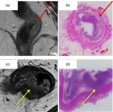
The degree of preservation of the submucosal layer seen on MRI as a hyperintense plane between tumour and MP (figure 1A,B) was recorded. A measurement of (≥1 mm) preserved submucosa was used as a definition for partial rather than full submucosal invasion and any such tumour would then be classified on MRI as T1 sm1 or sm2, depending on the maximal degree of preserva-tion of the submucosal layer in any one plane. If no hyperintense submucosal layer was identified between the tumour and the muscularis but the muscularis layer was completely preserved (figure1C,D), the tumour was staged as T1sm3/early T2.
Tumours with >1 mm preservation of MP were
classi-fied as T2, and those with <1 mm MP preservation or spread beyond the muscularis as T3a-b (depending on the depth of mesorectal invasion in millimetres).
Original pro-forma reports as discussed and presented at colorectal weekly MDT were analysed for the purposes of this study. The policy of the MDT was to offer primary surgery for ERC without any preoperative radiotherapy regardless of nodal status provided that no extramural venous invasion (EMVI) or circumferential resection margin (CRM) involvement was evident on MRI. However, there was no implemented policy to offer local excision based on MRI assessment during this audit period.
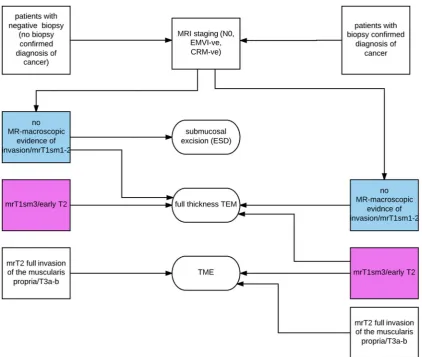
We subsequently evaluated the accuracy of MRI in defining endoscopic/surgical planes for ERC on the basis of the tumour depth invasion into the rectal wall comparing MRI results to final histopathology findings. We tested two models of possible endoscopic/surgical intervention based on preoperative MRI (figure 2 and
[image:3.595.115.481.54.414.2]table 1for two scenarios).
Figure 1 (A) Submucosa has a hyperintense signal on MRI (red arrow) which should be considered partially or fully preserved if at least 1 mm visible on high-resolution T2-WI. Findings confirmed on histopathology (B). (C) If no hyperintense signal present between the tumour invasion portion and muscularis propria, full invasion of the submucosal layer should be considered. Findings confirmed on histopathology (D).
copyright.
on May 30, 2020 by guest. Protected by
In thefinal analysis, we calculated MRI accuracy for two scenarios regardless of preoperative biopsyfindings (scen-ario 1: feasibility of MRI to identify tumours with minimal submucosal invasion (≤T1sm2); scenario 2: feasibility of MRI to identify tumours with no submucosal preservation but partial or full invasion of the MP (≤T2)).
In patients who had undergone total mesorectal resec-tion during the evaluaresec-tion period, we also compared the
accuracy of MRI in identifying patients with malignant lymph nodes.
The MRI scans were read by a second independent observer to measure interobserver agreement of MRI staging accuracy of ERC. Anonymised scans were scored by the observer 2 who was blinded to the histopathology results and results of the MRI reports from the observer 1. All of the scans were analysed during three sessions with 20±3 cases per session using the same reporting pro forma as by the observer 1. Scans were staged according to the TNM classification and then categorised into three groups (≤T1sm2,≤T2, >T2).
Statistical analysis
Data were tabulated and entered into a spreadsheet. All statistical analyses were performed using Microsoft Excel V.14.4.5 (Redmond, Washington, USA 2011) and IBM SPSS V.23.0 (IBM Corp, New York, USA, 2013).
The primary end point was the sensitivity, specificity and accuracy of MRI calculated for both scenarios.
[image:4.595.87.510.52.409.2]MRI accuracy for staging and defining endoscopic and surgical planes for local excision was calculated from histology data using cross tables. CIs for the sensitivity
Figure 2 Flow chart demonstrating potential treatment algorithms based on MRI-defined endoscopic and surgical planes. Blue colour indicates scenario 1 and pink colour indicates scenario 2.
Table 1 MRI vs histopathology in identifying rectal cancer with partial submucosal invasion (T1sm2 or less)
Histo≤T1sm2 Histo >T1sm2
(A) Scenario 1
MR≤T1sm2 True +ve False +ve MR >T1sm2 False−ve True−ve
≤Histo T2 Histo >T2 (B) Scenario 2
≤MR T2 (≥1 mm clear to MP)
True +ve False +ve
MR T2
(<1 mm clear to MP+)
False−ve True−ve
MP, muscularis propria.
copyright.
on May 30, 2020 by guest. Protected by
[image:4.595.45.286.605.737.2]and specificity for a binomial probability were calculated using the exact method.
For those patients treated by TME, sensitivity analysis was performed to evaluate MRI accuracy in assessing lymph node status.
The hypothetical effect of an MRI-directed assessment of likely suitability for a local excision pathway rather than the actual MDT decision based predominantly on clinical data was also calculated as a secondary end point. Differences in proportions of patients undergoing radical surgery according to clinical assessment and pro-posed having radical resection according to MRI staging were calculated using Newcombe method 10.12
For the interobserver agreement between the two radiol-ogists, Cohensκlevel was used to calculate results. A value of p<0.05 was chosen as the significance level for κ statis-tics. The value ofκ statistics was interpreted according to Altman.13Agreement lies between 0 and 1, where 0 is indi-cative of no agreement and 1 indicates complete agree-ment.‘Very good/near perfect’ agreement is considered as aκof 0.81–1.00;‘good’agreement as a κof 0.61–0.80; ‘moderate’agreement as aκof 0.41–0.60;‘fair’agreement as aκof 0.21–0.40; and‘poor’agreement as aκof <0.2.
RESULTS Participants
Overall, 65 patients with a median age of 69 (34–91)
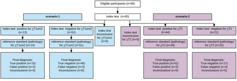
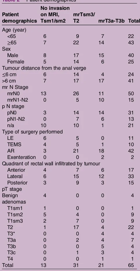
ful-filled the MRI staging inclusion criteria of mrT3b or less (figure 3 presents aflow chart of participants according to STARD); demographics are presented in thetable 2.
In 20% (13/65) of the patients, tumours were pre-operatively MR-categorised as lesions with no or minimal invasion of the submucosal layer (no invasion, T1sm1, T1sm2). Of these 13 patients, 10 were confirmed as such on pathology. Thirty-one patients (47%) had mr T1sm3-T2 stage with no MR-evidence of tumour breach-ing through the MP. Of these 31 patients, 24 were proven to be pT1sm3-T2 on histopathology, and 21 patients had mrT stage >T2 with 17 out of 21 confirmed to be pT3 disease.
Overall 44 out of 65 patients underwent radical surgery (table 3) and 22 were staged as T2 or less by MRI. Pathological nodal status was positive in 7 out of 22 patients, 4 of whom had been staged as node-positive on MRI. Thus 15 out of 18 patients were correctly predicted as pT2 or less and pathology node-negative. Among the 21 out of 65 (32%) patients undergoing local excision or TEM, 20 out of 21 were staged by MRI as T2 or less and confirmed as such by pathology. On follow-up, none had lymph node relapse. If the decision had been made to offer local excision on MRI TN staging rather than clinical assessment, a significant increase in organ pres-ervation surgery from 32% to 58% would have been observed (difference 26%, 95% CI 9% to 41%).
In one case, AP resection was performed for a benign large lower rectal adenoma, which was not proven to be malignant on the preoperative biopsy but was clinically too large and difficult for removal by local excision (the patient was counselled with regard to radical surgery for a benign lesion).
Test results
Results of MRI accuracy to predict safe resection plane According to thetable 4results, MRI accuracy in identi-fying a safe submucosal plane for non-invasive
aden-omas, T1sm1-sm2 tumours (scenario 1—tumours
≤T1sm2) was 89% (95% CI 63% to 87%), sensitivity 71%, specificity 94%, PPV 77% and NVP 92%.
When compared with histopathology data, MRI showed 89% (95% CI 79% to 95%) accuracy, 91% sensitivity, 85% specificity, 93% PPV and 81% NPV in identifying a safe MP plane, therefore differentiating tumour with full submucosal invasion but confined to MP versus tumour with spread beyond the MP (tumour≤T2—scenario 2).
Results of interobserver agreement
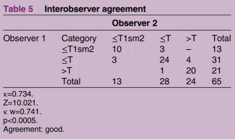
[image:5.595.63.532.56.217.2]Results of the κ statistic for the second observer when compared with the central reviewer (observe 1) can be seen in table 5. κ Agreement was calculated of value 0.734, with p<0.005 indicating a good agreement (weightedκwas 0.741).
Figure 3 Diagram reporting flow of participants though the study (according to STARD recommendations).
copyright.
on May 30, 2020 by guest. Protected by
Result of MRI accuracy to predict lymph node status
When compared with final histopathology in patients who underwent radical resection, MRI accuracy in pre-dicting lymph node status was 84% (95% CI 70% to 92%), PPV 71% and NPV 90%.
Overall MRI results to predict eligible patients for organ preservation treatment
Overall radical surgery was undertaken in 68% (44/65) of patients and 32% (21/65) patients were treated with local excision. According to MRI findings (T and N MR-staging), another 18 patients could potentially have
Implementation of bowel cancer screening has
increased the proportion of colorectal cancer identified as early stage. About 30% of screen-detected cancers are Dukes’ A14 compared with 10% of the non-screened population. Patients with ERC can only be offered the correct treatment modality if they are identified and accurately staged before resection. Organ preservation avoids the extra mortality of TME and abdominoperi-neal excision of rectum (APER) and morbidity such as permanent stoma, sexual dysfunction and a prolonged hospital stay and if appropriate patients can be safely identified, the option of organ preservation is a prefer-able option for the majority.15 Preoperative assessment of tumour depth invasion is crucially important in deter-mining endoscopic and surgical planes. Paris morph-ology, pit pattern and the lift signs are used by expert endoscopists to stratify the risk of submucosal invasion. However, the interobserver variability and accuracy of these tools in routine UK practice have not been audited or investigated. It is likely that other methods may well improve outcomes given that 33% of locally excised lesions in the TEM registry were unexpectedly malignant having been assessed by experienced endos-copists and that removal of such lesions was associated with a statistically significantly higher chance of an involved margin.16 17 Preoperative imaging, based on endorectal ultrasound (ERUS), has not been proven to be reliable in selecting the correct patients, for example, in a multicentre German study of over 3500 patients’ EUS, Ptok et al found an accuracy of 65% for T staging.18 At present, ERUS has not established itself in assessing ERC in UK practice, for example, of 487 patients undergoing TEM, only 148 were staged by ERUS with no difference in the rates of resection margin positivity or outcomes in patients having ERUS.3
MRI is the gold standard for advanced disease; however, it is not yet used as a staging tool for an early disease.
Internationally accepted standards suggest that only T1 rectal tumours with minimal invasion of the sub-mucosal layer—sm1 according to Kikuchi classification —should be considered eligible for local excision. However, the reality shows a different practice. Haboubi and Salmo19 previously reported that thickness of the submucosal layer varies greatly among the individuals
mr N Stage
mrN0 13 26 11 50
mrN1-N2 0 5 10 15
p N stage
pN0 3 14 14 31
pN1-N2 0 7 6 13
n/a 10 10 1 21
Type of surgery performed
LE 6 5 0 11
TEMS 4 5 1 10
AR 3 21 18 42
Exenteration 0 0 2 2
Quadrant of rectal wall infiltrated by tumour
Anterior 4 7 6 17
Lateral 6 15 12 33
Posterior 3 9 3 15
pT stage Benign adenomas
4 0 0 4
T1sm1 1 0 0 1
T1sm2 5 4 0 9
T1sm3 2 7 0 9
T2 1 17 4 22
T3* 0 0 4 4
T3a 0 2 4 7
T3b 0 0 5 4
T3c 0 1 3 4
T4 0 0 1 1
Total 13 31 21 65
*Missing histopathology data about the depth of tumour invasion into the mesorectum.
AR, anterior resection; LE, local excision; n/a, not applicable (patients were treated with local excision), TEMS, transanal endoscopic microsurgery.
copyright.
on May 30, 2020 by guest. Protected by
[image:6.595.45.287.67.535.2]and cannot be appropriately assessed in the absence of MP within the surgical specimen. Whenever the MP is lacking in the specimen, histopathological assessment of the submucosal depth invasion and differentiation of T1sm1/sm2 and even sm2/sm3 become challenging. It has also been stated that budding, lymphovascular inva-sion and tumour differentiation are prognostic factors that determine the need for either adjuvant therapy or subsequent completion/salvage surgery but the validity of these prognostic measures has been questioned because of the lack of reproducibility of these factors.
This has resulted in the review of the current standards of assessing such lesions. The emerging consensus is that rather than using a method with poor inter-observer agreement, absolute measures of depth and width are more reliable and therefore prognostically more secure.20 It has also been shown that depth and width of mucosal invasion predict the lymph node metastasis in ERC.21
[image:7.595.49.551.60.404.2]There is a paucity of good-quality data evaluating the performance of high-resolution MRI in staging ERC. Traditionally, it has not been relied on as a primary tool
Table 3 Type of surgery performed
pT stage Type of surgery
Total
AR LE TEMS Exenteration
Benign adenoma Nx
0 2 (2—mr no invasive
cancer)
1 (1—mr no invasive cancer)
0 3
Benign adenoma N0
1 (1—mr no invasive cancer) 0 0 0 1
T1sm1 0 0 1 (1—mrTsm2N0) 0 1
T1sm2Nx 0 3 (3—mrT1sm2-3N0) 3 (3—mrT1sm2-T2N0) 0 6
T1sm2N0 3 (3—mrT1sm2-3N0) 0 0 0 3
T1sm3Nx 0 5 (5—mrT1sm3N0) 1 (1—mrT1sm3N0) 0 6
T1sm3N+ 1 (1—mrT2N+) 0 0 0 1
T1sm3N0 2 (2—mrT1sm3N0) 0 0 0 2
T2Nx 0 1 (mrT1sm3N0) 3 (3—mrT1sm3-T2N0) 0 4
T2N+ 5 (2—mrT2N+, 1—mrT3bN+, 2 -mrT2N0)
0 0 0 5
T2N0 13 (9—mrT1sm3-T2N0; 4— mr>T2N0)
0 0 0 13
T3aN+ 1 (1—mrT2N0) 0 0 0 1
T3aN0 5 (5—mrT3a-bN0) 0 0 1 (1—
mrT3aN0) 6
T3bNx 0 0 1 (1—mrT3bN+) 0 1
T3bN+ 3 (3—mrT3bN+) 0 0 0 3
T3cN+ 1 (1—mrT2N+) 0 0 0 1
T3cN0 2 (2—mrT3bN+) 0 0 1 (1—
mrT3bN+) 3
T3N+ 1 (1—mrT3bN+) 0 0 0 1
T3N0 3 (2—mrT3N0; 1—mrT3bN+) 0 0 0 3
T4N+ 1 (mrT3bN+) 0 0 0 1
Total 42 11 10 2 65
*Depth of invasion into mesorectum not recorded by pathologist.
[image:7.595.47.554.645.744.2]AR, anterior resection; LE, local excision; TEMS, transanal endoscopic microsurgery; TxNx, patients who potentially could have less radical surgery according to mrTN stage.
Table 4 MRI accuracy in identifying patients with minimal and full invasion of the submucosal layer when compared with final histopathology
mrT stage
pT stage
Total
T0 T1sm1 T1sm2 T1sm3 T2 ≥T3a
Partial invasion of the submucosa (scenario 1)
mrT0,T1sm1,T1sm2 4 1 5 2 1 0 13
Full invasion of the submucosa, partially or fully spared MP (scenario 2)
mrT1sm3-T2 0 0 4 7 17 3 31
≥T3a-T3b 0 0 0 0 4 17 21
Total 4 1 9 9 22 20 65
copyright.
on May 30, 2020 by guest. Protected by
for staging ERC as it is perceived that MRI should not be used for staging ERC, with a tendency to overstage or inaccurately stage lesions as T1/2 without precisely speci-fying the depth of invasion. On imaging, differentiation between ‘sm’ subcategories remains challenging; never-theless we wanted to test the ability of MRI to identify potentially safe submucosal and MP planes. In this ana-lysis, we reviewed the hypothetical impact on patient treatment if an MRI-directed approach to local excision
had been followed. Therefore, a retrospective
hypothesis-generated study to determine if the criteria for identifying suitable patients are accurate was under-taken. Results have shown that 89% patients were cor-rectly identified for partial submucosal invasion and 89% were accurately staged as potentially suitable for full thickness excision ( patients with full submucosal invasion but spared MP); therefore, MRI shows better results than current standards that identify accurately only about a third of patients. Furthermore, the identifi -cation of patients suitable for primary local excision based on MRI node-negative prediction is such that only 3 out of 18 patients with node-positive status were missed but a further 15 patients were correctly identified as node-negative. This has implications for patient dis-cussions on the use of adjuvant radiotherapy in ERC rather than completion radical surgery. In our institu-tion, adjuvant radiotherapy is reserved only for those staged on final pathology as pTsm3 or more or where there is margin involvement and surveillance offered to the remainder. Following this policy, we have not observed disease recurrence after a minimum of 3.5 years follow-up.22 This paper suggests a new approach to MRI imaging of early disease and the fi nd-ings support that MRI may be able to accurately identify the safe resection plane for ERC.
One of the limitations of this study is that κ for the radiologists has been assessed among radiologists experi-enced in reporting rectal cancer as this is a single-centred study and this needs to be prospectively tested in order to determine whether this can change practice nationally and globally. However, there are studies under way and are in the prospective MINSTREL (MrI iN Staging RECTAL poLyps) study which is completing its recruitment using precisely these definitions. If it is
bearing in mind that 80% of patients with pT1sm2-sm3 tumours will have no metastases within the mesorectal lymph nodes or even less if there are no other adverse features. Furthermore, the potential to relapse within the mesorectum could then be monitored by MRI sur-veillance (which was not available in older TEM and local excision studies).
All these potential risks and benefits of organ pre-serving treatment should be discussed with patients and decision to treat should be based on patients’ choice as well.
It has been suggested that radiotherapy can be offered to all ERC defined as T2 or less prior to TME surgery because first, there is no reliable staging method to dis-tinguish between T1 and T2 tumours and second, com-plete responders following TME could avoid further surgery. If this policy had been followed, over 36 out of 44 patients with mrT2N0 or less would have been irra-diated for node-negative disease and potentially would have suffered a higher comorbidity rates associated with TEM defect healing in an irradiated rectum.23 As the SPECC programme has highlighted, the overtreatment of ERC may not be in the patients’ interest. This study, therefore, shows the use of preoperative imaging in pre-venting overtreatment and providing patients with robust prognostic information to make an informed choice about the decision and to understand the risk versus benefits of treatment options/surgical decisions.
The next phase is to determine whether this is repro-ducible in the multicentre setting and finally if it proves to be reproducible in the multicentre study (the MINSTREL study), then this will be used as a basis for offering more patients with ERC the possibility of organ preservation.
CONCLUSION
This analysis shows that high-resolution MRI assessment, with particular attention paid to the degree of preserva-tion of the submucosa and MP layers at the invasive margin of the tumour, is a reliable method of staging depth of invasion in ERC. If reproducible and
con-firmed in the prospective MINSTREL trial, there are very few resource barriers to prevent wider UK adoption of MRI as a standard of care for ERC.
copyright.
on May 30, 2020 by guest. Protected by
[image:8.595.45.286.58.201.2]AcknowledgementsAuthors thank Karen Thomas, Lead Statistician, The Royal Marsden Hospital, Sutton.
Contributors All authors have contributed significantly. SB was involved in conception of the study design, acquisition, analysis and interpretation of the data, and drafting the manuscript. JR and AW contributed to acquisition and interpretation of the data. SR, PT and DC revised the manuscript critically for the intellectual content. DT conceptualised the study design and revised the manuscript critically for the intellectual content. GB conceptualised the study design, revised the manuscript critically for the intellectual content, and approved the final version to be published.
Funding This study was supported by the Biomedical Research Centre. Competing interests None declared.
Patient consent Obtained.
Ethics approval Local research and development office.
Provenance and peer review Not commissioned; externally peer reviewed. Data sharing statement No additional data are available.
Open Access This is an Open Access article distributed in accordance with the Creative Commons Attribution Non Commercial (CC BY-NC 4.0) license, which permits others to distribute, remix, adapt, build upon this work non-commercially, and license their derivative works on different terms, provided the original work is properly cited and the use is non-commercial. See: http:// creativecommons.org/licenses/by-nc/4.0/
REFERENCES
1. Muto T, Bussey HJ, Morson BC. The evolution of cancer of the colon and rectum.Cancer1975;36:2251–70.
2. Greenaway KJH, Khatun S, Kuryba A,et al. National Bowel Cancer Audit Report 2015.
3. Ashraf S, Hompes R, Slater A,et al. A critical appraisal of endorectal ultrasound and transanal endoscopic microsurgery and
decision-making in early rectal cancer.Colorectal Dis2012;14: 821–6.
4. Williams JG, Pullan RD, Hill J,et al. Management of the malignant colorectal polyp: ACPGBI position statement.Colorectal Dis2013;15 (Suppl 2):1–38.
5. Videhult P, Smedh K, Lundin P,et al. Magnetic resonance imaging for preoperative staging of rectal cancer in clinical practice: high accuracy in predicting circumferential margin with clinical benefit. Colorectal Dis2007;9:412–19.
6. Kim NK, Kim MJ, Park JK,et al. Preoperative staging of rectal cancer with MRI: accuracy and clinical usefulness.Ann Surg Oncol 2000;7:732–7.
7. Giusti S, Buccianti P, Castagna M,et al. Preoperative rectal cancer staging with phased-array MR.Radiat Oncol2012;7:29.
8. Brown G, Radcliffe AG, Newcombe RG,et al. Preoperative assessment of prognostic factors in rectal cancer using high-resolution magnetic resonance imaging.Br J Surg 2003;90:355–64.
9. Taylor FG, Quirke P, Heald RJ,et al. Preoperative high-resolution magnetic resonance imaging can identify good prognosis stage I, II, and III rectal cancer best managed by surgery alone: a prospective, multicenter, European study.Ann Surg2011;253:711–9.
10. Taylor FG, Quirke P, Heald RJ,et al. One millimetre is the safe cut-off for magnetic resonance imaging prediction of surgical margin status in rectal cancer.Br J Surg2011;98:872–9.
11. Taylor FG, Swift RI, Blomqvist L,et al. A systematic approach to the interpretation of preoperative staging MRI for rectal cancer.AJR Am J Roentgenol2008;191:1827–35.
12. Newcombe RG. Interval estimation for the difference between independent proportions: comparison of eleven methods.Stat Med 1998;17:873–90.
13. Altman DG.Practical statistics for medical research. Boca Raton, FL: Chapman & Hall/CRC. xii, 611p, 1999.
14. Logan RF, Patnick J, Nickerson C,et al. Outcomes of the Bowel Cancer Screening Programme (BCSP) in England after the first 1 million tests.Gut2012;61:1439–46.
15. Garcia-Aguilar, J., Holt A. Optimal management of small rectal cancers: TAE, TEM, or TME?Surg Oncol Clin N Am2010;19:743–60. 16. Langer C, Liersch T, Süss M,et al. Surgical cure for early rectal
carcinoma and large adenoma: transanal endoscopic microsurgery (using ultrasound or electrosurgery) compared to conventional local and radical resection.Int J Colorectal Dis2003;18:222–9. 17. Moore JS, Cataldo PA, Osler T,et al. Transanal endoscopic
microsurgery is more effective than traditional transanal excision for resection of rectal masses.Dis Colon Rectum2008;51:1026–30; discussion 1030-1.
18. Marusch F, Ptok H, Sahm M,et al. Endorectal ultrasound in rectal carcinoma--do the literature results really correspond to the realities of routine clinical care?Endoscopy2011;43:425–31.
19. Haboubi N, Salmo E. Are we accurately measuring the depth of the submucosal invasion in early colorectal cancer by equating the Kikuchi submucosa levels with distances measured in fractions of a millimetre?Colorectal Dis2013;15:775–7.
20. Davenport A, Morris J, Pritchard SA,et al. Interobserver variability amongst gastrointestinal pathologists in assessing prognostic parameters of malignant colorectal polyps: a cause for concern. Tech Coloproctol2016;20:647–52.
21. Toh EW, Brown P, Morris E,et al. Area of submucosal invasion and width of invasion predicts lymph node metastasis in pT1 colorectal cancers.Dis Colon Rectum2015;58:393–400.
22. Balyasnikova S, Read J, Tait D,et al. The results of local excision with or without postoperative adjuvant chemoradiotherapy for early rectal cancer among patients choosing to avoid radical surgery. Colorectal Dis2017;19:139–47.
23. Habr-Gama A, Perez RO, Nadalin W,et al. Operative versus nonoperative treatment for stage 0 distal rectal cancer following chemoradiation therapy: long-term results.Ann Surg2004;240: 711–17; discussion 717-8.
copyright.
on May 30, 2020 by guest. Protected by