Identification of Group B Streptococcus
Capsule Type by Use of a Dual
Phenotypic/Genotypic Assay
Areej Alhhazmi,
a,cArmaan Pandey,
aGregory J. Tyrrell
a,bThe Division of Diagnostic and Applied Microbiology, Department of Laboratory Medicine and Pathology, University of Alberta, Edmonton, Alberta, Canadaa; The Provincial Laboratory for Public Health (Microbiology), Edmonton, Alberta, Canadab; Medical Laboratory Technology, College of Applied Medical Sciences, Taibah University, Madinah, Saudi Arabiac
ABSTRACT
The group B streptococcus (GBS) capsular polysaccharide (CPS) is an
im-portant virulence factor which is also used for GBS typing. There are 10 CPS types
(Ia, Ib, and II to IX). GBS that do not phenotypically type are considered
nontype-able. All genes required for CPS synthesis are found on the GBS
cps
operon, which
contains a highly variable CPS-determining region (
cpsG-cpsK
). The objective of this
study was development of an assay to detect sialic acid on the GBS cell surface,
fol-lowed by a genotypic PCR CPS typing assay. Sialic acid is located at the terminal
end of the side chain of all known GBS CPS types. Sialic acid can be bound to
com-mercially available lectins such as slug
Limax flavus
lectin. Biotinylated
L. flavus
-streptavidin-peroxidase complex was used in an enzyme immunoassay and dot blot
assay to detect sialic acid. This was followed by a PCR typing scheme that was
de-veloped to target the serotype-determining region of the
cps
locus for Ia, Ib, and II
to IX. Sialic acid from the CPS types Ia, Ib, and II to IX was detectable on the GBS
cell surfaces of all previously identified CPS-typed GBS strains assayed. This was
fol-lowed by the real-time PCR typing assay which successfully identified CPS Ia, Ib, and
II to IX types. The combination of phenotypic and genotypic assays provides an
ac-curate tool for detection of CPS expression and assignment of CPS typing. These
as-says have the potential to be used for CPS typing in large-scale epidemiological
studies.
KEYWORDS
RT-PCR, streptococcus,
Streptococcus agalactiae
, group B streptococcus,
lectin, serotyping, sialic acid
G
roup B streptococci (GBS) are recognized as a leading cause of neonatal invasive
disease, as well as invasive disease, in immunocompromised patients and in
elderly individuals (1–7). An important virulence factor of GBS is a capsular
polysac-charide (CPS); there are ten antigenic CPS variants designated Ia, Ib, and II to IX (8–11).
In North America and a number of European countries, five CPS types (Ia, Ib, II, III, and
V) cause the bulk of invasive disease cases, with CPS III causing a higher rate of disease
among neonates and CPS types Ia and V causing higher rates among adult patients
(12–23). Interestingly, recent studies have noted the emergence and circulation of CPS
IV, a previously uncommon serotype, as an important cause of both neonatal and adult
infections (14, 19, 24–28).
As attention is focused on invasive GBS (iGBS) disease with the emergence of new
strains and increased antibiotic resistance, the potential to prevent iGBS disease via
vaccination becomes more attractive (21, 29). If a GBS vaccine is developed for use that
is based on selected capsule types, it will become important to monitor distribution
patterns of CPS types in circulation in the target population using sensitive and specific
methods for determining CPS types.
Received18 February 2017Returned for modification10 April 2017Accepted8 June 2017
Accepted manuscript posted online14 June 2017
CitationAlhhazmi A, Pandey A, Tyrrell GJ. 2017. Identification of group B streptococcus capsule type by use of a dual phenotypic/ genotypic assay. J Clin Microbiol 55:2637–2650. https://doi.org/10.1128/JCM.00300-17. EditorRobin Patel, Mayo Clinic Copyright© 2017 American Society for Microbiology.All Rights Reserved. Address correspondence to Gregory J. Tyrrell, [email protected].
crossm
on May 16, 2020 by guest
http://jcm.asm.org/
Common phenotypic methods of GBS CPS type identification are based on
sero-logical assays such as capillary precipitin (30), immunodiffusion (30, 31), latex
aggluti-nation (32, 33), coagglutiaggluti-nation (34), or enzyme immunoassay (35), which have proven
invaluable for identifying CPS types. However, these assays may have complicated
interpretations resulting in a number of incorrect typing assignments due to poor
capsule expression, capsule operon mutations, rearrangements, or limited accuracy.
Molecular typing based on PCR assays has been already developed; however, these
assays target capsule gene detection rather than capsule expression. Moreover, they
tend to involve a combination of two different techniques, e.g., PCR plus sequencing
(36), PCR plus blot hybridization (37, 38), PCR plus enzymatic restriction (39), or
multiplex PCR plus agarose gel electrophoresis (40, 41). A real-time PCR assay is a more
attractive molecular method for assigning CPS type than conventional PCR since it is
usually more rapid.
The CPS type-specific epitopes of each GBS polysaccharide are created by different
arrangements of four component sugars (glucose, galactose,
N
-acetylglucosamine, and
sialic acid) into a unique repeating unit. Interestingly, all of these structures contain a
terminal sialic acid (Neu5Ac) bound to galactose in an
␣
2-3 linkage (40, 42, 43). The
conservation of Neu5Ac among all known GBS capsular types suggests that this
structural feature is essential to GBS capsular polysaccharide pathogenicity. We propose
that sialic acid on the surfaces of GBS capsule positive cells can be used as a universal
phenotypic method to ascertain capsule expression, followed by a CPS-specific
real-time PCR assay.
RESULTS
Detection of sialic acid from 10 GBS CPS types using a lectin enzyme
immu-noassay (EIA).
Understanding that sialylation is essential for full GBS capsule
biosyn-thesis and loss of sialylated capsule reduces the amount of CPS expressed on the
bacterial cell surface by 80% (43), we hypothesized that sialic acid could be used as a
recognition moiety for capsule expression. The commercially available biotinylated
lectin from the slug
Limax flavus
was selected to be used in our sialo-lectin binding
assay.
L. flavus
lectin was selected because it reacts with sialic acid in any linkage (44,
45), whereas other sialic acid-specific lectins recognize only specific glycosidic linkages
of sialic acid or other carbohydrate moieties (45–48). Sialic acid is conserved at the
terminus of the side chain of all GBS capsule types.
To examine the use of sialo-lectin binding to detect GBS CPS expression, 10 GBS
strains were selected representing all recognized GBS CPS types (Ia, Ib, and II to IX) for
CPS extraction (Table 1). Recognition of the immobilized sialic acid from the 10 assayed
CPS type bacterial isolates by biotinylated
L. flavus
lectin validated the presence of CPS.
It also provided a dose-dependent signal that exhibited a saturating signal at a lectin
concentration of 10
g/ml (Fig. 1). Therefore, the working concentration of biotinylated
[image:2.585.42.375.83.232.2]L. flavus
lectin with all the CPS types in the study was restricted to 10
g/ml. The
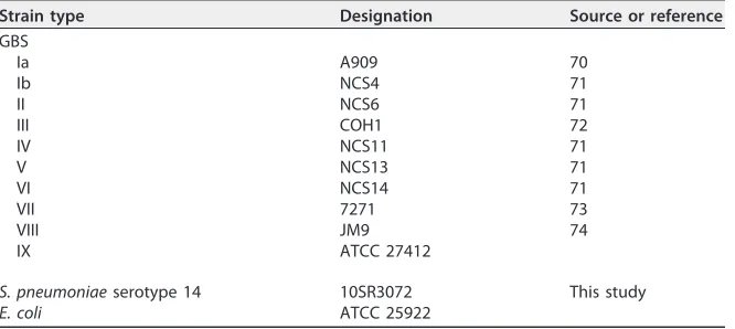
TABLE 1Strains used in study
Strain type Designation Source or reference GBS
Ia A909 70
Ib NCS4 71
II NCS6 71
III COH1 72
IV NCS11 71
V NCS13 71
VI NCS14 71
VII 7271 73
VIII JM9 74
IX ATCC 27412
S. pneumoniaeserotype 14 10SR3072 This study
E. coli ATCC 25922
on May 16, 2020 by guest
http://jcm.asm.org/
average absorbance at 450 nm for all CPS types was 0.85
⫾
0.079 with a 95%
confidence interval (CI) of 0.77 to 0.93 (Fig. 1). For the asialo-CPS (spn14) isolate with no
sialic acid in its capsule and the well with no capsule material, average absorbances of
0.45 nm (95% CI
⫽
0.44 to 0.45) and 0.022 nm (95% CI
⫽
0.02 to 0.03) were detected,
respectively. The upper absorbance limit of the 95% CI of the negative control (spn14)
preparation was 0.45. This value was used as a cutoff for detecting the presence of sialic
acid in further experiments.
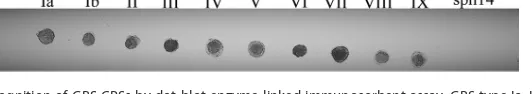
Detection of sialic acid from the 10 GBS CPS types using a lectin dot blot assay.
To verify that the sialo-lectin binding phenotypes from the 10 assayed CPS type isolates
by the lectin EIA was not due to endogenous sialic acids coextracted from the bacteria
during the capsule extraction process, whole bacteria were assayed in a lectin dot blot
assay. As shown in Fig. 2, sialic acid was uniformly expressed in all the assayed strains,
whereas no signal was detected on the spot corresponding to the asialo-CPS (spn14)
isolate. The results suggested that both the lectin EIA and the lectin dot blot assays
could be used as simple phenotypic assays to determine CPS expression for GBS.
GBS CPS typing assay. (i) Assignment of CPS types for types Ia, Ib, and II to IX
using a singleplex real-time PCR CPS typing assay.
Representative CPS DNA operon
sequences for CPS Ia, Ib, and II to VIII and a partial CPS DNA operon sequence for IX
were obtained from the NCBI website (
http://www.ncbi.nlm.nih.gov/
). The
se-quences of these strains were analyzed to generate
cps
-specific primer pairs and dually
labeled probes, which enabled the amplification of DNA amplicons to be easily
discriminated by specific probes. A CPS-specific gene scheme was developed based on
comparison analyses of
cps
genes. A unique region(s) of the
cps
gene(s) was identified
for each CPS type based on the nucleotide sequence comparison with other CPS types.
From this information, a set of primers for each CPS type, Ia, Ib, and II to IX, was
designed to uniquely target each of the 10 CPS types (Table 2). The identification of CPS
types Ia, Ib, and II to IX depends on the ability of the primer pairs to amplify and probe
FIG 1Recognition of GBS CPSs by the lectin EIA. Crude GBS CPS preparations (Ia, Ib, and II to IX) were incubated withL. flavuslectin specific for Neu5Ac. Three concentrations of biotin-L. flavuslectin (LFA) were used (10, 1, and 0.1g/ml), while 1g of HRP-labeled streptavidin/ml was used. Data are expressed as mean OD450values with the standard deviations (SD) for at least three independent experiments.
FIG 2Recognition of GBS CPSs by dot blot enzyme-linked immunosorbent assay. GBS type Ia, Ib, and II to IX whole bacteria (107CFU) were incubated withL. flavus. An spn14 isolate was used as a negative
control.
on May 16, 2020 by guest
http://jcm.asm.org/
[image:3.585.104.308.70.233.2] [image:3.585.73.339.659.706.2]to identify a specific fragment for the following
cps
genes:
cpsH
,
cpsJ
,
cpsK
,
cpsG
,
cpsN
,
cpsO-V
,
cpsI
,
cpsM
,
cpsR
, and
cpsO
-IX, respectively.
To verify the specificity of the designed primer pairs and probes, they were BLAST
searched against the NCBI nonredundant sequence database to confirm the absence of
serendipitous similarities. Based on this analysis, the designed primer pairs and probes
were determined to have 100% specificity for the identification of GBS CPS types,
except for CPS type III. The primers and probe targeting
cpsG
were similar to CPS types
V and VI. However, the primers for CPS V and VI are specific for the identification of
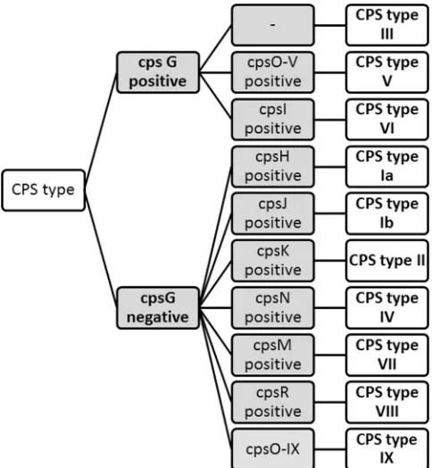
these CPS types. An algorithm was constructed to allow identification of the 10 CPS
types, as shown in Fig. 3. The specificity and efficiency of each primer pair used
separately were determined by singleplex reverse transcription-PCR (RT-PCR) with DNA
extracted from 10 GBS strains representing all GBS recognized CPS types (Table 3) that
were used as reference strains. Specific and characteristic PCR patterns were obtained
with all primer pairs and probes for each CPS type (Table 4). No signal was obtained for
the negative controls, isolates spn14 and
Escherichia coli
ATCC 2592 (data not shown).
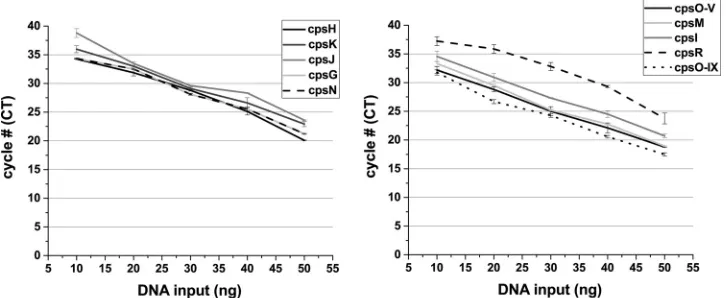
[image:4.585.40.370.85.419.2]The standard curve and efficiency of each primer pair was determined by using a
series of diluted genomic DNA extracts from the recognized CPS types to determine the
smallest amount of DNA detected while maintaining a desirable efficiency. The
mini-mum concentration of purified genomic DNA required for the cpsH, cpsK, cpsJ, cpsN,
and cpsI primer pairs was 30 ng, whereas for the cpsG, cpsM, cpsO-V, and cpsO-IX
primers 20 ng was required (Fig. 4). For the cpsR primer set, 40 ng was required for the
detection. The designed primers have high amplification efficiency ranging between
90.9 and 99.9%, as shown in Table 3, indicating that the assay is robust and
reproduc-ible.
TABLE 2Primer pairs and probes used in the singleplex and multiplex RT-PCR assays
Identifier CPS type Sequence (5=–3=)
Duplex reaction Primer
cpsO-F V AACAGAGGCCAATCAGTTGCA 1
cpsO-R CGGCATTGGTAGCTTTCTGTATG
cpsI-F VI TTCACCTTCTGCCATCTCAA 1
cpsI-R AAGGGATAGTCGCGTAAAAGTC
cpsG-F III AAACGGGTTACTCAGACTTCG 2
cpsG-R TCACCAAACTGCTTTCTCCTAG
cpsK-F II GCTATTCCCTACATGGAAGATGG 2
cpsK-R TTACTGAAGCCATGATATCGGG
cpsM-F VII CCTTTGAGAGTTCATAACTGTT 3
cpsM-R GTCCTCTAATTGCACCAATAAT
cpsR-F VIII CCAGATGGGCATGAGTGGTTAC 3
cpsR-R CAGTCCCATAGGCGATGTAGG
cpsN-F IV GTATGCTTTCGTGTCTGATTATGC 4
cpsN-R ATTGATCCAAAACCCAAACCTG
cpsH-F Ia TTAATTTGCGATCCGGGAGTAG 4
cpsH-R GCAGGCCACTTTTGTAGAAATAG
cpsJ-F Ib TGGGATATAGAGATTTAGTACCTGTTG 5
cpsJ-R ATTGGTTTGTGATATTCCATTCTCG
cpsO-F IX CTGATGATCTTTGTTCGCCATTT 5
cpsO-R ACAAGGGTGATCCTCAATTCC
Probe
cpsO V Cal Fluor 560-CAACGGAGTACTTAGGTGTACAGGAGA-BHQ1 1
cpsI VI FAM-ACAATGGAGGTGCATCATCAGCA-BHQ1 1
cpsK II Cal Fluor 560-TCTTGTCACAAAGACCATCTGGAGCG-BHQ1 2
cpsG III FAM-ATTGTTATCACACATGGCGGCCC-BHQ1 2
cpsM VII Cal Fluor 540-CTAGGGAGTTAAGTTATGATGTGA-BHQ1 3 cpsR VIII FAM-TGGAGTATTCCTGTACGTCGCTATTGGA-BHQ1 3 cpsN IV Cal Fluor 560-CCTCTCCAGGTAGCTCACAAGCAAA-BHQ1 4
cpsH Ia FAM-TTGAATGCGACCCCAAAGGGAGA-BHQ1 4
cpsJ Ib FAM-CCGATTTTGAAATCAGCCAGAGCTCCT-BHQ1 5
cpsO IX Cal Fluor CAACATGAAACTGGTGCTGACCTTGT-BHQ1
on May 16, 2020 by guest
http://jcm.asm.org/
(ii) Assignment of CPS types using a duplex RT-PCR GBS typing assay.
Five
duplex reactions (1 to 5) (Table 2) containing primer pairs and probes specific for CPS
types V/VI, III/II, VII/VIII, Ia/IV, and Ib/IX were used in a real-time PCR platform. The 10
GBS CPS reference strains previously assayed in the singleplex PCR typing assay were
analyzed using the duplex real-time PCR assay to assign CPS type. The duplex real-time
PCR assay allowed identification of the CPS type for the GBS strains shown in Table 5.
A collection of 70 previously serotyped GBS clinical isolates representing all
recog-nized CPS types were analyzed to determine the reliability of the duplex real-time PCR
typing with the exception of CPS VII. No CPS type VII except for the reference strain was
present in our collection; therefore, no clinical CPS VII isolates were assayed (15). We
compared the CPS typing by the duplex RT-PCR assay against serotyping by the
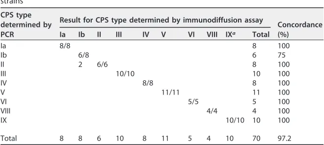
immunodiffusion assay for CPS types Ia, Ib, and II to VIII or a previously described PCR
assay (40) for CPS type IX. A concordance of 97.2% for the duplex RT-PCR GBS typing
and the phenotypic serotyping assay (immunodiffusion) was observed. No discordant
results were obtained between the molecular assay and the serological assay for all CPS
types, with the exception of CPS type Ib. Two isolates were typed by the serological
assay as CPS type Ib, and yet the molecular assay assigned CPS type II for these isolates
[image:5.585.85.327.71.333.2]FIG 3Algorithm to identify GBS CPS types Ia, Ib, and II to IX. Gray boxes arecpstarget genes, and white boxes are GBS CPS types.
TABLE 3Efficiency calculated for primers for real-time PCR GBS CPS typing
cpsgene target Efficiency (%)
cpsH 92.60
cpsK 99.87
cpsJ 90.94
cpsG 92.31
cpsN 99.79
cpsO-V 98.56
cpsI 96.14
cpsM 91.84
cpsR 98.60
cpsO-IX 96.68
on May 16, 2020 by guest
http://jcm.asm.org/
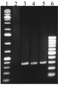
[image:5.585.42.371.627.740.2](Table 6). To confirm CPS genotyping of these two isolates as CPS type II, a previously
described conventional PCR assay (41) was performed to distinguish between CPS
types II and Ib. The PCR assay verified these isolates as CPS type II (Fig. 5).
Assignment of CPS type to a collection of clinical NT isolates using lectin
EIA/lectin dot assays and a duplex RT-PCR GBS typing assay.
To determine the
robustness of our assay algorithm, we assayed 159 GBS clinical isolates from Alberta,
Canada, collected from 2003 to 2013 that were determined to be nontypeable (NT) in
a double immunodiffusion assay. These GBS isolates were assayed for the presence of
sialo-CPS using the lectin EIA after CPS extraction. A total of 47.2% (75/159) of the
capsule preparations from the NT isolates reacted positively with
L. flavus
lectin,
suggesting the presence of capsule (data are not shown). The lectin dot blot assay
supported the results obtained from the lectin EIA since all 75 isolates spotted in the
lectin dot blot assay displayed spots darker than the spots from isolates with an optical
density (OD) below 0.45 in the lectin EIA (Fig. 6), suggesting the presence of sialic acid.
To prove the stability and veracity of the duplex RT-PCR typing assay, the capsule
types of the sialo-CPS-positive NT strains were assayed. DNA was extracted, and PCR
amplification with the
cfb
primer pair was performed. All strains yielded the expected
[image:6.585.45.551.82.210.2]PCR product, which confirmed that the DNA preparations were devoid of PCR inhibitors
and that the corresponding strains were GBS (data not shown). A duplex RT-PCR CPS
typing assay was then performed to identify CPS genotypes. A capsular genotype was
assigned for 71 of 75 isolates (94.7%). The majority of the strains were typed as CPS type
TABLE 4Singleplex real-time PCR for GBS typinga
Gene target
MeanCTⴞSD for CPS type:
Ia Ib II III IV V VI VII VIII IX
cpsH 23.9⫾0.2
cpsJ 24.1⫾0.5
cpsK 26.8⫾1.5
cpsG 18.4⫾1.6 23.2⫾2.2 26.4⫾2.7
cpsN 18.5⫾0.0
cpsO-V 20.0⫾1.4
cpsI 21.5⫾0.6
cpsM 26.2⫾2.3
cpsR 23.5⫾1.1
cpsO-IX 17.6⫾0.1
aData are expressed as meanC
Tvalues for at least three independent experiments.
FIG 4Standard curves and efficiencies calculated for primers for real-time PCR GBS CPS typing. Tenfold dilutions of DNA template (10, 20, 30, 40, and 50 ng) were plotted against theCTvalue for each dilution to generate a standard curve. From this standard curve, the smallest amount of DNA detected and theCTslope were determined for the primers cpsH, cpsK, cpsJ, cpsG, cpsN, cpsO-V, cpsI, cpsM, cpsR, and cpsO-IX to identify CPS types Ia, Ib, II, III, IV, V, VI, VII, VIII, and IX, respectively. The efficiency values were measured for each primer pair using theCTslope method. The amplification efficiency was calculated according to the following equation: Ex⫽10(⫺1/slope)⫺1, where Ex is the efficiency. Data are expressed as meanCTvalues with the SD for at least three independent experiments.
on May 16, 2020 by guest
http://jcm.asm.org/
[image:6.585.41.404.515.664.2]V (53.3%, 40/75). A total of 14.7% (11/75) of the isolates were typed as CPS type III, 8%
(6/75) as type II, 5% (4/75) as type Ia, 5% (4/75) as Ib, 4% (3/75) as type VI, 2.7% (2/75)
as type IV, and 1.3% (1/75) as type IX. Four isolates failed to be assigned a CPS type.
To test the ability of the duplex PCR assay to assign CPS types to GBS isolates, we
randomly selected 32 of the remaining 84 GBS isolates that did not react with
L. flavus
lectin in EIAs and dot blot assays and assayed them in the duplex real-time PCR assay.
The capsule types identified were CPS types Ia (3.1%, 1/32), Ib (31.3%, 10/32), II (3.1%,
1/32), III (6.3%, 2/32), V (18.3%, 6/32), VI (3.1%, 1/32), and IX (28.1%, 9/32). Four isolates
failed to be assigned a capsule type. This demonstrated the ability of the duplex RT-PCR
assay to genotypically identify CPS types which failed to express sialic acid on their
surfaces, suggesting the absence of capsule.
DISCUSSION
GBS CPS is a well-known protective antigen against GBS (29, 49). Past efforts to
develop GBS vaccines have focused primarily on the use of capsular polysaccharides
from more common types associated with GBS disease (29, 49). For this avenue of
vaccine development to be effective requires inclusion of the most relevant CPS types
in the target population (29, 49). Here, we present a dual GBS typing system that
provides information regarding capsule expression and identification of capsule type.
The first step of the proposed GBS typing system involves the detection of sialic acid
and confirmation of capsule expression. GBS CPS is terminally linked to sialic acid.
Sialylation is an essential process for full GBS CPS biosynthesis and expression. A past
study reported that sialylation of GBS capsular polysaccharide is required for full
synthesis of CPS by GBS (43). These investigators found an 80% reduction in surface
associated CPS produced by asialo mutant strains that had a deletion in the
cpsK
gene
encoding sialyltransferase compared to the parental strain (43). This group also
re-TABLE 5Duplex real-time PCR for GBS CPS typinga
Reaction cpsgene CPS type MeanCTⴞSD
1 cpsO V 18.04⫾1.33
cpsI VI 18.07⫾0.07
2 cpsG III 16.77⫾0.70
cpsK II 24.88⫾0.50
3 cpsM VII 19.70⫾1.34
cpsR VIII 22.44⫾0.22
4 cpsH Ia 24.55⫾0.04
cpsN IV 18.12⫾0.02
5 cpsJ Ib 19.97⫾1.41
cpsO IX 17.84⫾0.06
aData are expressed as meanC
[image:7.585.39.376.83.193.2]Tvalues for at least three independent experiments.
TABLE 6Comparison of typing results obtained with the immunodiffusion assay (the serological method) and the duplex real-time PCR assay using a collection of GBS clinical strains
CPS type determined by PCR
Result for CPS type determined by immunodiffusion assay
Concordance (%)
Ia Ib II III IV V VI VIII IXa Total
Ia 8/8 8 100
Ib 6/8 6 75
II 2 6/6 8 100
III 10/10 10 100
IV 8/8 8 100
V 11/11 11 100
VI 5/5 5 100
VIII 4/4 4 100
IX 10/10 10 100
Total 8 8 6 10 8 11 5 4 10 70 97.2
aCPS type IX isolates were typed by a PCR assay as previously described (40).
on May 16, 2020 by guest
http://jcm.asm.org/
[image:7.585.41.371.582.730.2]ported the same scenario with an asialo mutant with a deletion in
neuA
encoding
CMP-sialic acid synthase (50). The lectin from
L. flavus
agglutinin was chosen in our
study to detect sialic acid from GBS CPS types for two reasons. First,
L. flavus
lectin binds
to Neu5Ac regardless of its linkage. This was optimal for our assay since sialic acid in the
capsule among the known GBS CPS types has different linkages. Second, to date,
nonspecific binding has not been identified for
L. flavus
lectin (51). For these reasons,
biotinylated
L. flavus
lectin was selected.
Serological CPS typing is the most common method for GBS CPS assignment, but
the proportion of NT isolates is significant and has increased over time (11, 37, 52). This
leads to misrepresentation of some of the CPS types. The sensitivity of the serological
assays used to detect GBS capsule polysaccharide can vary. These methods depend on
the quality of the antibodies, the technical experience of the operator, and the
expression of a detectable amount of capsule (53). A study by Kilian and coworkers (54)
identified limitations associated with GBS latex agglutination serotyping. The
aggluti-nation assay was found to be less sensitive than the flow cytometric assay.
Approxi-mately one-half of the strains assigned as NT in the agglutination test were found to
express type-specific polysaccharides by the flow cytometric method (54). To
over-come these limitations, we developed a sialo-lectin assay for which sialic acid could
be used as a universal approach to detect capsule expression without the need for
FIG 5GBS PCR typing of the two isolates that displayed a discrepancy between the immunodiffusion assay (CPS type Ib) and duplex real-time PCR typing (CPS type II). To identify CPS type II, a previously described PCR assay (41) was used. Lanes: 1, 1-kb DNA ladder; 2, CPS type Ib genomic DNA (gDNA); 3, CPS type II gDNA; 4, 04SR421 gDNA; 5, 13SR567 gDNA; 6, 100-bp DNA ladder.
FIG 6A lectin dot blot assay was performed for GBS isolates that failed to be assigned a CPS type by the immunodiffusion assay (NT isolates). Underlined dots are positive for sialic acid. There are 75 positive spots. GBS whole bacteria (107CFU) were incubated withL. flavus; the positive control was CPS type III GBS strain COH1. The
negative control was the spn14 isolate.
on May 16, 2020 by guest
http://jcm.asm.org/
[image:8.585.145.266.70.247.2] [image:8.585.43.394.563.701.2]antibodies. Moreover, the use of biotinylated lectin in a streptavidin-horseradish
peroxidase (HRP) detection and amplification system has the advantage of detecting
a small amount of sialylated capsule (43, 46, 55, 56). Two assays, lectin EIAs and lectin
dot blot assays, were described in our work to identify isolates with capsule. We also
used these assays to characterize isolates in our collection that were identified as NT by
immunodiffusion assay. Based on our results, there were 52.8% (84/159) NT isolates that
lacked sialic acid expression on the bacterial surface. However, 47.2% (75/159) of our
serologically NT isolates expressed sialic acid on the surface, suggesting the presence
of the capsule. The immunodiffusion assay is less sensitive than the sialo-lectin
meth-ods, as confirmed by our observation that 47.2% of the strains assigned as NT in this
assay were found to express sialylated capsule polysaccharide when examined by the
sialo-lectin assays. The 84 sialic acid negative isolates in the lectin EIAs and lectin dot
blot assays presumably do not express capsular polysaccharides. Mechanisms of
cap-sule loss could potentially be caused by genetic alterations such as the insertion or
deletion of DNA fragments in the
cps
operon or mutations in specific conserved genes,
such as
cpsE
,
cpsF
,
cpsA
, and
cpsG
, located in the
cps
locus (37, 57, 58).
The second step of our proposed GBS typing system is identification of the CPS type
by PCR. Several molecular typing-based PCR assays have been developed (36, 38, 40,
41, 59). These assays target
cps
-determining genes in the
cps
operon. PCR assays are an
attractive approach to GBS CPS typing because of their high discriminatory power for
identifying CPS types. Such PCR assays were able to assign a CPS type to isolates that
failed to be identified by serological assay methods (36, 40, 41, 59). Although PCR-based
assays for typing GBS work well, a number of limitations have been identified.
Gener-ally, these assays target capsule genes rather than expression; hence, the
cps
locus
identified by the PCR-based assays does not confirm whether the CPS is expressed or
not. Moreover, to identify CPS types by the available PCR-based assays, two different
techniques are required, such as PCR plus sequencing (36), PCR plus blot hybridization
(37, 38), PCR plus enzymatic restriction (39), or multiplex PCR plus agarose gel
electro-phoresis (40, 41). The involvement of two molecular techniques to identify CPS types
only at the genotypic level can consume time and may involve increased expense when
techniques such as sequencing and blot hybridization are used. Also, these assays may
not always be accurate. For example, the multiplex PCR assay described by Imperi et al.
(40) that was designed to distinguish among types Ia, Ib, and II to IX was found to
misidentify some CPS type Ib and IV strains as CPS type Ia (54, 60). In addition, a two-set
multiplex PCR assay (set 1, types Ia, Ib, II, III, and IV; set 2, types V, VI, VII, and VIII)
developed by Poyart et al. (41) failed to distinguish between CPS types VII and IX (54).
Also, the CPS-specific PCR assay described by Kong et al. (36, 38) did not include a PCR
specific for CPS type II, VII, or VIII (54). To address these issues, we developed a duplex
real-time PCR assay that identifies CPS type based on detecting the presence of
capsular genes. The real-time PCR technique used in our typing system is a more
attractive approach to assigning CPS types than conventional PCR. The main
advan-tages of the TaqMan real-time PCR assay are its high sensitivity and reliability (one step
and gel free) with a specific set of primers and probes to identify each CPS type. The
assay is easy to perform and does not require the use of antisera. The method also has
time advantages over previously described molecular methods (36, 40, 41, 59) because
it requires only a single PCR for amplification and detection and can be completed
within 1 h. To overcome GBS typing inaccuracies using PCR, previous investigators
have developed a GBS CPS typing algorithm that includes three previously described
PCR assays (36, 38, 40, 41). This approach resulted in a GBS CPS typing accuracy of 99%
(60). Our duplex real-time PCR assay had a level of accuracy of 100%. We believe that
our assay would be ideally suited for high-throughput GBS epidemiological studies in
which a large collection of GBS isolates requires CPS typing. A limitation of our assay
was the inability to type a small subset of isolates. Four isolates of the 75 isolates that
were sialic acid positive and 2 isolates of the 32 randomly selected sialic acid-negative
isolates failed to be typed by our PCR assay. Possible reasons for this may be the
presence of an insertion(s) or mutation(s) impairing primer and/or probe binding.
on May 16, 2020 by guest
http://jcm.asm.org/
Alternatively, these isolates may represent new CPS types resulting from capsular
switching events, in which GBS has acquired new capsule genes by horizontal gene
transfer, followed by genetic recombination. Recently, a short report by Breeding et al.
(61) described a real time-PCR CPS typing assay for GBS. These authors examined a
collection of 21 clinical GBS isolates using their real-time PCR assay compared to latex
agglutination and found 100% concordance for their assay, suggesting that the
real-time PCR assay performs well. Our algorithm differs in that the presence of capsule is
also determined phenotypically and assignment of CPS type is done by real-time PCR.
Nonetheless, the assay described by Breeding et al. and our assay algorithm
demon-strate the strength of the real-time PCR for GBS CPS typing.
In summary, we propose a new GBS typing system for which the first step is to
confirm capsule expression using sialo-lectin assays, followed by duplex real-time PCR
typing that identifies the
cps
genotype. The combination of sialo-CPS lectin binding and
duplex real time-PCR typing assays provides a simple and reliable tool for CPS
expres-sion confirmation and GBS CPS typing, respectively. This sensitive and specific method
enables the characterization of GBS CPS types Ia, Ib, and II to IX, thereby reducing the
rate of detection of NT isolates. It is therefore particularly well adapted for GBS CPS
typing in large-scale epidemiological studies.
MATERIALS AND METHODS
Bacterial strains used.Ten GBS strains with known CPS types were used as reference strains for CPS typing (Table 1). Seventy GBS clinical isolates previously typed by the phenotypic immunodiffusion assay and 159 GBS clinical isolates that previously failed to be typed (nontypeable [NT]) by the phenotypic immunodiffusion assay were included in this study (15). AStreptococcus pneumoniaeserotype 14 isolate (spn14) andEscherichia coliATCC 25922 were used as control strains (Table 1) (62, 63). GBS isolates were cultured on Columbia blood agar plates (Dalynn Biologicals, Canada) containing 5% sheep blood overnight at 37°C. They subsequently were inoculated into Todd-Hewitt broth (Becton Dickinson, USA) and incubated overnight at 37°C for use in the experiments described here.
Identification of GBS.Isolates were identified on the basis of colony morphology, beta-hemolysis, Gram stain, and Lancefield grouping with type B antisera (Oxoid, Canada) (64). GBS isolates were additionally confirmed as GBS using a conventional PCR-based assay targeting thecfbgene that encodes the Christie-Atkins-Munch-Petersen factor (65).
GBS CPS typing. (i) Double immunodiffusion assay.Phenotypic CPS typing was performed using a Lancefield heat-acid extraction assay, followed by a double immunodiffusion method described previously (30, 31). The immunodiffusion assay of GBS CPS typing used for this study was based on reactions with antisera raised against CPS types Ia, Ib, and II to VIII. A type-specific antiserum panel was prepared in rabbits as previously described (30, 31).
(ii) CPS type IX identification.PCR CPS typing was used for the identification of CPS IX using the primers cpsI-7-9-F (5=-CTGTAATTGGAGGAATGTGGATCG) and cpsI-9-R (5=-AATCATCTTCATAATTTATCTCC CATT), which amplify target regions specific to CPS IX as previously described (40).
(iii) CPS type II identification.To identify CPS type II, a previously described PCR assay (41) was performed using the primers II-F (5=-TCCGTACTACAACAGACTCATCC) and II-R (5=-TTCTCTAGGAAATCAA ATAATTCTATAGGG), which amplify target region specific for CPS type II (397 bp).
Genomic DNA extraction.Genomic DNA extraction was performed as follows. Overnight broth cultures (1.5 ml) were centrifuged for 10 min at 3,000⫻g. The pellet was resuspended in 500l of 1⫻ phosphate-buffered saline (PBS; 8 g of NaCl, 0.2 g of KCl, 1.44 g of Na2HPO4, 0.24 g of KH2PO4, and 1,000
ml of H2O [pH 7.2]) and washed two times with PBS. Genomic DNA was extracted by using a DNA
Mericon kit (Qiagen, Germany). Extracted genomic DNA was concentrated and dissolved in 30l of Qiagen elution buffer or water and then stored at ⫺20°C. RNase pretreatment was done prior to quantification of genomic DNA.
Isolation of CPS. CPS from GBS strains was isolated as previously described (43, 66–68), with modifications. Bacteria were grown overnight in 200 ml of Todd-Hewitt broth (THB) with 3% neopeptone for 24 h at 35°C, diluted to 2 liters in fresh THB, and grown to an optical density at 600 nm (OD600) of
0.7. The cultures were chilled on ice, and the cells were pelleted and washed twice with ice-cold PBS. The cells were then resuspended in 200 ml of lysis buffer (25 mM sodium phosphate buffer, 10 mM MgCl2,
40% [wt/vol] sucrose, 13.3 U/ml mutanolysin [Sigma-Aldrich, Canada]; pH 7.0), followed by incubation for 19 h at 37°C with end-over-end mixing. Protoplasts were removed by centrifugation, and the mutano-lysin extract was treated with DNase buffer (3.5 ml, 400 mM Tris, 60 mM MgCl2, 20 mM CaCl2, pH 7.5),
along with sodium azide to a 0.05% final concentration. DNase (300 U) and RNase (200g) were added, and the sample was incubated for 24 h at 37°C with rocking. After centrifugation at 3,200⫻gfor 30 min at 4°C to pellet precipitated material, pronase (0.5 mg predigested for 2 h at 50°C to destroy glycosidases in the preparation) was added, along with 0.1 ml CaCl2(10 mM final concentration), and the sample was
incubated for 17 h at 37°C with rocking. The remaining insoluble cell wall fragments and cell bodies were removed by centrifugation. The supernatant was collected and stored at 4°C.
on May 16, 2020 by guest
http://jcm.asm.org/
Lectin enzyme immunoassay (lectin EIA).Capsule extract (200l), along with 0.15 M sodium carbonate, was used to coat the wells of 96-well EIA plates (MP Biomedicals, USA) at 4°C overnight. The detection method was based on biotinylated lectin extracted fromL. flavusslug agglutinin (EY Labora-tories, USA). The amount of bound biotinylatedL. flavuswas quantified by using HRP-labeled streptavidin (Vector Laboratories, USA). Streptavidin was selected as a detection method because it had no carbo-hydrate groups to which lectins may incidentally bind, and it provided an amplification step to detect small amounts of sialylated CPS (43, 46, 55, 56). The plates were washed with PBS for 15 min and blocked with 1% bovine serum albumin (BSA) in PBS for 4 h at 37°C, and then 50-l portions of three dilutions (10, 1, and 0.1g/ml) of biotinylatedL. flavusin PBS with 1% BSA and 0.05% Tween 20 (PBSAT) were added to the wells. After incubation for 1 h at room temperature, the plates were washed with PBS for 15 min, and 50l of HRP conjugated to streptavidin (1g/ml) in PBSAT was added to each well. After incubation for 1 h at room temperature, the plates were washed with PBS for 15 min. Then, 50l of HRP substrate (1-Step Ultra TMB-ELISA; Thermo Scientific, USA) was added to each well, followed by incubation at 37°C for 15 min. The absorbance was read on a microplate reader (Dynex Technologies, USA) at 450 nm. To identify isolates that were sialic acid positive, the average absorbance (95% CI) value was calculated for the negative-control spn14 (no sialic acid in its capsule), and this value was subtracted from the absorbance values of the assayed GBS isolates.
Lectin dot blot assay.Late exponentially growing bacteria were washed with PBS and resuspended in PBS to give an OD600of approximately 2. The bacterial suspension was spotted onto nitrocellulose
membrane (20 l/spot) using a Biodot apparatus (Bio-Rad, USA) and dried for 30 min at room temperature. The membranes were washed for 15 min with TBS (6.05 g of Tris and 8.76 g of NaCl in 1,000 ml of H2O [pH 7.5]) and then incubated with blocking buffer (5% skim milk and 0.1% Tween 20 in TBS)
for 1 h at 37°C. Membranes were subsequently washed with TBS for 15 min, incubated with biotinylated
L. flavuslectin (10g/ml in blocking buffer) for 1 h, washed three times (15 min each time) with TBS, and then incubated with HRP-conjugated streptavidin (1g/ml in blocking buffer). After incubation for 1 h at room temperature, the membranes were washed with TBST three times (15 min each time) and then twice with TBS (15 min each time). Detection was performed using 4-chloro-1-naphthol solution (Sigma-Aldrich, Canada). A positive spot was identified as a clearly defined spot at the site where the bacteria were applied, and a negative result was identified as a trace reaction or the absence of any reaction.
Primer design for real-time PCR GBS typing assay.Representative isolates of GBS CPS types with completecpsoperon sequences available in GenBank were included in this study (Ia,CP000114; Ib,
AB050723; II,AY375362.1; III,HG939456.1; IV,AF355776.1; V,AE009948.1; VI,AF337958.1; VII,AY376403; and VIII,AY375363.1). The deduced nucleotide sequences of GBS capsular genescpsG, -H, -I, -J, -K, -M, -N, and -Oavailable for serotypes Ia, Ib, and II to VIII in the data bank were aligned by the Muscles program (http://www.ebi.ac.uk/Tools/msa/muscle/help/) (69), and then distinctive genes or regions were chosen to infer the primer and probe sequences listed in Table 2. The partial CPS IX sequence was also included in the multiple-sequence alignment (GenBank accession no.GQ499301). CPS types Ia, Ib, and II to IX were identified based on the ability of the primer pairs to amplify an amplicon and the probe to bind thecps
genescpsH,cpsJ,cpsK,cpsG,cpsN,cpsO-V,cpsI,cpsM,cpsR, andcpsO-IX. Probes used in the singleplex and duplex real-time PCR assays are labeled with a fluorescent reporter dye and a quencher dye. The sequences of the 10 primer pairs and 10 TaqMan probes used are given in Table 2.
Singleplex real-time PCR assay.A real-time PCR mixture was prepared in a final volume of 20l. TaqMan Fast Universal PCR master mix (2⫻) (Applied Biosystems, USA) was used. Reaction mixtures included 0.18 M reverse primer, 0.18 M forward primer, and 0.5 mM probe. Two microliters of extracted DNA was used for each reaction. An RT-PCR assay was performed in a TaqMan RT-PCR Applied Biosystems 7500. The cycling conditions were denaturation at 95°C for 1 min, followed by 40 cycles of amplification at 95°C for 3 s and 60°C for 30 s. The rate of temperature increase was 1°C/s (or 0.5°C/s), and the fluorescence was acquired once.
Duplex RT-PCR assay.Two fluorogenic probes were utilized in each duplex reaction as in Table 2. The first probe was covalently labeled at the 5=-terminal nucleotide with the FAM (6-carboxyfluorescein) reporter dye and at the 3=-terminal nucleotide with the BHQ1 (Black Hole Quencher 1) quencher. The second probe was labeled with Cal Fluor 540 or 560 (Integrated DNA Technologies [IDT], USA) reporter dye at the 5=-terminal nucleotide and again with the BHQ1 quencher dye at the 3=-terminal nucleotide. The duplex RT-PCR mixture was prepared in a final volume of 20l. TaqMan Fast Universal PCR master mix (2⫻; Applied Biosystems) was used, and a PCR was performed in the TaqMan RT-PCR Applied Biosystems 7500 as in the singleplex reaction. However, the duplex mixtures include two reverse primers (0.18M), two forward primers (0.18M), and two probes (0.5 mM) that target twocps-specific regions. Four microliters of extracted DNA was used for each reaction. The cycling conditions were denaturation at 95°C for 1 min, followed by 40 cycles of amplification at 95°C for 3 s and 60°C for 30 s. The rate of temperature increase was 1°C/s (or 0.5°C/s), and the fluorescence was acquired once.
Standard curve and efficiency measurements.A standard curve was established for each primer pair and probe targetingcpsgenes (cpsH,cpsJ,cpsK,cpsG,cpsN,cpsO-V,cpsI,cpsM,cpsR, andcpsO-IX) in the PCR assay. Tenfold dilutions of the template were generated and a plot of the threshold cycle (CT) versus the DNA concentration was constructed. From this standard curve, information about the smallest amount of DNA detected and theCTslope were determined. The efficiency values were measured for each primer pair using theCTslope method. The amplification efficiency was calculated according to the following equation: Ex⫽10(⫺1/slope)⫺1, where Ex is the efficiency.
on May 16, 2020 by guest
http://jcm.asm.org/
ACKNOWLEDGMENTS
We gratefully acknowledge the diagnostic laboratories in Alberta that identified and
submitted for CPS typing group B streptococcal isolates from cases of invasive disease.
This study was supported by Alberta Health.
All authors have submitted the ICMJE Form for Disclosure of Potential Conflicts of
Interest. Conflicts relevant to the content of the manuscript have been disclosed.
REFERENCES
1. Schuchat A. 1999. Group B streptococcus. Lancet 353:51–56.https://doi .org/10.1016/S0140-6736(98)07128-1.
2. Baker CJ, Barrett FF. 1973. Transmission of group B streptococci among parturient women and their neonates. J Pediatr 83:919 –925.https://doi .org/10.1016/S0022-3476(73)80524-4.
3. Baker CJ, Barrett FF. 1974. Group B streptococcal infections in infants: the importance of the various serotypes. JAMA 230:1158 –1160.https:// doi.org/10.1001/jama.1974.03240080040025.
4. Berardi A, Rossi C, Lugli L, Creti R, Bacchi Reggiani ML, Lanari M, Memo L, Pedna MF, Venturelli C, Perrone E, Ciccia M, Tridapalli E, Piepoli M, Contiero R, Ferrari F, GBS Prevention Working Group. 2013. Group B streptococcus late-onset disease: 2003-2010. Pediatrics 131:e361– e368.
https://doi.org/10.1542/peds.2012-1231.
5. Campbell JR, Hillier SL, Krohn MA, Ferrieri P, Zaleznik DF, Baker CJ. 2000. Group B streptococcal colonization and serotype-specific immunity in pregnant women at delivery. Obstet Gynecol 96:498 –503.https://doi .org/10.1016/S0029-7844(00)00977-7.
6. Edmond KM, Kortsalioudaki C, Scott S, Schrag SJ, Zaidi AK, Cousens S, Heath PT. 2012. Group B streptococcal disease in infants aged younger than 3 months: systematic review and meta-analysis. Lancet 379: 547–556.https://doi.org/10.1016/S0140-6736(11)61651-6.
7. Franciosi RA, Knostman JD, Zimmerman RA. 1973. Group B streptococcal neonatal and infant infections. J Pediatr 82:707–718.https://doi.org/10 .1016/S0022-3476(73)80604-3.
8. Doran KS, Nizet V. 2004. Molecular pathogenesis of neonatal group B streptococcal infection: no longer in its infancy. Mol Microbiol 54:23–31.
https://doi.org/10.1111/j.1365-2958.2004.04266.x.
9. Herbert MA, Beveridge CJ, Saunders NJ. 2004. Bacterial virulence factors in neonatal sepsis: group B streptococcus. Curr Opin Infect Dis 17: 225–229.https://doi.org/10.1097/00001432-200406000-00009. 10. Rubens CE, Wessels MR, Kuypers JM, Kasper DL, Weiser JN. 1990.
Mo-lecular analysis of two group B streptococcal virulence factors. Semin Perinatol 14:22–29.
11. Slotved HC, Kong F, Lambertsen L, Sauer S, Gilbert GL. 2007. Serotype IX, a proposed new Streptococcus agalactiae serotype. J Clin Microbiol 45:2929 –2936.https://doi.org/10.1128/JCM.00117-07.
12. Davies HD, Raj S, Adair C, Robinson J, McGeer A, Alberta GBS Study Group. 2001. Population-based active surveillance for neonatal group B streptococcal infections in Alberta, Canada: implications for vaccine formulation. Pediatr Infect Dis J 20:879 – 884.https://doi.org/10.1097/ 00006454-200109000-00011.
13. Phares CR, Lynfield R, Farley MM, Mohle-Boetani J, Harrison LH, Petit S, Craig AS, Schaffner W, Zansky SM, Gershman K, Stefonek KR, Albanese BA, Zell ER, Schuchat A, Schrag SJ. 2008. Epidemiology of invasive group B streptococcal disease in the United States, 1999-2005. JAMA 299: 2056 –2065.https://doi.org/10.1001/jama.299.17.2056.
14. Teatero S, McGeer A, Low DE, Li A, Demczuk W, Martin I, Fittipaldi N. 2014. Characterization of invasive group B streptococcus from the greater Toronto area, Canada. J Clin Microbiol 52:1441–1447.https://doi .org/10.1128/JCM.03554-13.
15. Alhhazmi A, Hurteau D, Tyrrell GJ. 2016. Epidemiology of invasive group B streptococcal disease in Alberta, Canada 2003-2013. J Clin Microbiol 54:1774 –1781.https://doi.org/10.1128/JCM.00355-16.
16. Barcaite E, Bartusevicius A, Tameliene R, Kliucinskas M, Maleckiene L, Nadisauskiene R. 2008. Prevalence of maternal group B streptococcal colonisation in European countries. Acta Obstet Gynecol Scand 87:260 –271.
https://doi.org/10.1080/00016340801908759.
17. Blumberg HM, Stephens DS, Modansky M, Erwin M, Elliot J, Facklam RR, Schuchat A, Baughman W, Farley MM. 1996. Invasive group B strepto-coccal disease: the emergence of serotype V. J Infect Dis 173:365–373.
https://doi.org/10.1093/infdis/173.2.365.
18. Figueira-Coelho J, Ramirez M, Salgado MJ, Melo-Cristino J. 2004.
Strep-tococcus agalactiaein a large Portuguese teaching hospital: antimi-crobial susceptibility, serotype distribution, and clonal analysis of macrolide-resistant isolates. Microb Drug Resist 10:31–36.https://doi .org/10.1089/107662904323047772.
19. Florindo C, Damiao V, Silvestre I, Farinha C, Rodrigues F, Nogueira F, Martins-Pereira F, Castro R, Borrego MJ, Santos-Sanches I, Group for the Prevention of Neonatal GBS Infection. 2014. Epidemiological surveillance of colonising group B Streptococcus epidemiology in the Lisbon and Tagus Valley regions, Portugal (2005 to 2012): emergence of a new epidemic type IV/clonal complex 17 clone. Euro Surveill 19:pii20825. 20. Gherardi G, Imperi M, Baldassarri L, Pataracchia M, Alfarone G, Recchia S,
Orefici G, Dicuonzo G, Creti R. 2007. Molecular epidemiology and distri-bution of serotypes, surface proteins, and antibiotic resistance among group B streptococci in Italy. J Clin Microbiol 45:2909 –2916.https://doi .org/10.1128/JCM.00999-07.
21. Harrison LH, Elliott JA, Dwyer DM, Libonati JP, Ferrieri P, Billmann L, Schuchat A. 1998. Serotype distribution of invasive group B streptococ-cal isolates in Maryland: implications for vaccine formulation. J Infect Dis 177:998 –1002.
22. Lamagni TL, Keshishian C, Efstratiou A, Guy R, Henderson KL, Broughton K, Sheridan E. 2013. Emerging trends in the epidemiology of invasive group B streptococcal disease in England and Wales, 1991-2010. Clin Infect Dis 57:682– 688.https://doi.org/10.1093/cid/cit337.
23. Persson E, Berg S, Trollfors B, Larsson P, Ek E, Backhaus E, Claesson BE, Jonsson L, Radberg G, Ripa T, Johansson S. 2004. Serotypes and clinical manifestations of invasive group B streptococcal infections in western Sweden 1998-2001. Clin Microbiol Infect 10:791–796.https://doi.org/10 .1111/j.1469-0691.2004.00931.x.
24. Ferrieri P, Lynfield R, Creti R, Flores AE. 2013. Serotype IV and invasive group B streptococcus disease in neonates, Minnesota, USA, 2000-2010. Emerg Infect Dis 19:551–558.https://doi.org/10.3201/eid1904.121572. 25. Kiely RA, Cotter L, Mollaghan AM, Cryan B, Coffey A, Lucey B. 2011.
Emergence of group B streptococcus serotype IV in women of child-bearing age in Ireland. Epidemiol Infect 139:236 –238.https://doi.org/10 .1017/S0950268810001275.
26. Diedrick MJ, Flores AE, Hillier SL, Creti R, Ferrieri P. 2010. Clonal analysis of colonizing group B streptococcus, serotype IV, an emerging pathogen in the United States. J Clin Microbiol 48:3100 –3104.https://doi.org/10 .1128/JCM.00277-10.
27. Amin A, Abdulrazzaq YM, Uduman S. 2002. Group B streptococcal serotype distribution of isolates from colonized pregnant women at the time of delivery in United Arab Emirates. J Infect 45:42– 46.https://doi .org/10.1053/jinf.2001.0990.
28. Teatero S, Athey TB, Van Caeseele P, Horsman G, Alexander DC, Melano RG, Li A, Flores AR, Shelburne SA, McGeer A, Demczuk W, Martin I, Fittipaldi N. 2015. Emergence of serotype IV group B streptococcus adult invasive disease in Manitoba and Saskatchewan, Canada, is driven by clonal sequence type 459 strains. J Clin Microbiol 53:2919 –2926.https:// doi.org/10.1128/JCM.01128-15.
29. Johri AK, Paoletti LC, Glaser P, Dua M, Sharma PK, Grandi G, Rappuoli R. 2006. Group B streptococcus: global incidence and vaccine development. Nat Rev Microbiol 4:932–942.https://doi.org/10.1038/nrmicro1552. 30. Johnson DR, Ferrieri P. 1984. Group B streptococcal Ibc protein antigen:
distribution of two determinants in wild-type strains of common sero-types. J Clin Microbiol 19:506 –510.
31. Lancefield RC, McCarty M, Everly WN. 1975. Multiple mouse-protective antibodies directed against group B streptococci: special reference to antibodies effective against protein antigens. J Exp Med 142:165–179.
https://doi.org/10.1084/jem.142.1.165.
32. Slotved HC, Elliott J, Thompson T, Konradsen HB. 2003. Latex assay for serotyping of group B streptococcus isolates. J Clin Microbiol 41: 4445– 4447.https://doi.org/10.1128/JCM.41.9.4445-4447.2003.
on May 16, 2020 by guest
http://jcm.asm.org/
33. Park CJ, Vandel NM, Ruprai DK, Martin EA, Gates KM, Coker D. 2001. Detection of group B streptococcal colonization in pregnant women using direct latex agglutination testing of selective broth. J Clin Micro-biol 39:408 – 409.https://doi.org/10.1128/JCM.39.1.408-409.2001. 34. Ha˚kansson S, Burman LG, Henrichsen J, Holm SE. 1992. Novel
coagglu-tination method for serotyping group B streptococci. J Clin Microbiol 30:3268 –3269.
35. Arakere G, Flores AE, Ferrieri P, Frasch CE. 1999. Inhibition enzyme-linked immunosorbent assay for serotyping of group B streptococcal isolates. J Clin Microbiol 37:2564 –2567.
36. Kong F, Gowan S, Martin D, James G, Gilbert GL. 2002. Serotype identi-fication of group B streptococci by PCR and sequencing. J Clin Microbiol 40:216 –226.https://doi.org/10.1128/JCM.40.1.216-226.2002.
37. Kong F, Lambertsen LM, Slotved HC, Ko D, Wang H, Gilbert GL. 2008. Use of phenotypic and molecular serotype identification methods to char-acterize previously nonserotypeable group B streptococci. J Clin Micro-biol 46:2745–2750.https://doi.org/10.1128/JCM.00189-08.
38. Kong F, Ma L, Gilbert GL. 2005. Simultaneous detection and serotype identification ofStreptococcus agalactiaeusing multiplex PCR and re-verse line blot hybridization. J Med Microbiol 54:1133–1138.https://doi .org/10.1099/jmm.0.46244-0.
39. Sellin M, Olofsson C, Ha˚kansson S, Norgren M. 2000. Genotyping of the capsule gene cluster (cps) in nontypeable group B streptococci reveals two major cps allelic variants of serotypes III and VII. J Clin Microbiol 38:3420 –3428.
40. Imperi M, Pataracchia M, Alfarone G, Baldassarri L, Orefici G, Creti R. 2010. A multiplex PCR assay for the direct identification of the capsular type (Ia to IX) of Streptococcus agalactiae. J Microbiol Methods 80:212–214.
https://doi.org/10.1016/j.mimet.2009.11.010.
41. Poyart C, Tazi A, Réglier-Poupet H, Billoët A, Tavares N, Raymond J, Trieu-Cuot P. 2007. Multiplex PCR assay for rapid and accurate capsular typing of group B streptococci. J Clin Microbiol 45:1985–1988.https:// doi.org/10.1128/JCM.00159-07.
42. Cieslewicz MJ, Chaffin D, Glusman G, Kasper D, Madan A, Rodrigues S, Fahey J, Wessels MR, Rubens CE. 2005. Structural and genetic diversity of group B streptococcus capsular polysaccharides. Infect Immun 73: 3096 –3103.https://doi.org/10.1128/IAI.73.5.3096-3103.2005.
43. Chaffin DO, Mentele LM, Rubens CE. 2005. Sialylation of group B strep-tococcal capsular polysaccharide is mediated bycpsKand is required for optimal capsule polymerization and expression. J Bacteriol 187: 4615– 4626.https://doi.org/10.1128/JB.187.13.4615-4626.2005. 44. Knibbs RN, Osborne SE, Glick GD, Goldstein IJ. 1993. Binding
determi-nants of the sialic acid-specific lectin from the slugLimax flavus. J Biol Chem 268:18524 –18531.
45. Wasylnka JA, Simmer MI, Moore MM. 2001. Differences in sialic acid density in pathogenic and nonpathogenicAspergillusspecies. Microbi-ology 147:869 – 877.https://doi.org/10.1099/00221287-147-4-869. 46. Rogerieux F, Belaise M, Terzidis-Trabelsi H, Greffard A, Pilatte Y, Lambré
CR. 1993. Determination of the sialic acid linkage specificity of sialidases using lectins in a solid-phase assay. Anal Biochem 211:200 –204.https:// doi.org/10.1006/abio.1993.1257.
47. Imberty A, Gautier C, Lescar J, Pérez S, Wyns L, Loris R. 2000. An unusual carbohydrate binding site revealed by the structures of twoMaackia amurensislectins complexed with sialic acid-containing oligosaccha-rides. J Biol Chem 275:17541–17548. https://doi.org/10.1074/jbc .M000560200.
48. Shibuya N, Goldstein IJ, Broekaert WF, Nsimba-Lubaki M, Peeters B, Peumans WJ. 1987. The elderberry (Sambucus nigraL.) bark lectin rec-ognizes the Neu5Ac(␣2-6)Gal/GalNAc sequence. J Biol Chem 262: 1596 –1601.
49. Rodriguez-Granger J, Alvargonzalez JC, Berardi A, Berner R, Kunze M, Hufna-gel M, Melin P, Decheva A, Orefici G, Poyart C, Telford J, Efstratiou A, Killian M, Krizova P, Baldassarri L, Spellerberg B, Puertas A, Rosa-Fraile M. 2012. Prevention of group B streptococcal neonatal disease revisited. The DEVANI European Project. Eur J Clin Microbiol Infect Dis 31:2097–2104.https://doi .org/10.1007/s10096-012-1559-0.
50. Wessels MR, Haft RF, Heggen LM, Rubens CE. 1992. Identification of a genetic locus essential for capsule sialylation in type III group B strep-tococci. Infect Immun 60:392– 400.
51. Geisler C, Jarvis DL. 2011. Effective glycoanalysis withMaackia amurensis
lectins requires a clear understanding of their binding specificities. Glyco-biology 21:988 –993.https://doi.org/10.1093/glycob/cwr080.
52. Jones N, Bohnsack JF, Takahashi S, Oliver KA, Chan MS, Kunst F, Glaser P, Rusniok C, Crook DW, Harding RM, Bisharat N, Spratt BG. 2003.
Multilocus sequence typing system for group B streptococcus. J Clin Microbiol 41:2530 –2536. https://doi.org/10.1128/JCM.41.6.2530-2536 .2003.
53. Afshar B, Broughton K, Creti R, Decheva A, Hufnagel M, Kriz P, Lam-bertsen L, Lovgren M, Melin P, Orefici G, Poyart C, Radtke A, Rodriguez-Granger J, Sørensen UB, Telford J, Valinsky L, Zachariadou L, Efstratiou A, The DEVANI Study Group. 2011. International external quality assurance for laboratory identification and typing of Streptococcus agalactiae
(group B streptococci). J Clin Microbiol 49:1475–1482.https://doi.org/10 .1128/JCM.02365-10.
54. Yao K, Poulsen K, Maione D, Rinaudo CD, Baldassarri L, Telford JL, Sorensen UB, Kilian M. 2013. Capsular gene typing ofStreptococcus agalactiae com-pared to serotyping by latex agglutination. J Clin Microbiol 51:503–507.
https://doi.org/10.1128/JCM.02417-12.
55. Duk M, Lisowska E, Wu JH, Wu AM. 1994. The biotin/avidin-mediated microtiter plate lectin assay with the use of chemically modified glyco-protein ligand. Anal Biochem 221:266 –272.https://doi.org/10.1006/abio .1994.1410.
56. Diamandis EP, Christopoulos TK. 1991. The biotin-(strept)avidin system: principles and applications in biotechnology. Clin Chem 37:625– 636. 57. Creti R, Imperi M, Pataracchia M, Alfarone G, Recchia S, Baldassarri L.
2012. Identification and molecular characterization of aStreptococcus agalactiaestrain lacking the capsular locus. Eur J Clin Microbiol Infect Dis 31:233–235.https://doi.org/10.1007/s10096-011-1298-7.
58. Ramaswamy SV, Ferrieri P, Madoff LC, Flores AE, Kumar N, Tettelin H, Paoletti LC. 2006. Identification of novelcpslocus polymorphisms in nontypeable group B streptococcus. J Med Microbiol 55:775–783.
https://doi.org/10.1099/jmm.0.46253-0.
59. Wen L, Wang Q, Li Y, Kong F, Gilbert GL, Cao B, Wang L, Feng L. 2006. Use of a serotype-specific DNA microarray for identification of group B streptococcus (Streptococcus agalactiae). J Clin Microbiol 44:1447–1452.
https://doi.org/10.1128/JCM.44.4.1447-1452.2006.
60. Brigtsen AK, Dedi L, Melby KK, Holberg-Petersen M, Radtke A, Lyng RV, Andresen LL, Jacobsen AF, Fugelseth D, Whitelaw A. 2015. Comparison of PCR and serotyping of group B streptococcus in pregnant women: the Oslo GBS Study. J Microbiol Methods 108:31–35.https://doi.org/10.1016/ j.mimet.2014.11.001.
61. Breeding KM, Ragipani B, Lee KD, Malik M, Randis TM, Ratner AJ. 2016. Real-time PCR-based serotyping ofStreptococcus agalactiae. Sci Rep 6:38523.https://doi.org/10.1038/srep38523.
62. Kolkman MA, Morrison DA, Van Der Zeijst BA, Nuijten PJ. 1996. The capsule polysaccharide synthesis locus of Streptococcus pneumoniae
serotype 14: identification of the glycosyl transferase gene cps14E. J Bacteriol 178:3736 –3741. https://doi.org/10.1128/jb.178.13.3736-3741 .1996.
63. Kolkman MA, Wakarchuk W, Nuijten PJ, van der Zeijst BA. 1997. Capsular polysaccharide synthesis inStreptococcus pneumoniaeserotype 14: mo-lecular analysis of the complete cps locus and identification of genes encoding glycosyltransferases required for the biosynthesis of the tet-rasaccharide subunit. Mol Microbiol 26:197–208. https://doi.org/10 .1046/j.1365-2958.1997.5791940.x.
64. Facklam RR, Padula JF, Wortham EC, Cooksey RC, Rountree HA. 1979. Presumptive identification of group A, B, and D streptococci on agar plate media. J Clin Microbiol 9:665– 672.
65. Ke D, Ménard C, Picard FJ, Boissinot M, Ouellette M, Roy PH, Bergeron MG. 2000. Development of conventional and real-time PCR assays for the rapid detection of group B streptococci. Clin Chem 46:324 –331. 66. Calandra GB, Cole RM. 1980. Lysis and protoplast formation of group B
streptococci by mutanolysin. Infect Immun 28:1033–1037.
67. Cieslewicz MJ, Kasper DL, Wang Y, Wessels MR. 2001. Functional analysis in type Ia group B streptococcus of a cluster of genes involved in extracel-lular polysaccharide production by diverse species of streptococci. J Biol Chem 276:139 –146.https://doi.org/10.1074/jbc.M005702200.
68. Wessels MR, Benedí WJ, Jennings HJ, Michon F, DiFabio JL, Kasper DL. 1989. Isolation and characterization of type IV group B streptococcus capsular polysaccharide. Infect Immun 57:1089 –1094.
69. Edgar RC. 2004. MUSCLE: a multiple sequence alignment method with reduced time and space complexity. BMC Bioinformatics 5:113.https:// doi.org/10.1186/1471-2105-5-113.
70. Madoff LC, Michel JL, Kasper DL. 1991. A monoclonal antibody identifies a protective C-protein alpha-antigen epitope in group B streptococci. Infect Immun 59:204 –210.
71. Tyrrell GJ, Kennedy A, Shokoples SE, Sherburne RK. 2002. Binding and
on May 16, 2020 by guest
http://jcm.asm.org/
invasion of HeLa and MRC-5 cells byStreptococcus agalactiae. Microbi-ology 148:3921–3931.https://doi.org/10.1099/00221287-148-12-3921. 72. Rubens CE, Heggen LM, Haft RF, Wessels MR. 1993. Identification ofcpsD,
a gene essential for type III capsule expression in group B streptococci. Mol Microbiol 8:843– 855. https://doi.org/10.1111/j.1365-2958.1993 .tb01631.x.
73. Perch B, Kjems E, Henrichsen. 1979. New serotypes of group B streptococci isolated from human sources. J Clin Microbiol 10: 109 –110.
74. Wagner M, Murai T, Wagner B, Günther E, Jelinkova J. 1994. JM9 strains, a new type of group B streptococci from Japan. Zentralbl Bakteriol 280:488 – 496.https://doi.org/10.1016/S0934-8840(11)80508-6.