http://dx.doi.org/10.4236/wjcd.2015.510032
How to cite this paper: Mpembe, B.C., Lepira, F.B., Mbutiwi, F.I., Makulo, J.R., Kintoki, E.V., Bayauli, M.P. and M’Buyamba- Kabangu, J.R. (2015)Left Ventricular Structure, Geometry and Systolic Function among Hypertensive Black Patients with Reduced Kidney Function. World Journal of Cardiovascular Diseases, 5, 287-295.
http://dx.doi.org/10.4236/wjcd.2015.510032
Left Ventricular Structure, Geometry and
Systolic Function among Hypertensive Black
Patients with Reduced Kidney Function
B. C. Mpembe1, F. B. Lepira2*, F. I. Mbutiwi2, J. R. Makulo2, E. V. Kintoki1, M. P. Bayauli3, J. R. M’Buyamba-Kabangu1
1Division of Cardiology and Hypertension, Kinshasa School of Medicine, University of Kinshasa, Kinshasa,
Democratic Republic of the Congo
2Division of Nephrology, Kinshasa School of Medicine, University of Kinshasa, Kinshasa,Democratic Republic of
the Congo
3Division of Endocrinology and Metabolic Diseases, Kinshasa School of Medicine, University of Kinshasa,
Kinshasa,Democratic Republic of the Congo Email: *[email protected]
Received 18 May 2015; accepted 17 October 2015; published 20 October 2015 Copyright © 2015 by authors and Scientific Research Publishing Inc.
This work is licensed under the Creative Commons Attribution International License (CC BY).
http://creativecommons.org/licenses/by/4.0/
Abstract
Objective: To assess the LV mass, geometry and systolic function in hypertensive patients with reduced kidney function. Methods: According to ASE guidelines, we estimated LV ventricular mass, geometry and systolic function in 155 consecutive hypertensive patients [51% women, mean age 51 ± 12 years, median duration of hypertension 7 years] with reduced kidney function (eGFR < 60 ml/min/1.73 m2or dipstick proteinuria ≥ 1+). LVH was defined as LVMI >125 g/m2in men, >110 g/m2 in non obese women or >51 g/m2.7 for obese men or women. Where appropriate, we used Student t, Mann Whitney, one way ANOVA or Chi square tests. A P value of 0.05 or less was consi-dered significant. Results: Seventy four patients in the series (48%) had reduced kidney function (eGFR 30 ± 15 ml/min/1.73 m2). Compared to patients with relatively normal kidney function, non obese and obese patients with reduced kidney function had significantly greater LVM [271 (198 - 348) vs 276 (175 - 284) g/m2, p = 0.008] for non obese; LVM 72 (47 - 88) vs 54 (44 - 73) g/m2.7, p = 0.007 for obese] and lower EF (60 ± 14 vs 68 ± 13%, p < 0.001) was significantly lower. LVH of mainly concentric geometric pattern was present in 68 patients with reduced kidney function (92%). Conclusion: In the present case series, reduced kidney function was associated with in-creased LVM, concentric geometric pattern and impaired systolic function.
Keywords
Echocardiographic-LVH, Reduced Kidney Function, Hypertension, Black Africans
1. Introduction
Left ventricular hypertrophy (LVH) and abnormal LV geometry are important markers of cardiovascular risk in hypertensive as well as chronic kidney disease (CKD) patients [1] [2]. They are associated with increased car-diovascular morbidity due to progressive ischaemic compromise, systolic and/or diastolic dysfunction, arrhyth-mias and sudden cardiac death [3]. LVH in hypertension is also associated with increased prothrombotic state, microalbuminuria, higher systolic hypertension, increased body mass index, fasting serum lipids and blood sug-ar levels [4]. Adaptation of the LV to increasing wall tension, pressure and volume changes in hypertension may result in four LV geometric patterns (normal, concentric remodeling, eccentric hypertrophy and concentric hy- pertrophy) based on relative wall thickness and LV mass [1] [2]. These patterns bear potential significant impact on systolic and/or diastolic function of the left ventricle [1] [2] and hence, could help to identify subjects at high risk for cardiovascular disease (CVD) who can benefit from preventive measures [5].
In the Democratic Republic of the Congo (DRC), the prevalence of hypertension and CKD has been estimated to be of 30% and 12%, respectively [6]-[8]. LVH and CKD are target organ damage (TOD) commonly seen in hypertensive patients [9] [10]. While LVH and its geometric patterns have already been described in type 2 di-abetic patients with CKD [11], the exploration has not yet been done among hypertensive patients with CKD. Therefore, the aim of the present work was to study the pattern and clinical correlates of LVH and geometry among hypertensive patients with reduced kidney function (RKF).
2. Methods
Treated and naïve consecutive hypertensive patients (n = 155; 51% females) referred for echocardiography to the Division of Cardiology of the University of Kinshasa Hospital between January 6, 2012 and January 10, 2013 were enrolled in the present study. We collected information on age, gender, duration of hypertension, lifestyle habits (alcohol intake, smoking, physical activity), history of diabetes and, current medication for chronic diseases and measured body weight, height, waist circumference and seated blood pressure. Blood pressure was recorded using an electronic device (Zydus, Andon Health Co, Tianjin, China) with appropriate cuff secured on the left arm after at least five minutes at rest. Hypertension was systolic/diastolic blood pressure ≥ 140/90 mmHg on two separate hospital visits at one week interval or use of antihypertensive therapy [12]. Hypertension duration was the time elapsed from diagnosis to the present procedure. We calculated body mass index (BMI) as weight (in Kg) divided by the square of height (in m) and defined overweight and obesity as BMI ≥ 25 and ≥ 30 Kg/m2
, respectively [13]. We obtained measurements of blood hemoglobin, hematocrit, fasting plasma glucose (FPG), lipids and serum creatinine and a dipistick qualitative proteinuria. Anemia was blood hemoglobin <12 g/dl in men or < 11 g/dl in women [14]. Diabetes was fasting plasma glucose ≥ 126 mg/dl or use of antidiabetic drugs [15]. We estimated the glomerular filtration rate (GFR, ml/min/1.73 m2) using the abbreviated Modification of Diet in Renal Disease equation (MDRD) [16]. Reduced kidney function (RKF) was defined as GFR < 60 ml/min/1.73 or dipstick proteinuria ≥ 1+ and was stratified according to Kidney Dis-ease Outcome Quality Initiative (KDOQI) [17]. We used NCEP-ATP III criteria to define metabolic syndrome (MetS) [18].
short-ening (SF) fractions1; the LV systolic dysfunction was EF < 50% or SF < 28% [1].
Statistical Analyses
Data are presented as means ± SD for normally distributed variables, median and interquartile range (IQR) for skewed variables and frequencies and percentages for categorical variables. Student t test and Mann Whitney Wilcoxon test were used to compare continuous variables in patients with and without RKF and one-way analy-sis of variance (ANOVA) in patients at different stages of RKF. Comparison of categorical variables was done using Chi square test. P value < 0.05 defined the level of statistical significance.
3. Results
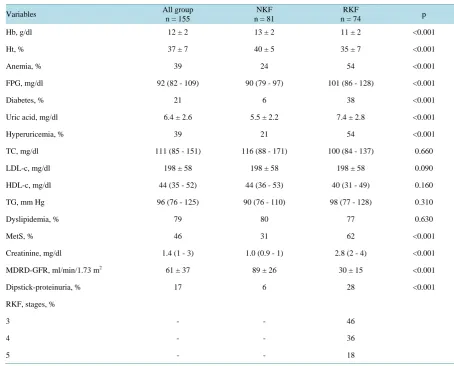
Main clinical and biological characteristics of the study population and difference according to renal function status are shown inTable 1 andTable 2. RKF was observed in 74 hypertensive patients (53% females, mean age 59 ± 12 years), 34 (46%), 27 (36%) and 13 (18%) of whom being on stage 3, 4 and 5, respectively. Com-pared to patients with relatively normal kidney function (NKF), those with RKF had a longer duration of hyper-tension [8 (4 - 12) vs 6 (1 - 13) years; p = 0.004), a greater BMI (27 vs 25 Kg/m2; p = 0.04) and WC (95 ± 14 vs 95 ± 14 cm; p = 0.03). The proportions of RKF patients receiving one, two or more antihypertensive drugs, amounted to 45 (61%), 20 (27%) and 4 (5%) patients, respectively. Uncontrolled hypertension predominated among patients with RKF (77 vs 62%; p = 0.04) who also had higher FPG [101 (86 - 126) mg/dl], uric acid (7.4 ± 2.8 vs 5.5 ± 2.2 mg/dl; p < 0.001) but lower hemoglobin (11 ± 2 vs 13 ± 2 g/dl; p < 0.001) or hematocrit (35% vs 40%; p < 0.001). Patients with RKF had a higher proportion of subjects with global obesity (30% vs 10%; p = 0.002), central obesity (55% vs 39%; p = 0.04), diabetes (38% vs 6%; p < 0.001), hyperuricemia (54% vs 21%;
Table 1. Clinical characteristics of the study population as a whole and according to kidney function status.
Variables All group
n = 155
NKF n = 81
RKF
n = 74 p
Gender, % M 49 51 47 0.681
F 51 49 53
Age, years 58 ± 12 58 ± 12 59 ± 12 0.732
DHT, years 7 (2 - 13) 6 (1 - 13) 8 (4 - 12) 0.040
Smoking, % 20 22 18 0.470
Physical activity, % 60 66 54 <0.001
BMI, Kg/m2 26 ± 5 25 ± 4 27 ± 5 0.047
Obesity, % 19 10 30 0.002
WC, cm 93 ± 14 90 ± 13 95 ± 14 0.031
Central obesity, % 47 39 55 0.048
SBP, mm Hg 158 ± 24 157 ± 24 159 ± 25 0.510
DBP, mm Hg 93 ± 17 94 ± 15 93 ± 18 0.890
PP, mm Hg 65 ± 20 63 ± 18 68 ± 22 0.180
Heart rate, bpm 84 ± 14 83 ± 15 84 ± 14 0.600
AntiHT regimen, % 0.530
- Monotherapy 64 64 63
- Bitherapy 28 28 27
- ≥ 3 drugs 3 1 5
Uncontrolled BP, % 69 62 77 0.040
Table 2. Biological characteristics of the study population as a whole and according to kidney function status.
Variables All group
n = 155
NKF n = 81
RKF
n = 74 p
Hb, g/dl 12 ± 2 13 ± 2 11 ± 2 <0.001
Ht, % 37 ± 7 40 ± 5 35 ± 7 <0.001
Anemia, % 39 24 54 <0.001
FPG, mg/dl 92 (82 - 109) 90 (79 - 97) 101 (86 - 128) <0.001
Diabetes, % 21 6 38 <0.001
Uric acid, mg/dl 6.4 ± 2.6 5.5 ± 2.2 7.4 ± 2.8 <0.001
Hyperuricemia, % 39 21 54 <0.001
TC, mg/dl 111 (85 - 151) 116 (88 - 171) 100 (84 - 137) 0.660
LDL-c, mg/dl 198 ± 58 198 ± 58 198 ± 58 0.090
HDL-c, mg/dl 44 (35 - 52) 44 (36 - 53) 40 (31 - 49) 0.160
TG, mm Hg 96 (76 - 125) 90 (76 - 110) 98 (77 - 128) 0.310
Dyslipidemia, % 79 80 77 0.630
MetS, % 46 31 62 <0.001
Creatinine, mg/dl 1.4 (1 - 3) 1.0 (0.9 - 1) 2.8 (2 - 4) <0.001
MDRD-GFR, ml/min/1.73 m2 61 ± 37 89 ± 26 30 ± 15 <0.001
Dipstick-proteinuria, % 17 6 28 <0.001
RKF, stages, %
3 - - 46
4 - - 36
5 - - 18
Data are expressed as mean ± standard deviation, median (range) or relative frequency in percent. Abbreviations: RKF & NKF, reduced and relatively normal kidney function Hb, hemoglobin Ht, hematocrit FBG, fasting plasma glucose TC, total cholesterol LDL-c, low-density lipoprotein cholesterol HDL-c, high-density lipoprotein cholesterol TG, triglycerides MetS, metabolic syndrome MDRD, modification of diet in renal disease GFR, glome-rular filtration rate.
p < 0.001), proteinuria (28% vs 6%; p < 0.001), MetS (62% vs 31%; p < 0.001), anemia (54% vs 24%; p < 0.001) and lower proportion of physically active individuals (66% vs 54%; p < 0.001).
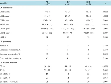
Echocardiographic LV dimensions are shown in Table 3 for the whole study population and according to renal function status. In comparison to those with relatively NKF, RKF patients had greater LVIDd (51 ± 8 vs 47 ± 7 mm; p < 0.001), LVIDs (34 ± 9 vs 29 ± 7 mm; p < 0.001), IVSTd [12 (10 - 13) vs 11 (0.9 - 12) mm; p = 0.003], PWTd [12 (10 - 13) vs 10 (0.8 - 12) mm; p < 0.001], LVMI (BSA) [276 (198 - 348) vs 216 (175 - 284); p = 0.008] and LVMI (height2.7) [72 (47 - 88) vs 24 (44 - 73) g/m2.7; p = 0.007]. LVH was present in 68 RKF patients (92%) of whom 50% and 42% had concentric and eccentric geometric patterns, respectively. Concentric pattern of LVH was present in 47%, 63% and 31% of patients with RKF stage 3, 4 and 5, respectively; these proportions were 41%, 33% and 61% for the eccentric pattern; however, the differences observed between RKF stages were not statistically significant.
Table 3. Echocardiographic left ventricular dimensions, mass and systolic function of the study population as a whole and according to kidney function status.
Variables All
n = 155
NKF n = 81
RKF
n = 74 p
LV dimensions
LVIDd, mm 49 ± 8 47 ± 7 51 ± 8 <0.001
LVIDs, mm 31 ± 9 29 ± 7 34 ± 9 <0.001
IVSTd, mm 12 (7 - 13) 11 (0.9 - 12) 12 (10 - 13) 0.003
PWTd, mm 11 (0.9 - 13) 10 (0.8 - 12) 12 (10 - 13) <0.001
LVMI, g/m2 239 (178 - 325) 216 (175 - 284) 276 (198 - 348) 0.008
LVMI, g/m2.7 63 (45 - 80) 54 (44 - 73) 72 (47 - 88) 0.007
LVH, % 91 92 92 0.280
LV geometry
Normal, % 6 7 4 0.370
Concentric remodeling, % 3 1 4 0.350
Eccentric hypertrophy, % 46 51 42 0.250
Concentric hypertrophy, % 45 41 50 0.280
LV systolic function
EF, % 64 ± 14 68 ± 13 60 ± 14 <0.001
SF, % 36 ± 11 39 ± 10 34 ± 11 0.005
EF < 50%, % 15 10 22 0.043
SF < 28%, % 20 12 28 0.013
EF < 50% + < 28%, % 21 12 30 0.008
Data are expressed as mean ± standard deviation, median (range) or relative frequency in percent. Abbreviations: RKF & NKF, reduced and relatively normal kidney function LVIDD, left ventricular internal diameter diastolic LVIDS, left ventricular internal diameter systolic, IVSD, interventricular septum diastolic PWT, posterior wall thickness diastolic LVMI, left ventricular mass index LVH, left ventricular hypertrophy EF, ejection fraction SF, shortening fraction.
combination of both criteria. However, the differences observed between RKF stages were not statistically sig-nificant.
4. Discussion
The main findings of the present study are as follows: first, LV dimensions and mass were higher in patients with RKF 92% of whom showing the concentric geometric pattern of LVH. Second, LV systolic dysfunction was more frequent in patients with RKF.
The observation of greater LV dimensions and mass in patients with RKF agrees with previous studies re-porting an increase in LVM among hypertensive patients with progressive decline in GFR [24] [25]. Nardi et al.
[24] found that values of LVM progressively increased from CKD stage 2 to 5. An inverse association between LVM and GFR was also reported by Redon et al. [25]
In Sub-Saharan Africa [20] [26]-[28], the prevalence of LVH in hypertensive patients with RKF has generally been reported to be elevated. The frequency of LVH in our study is quite similar to the 95.5% reported by Ulasi
proportion of subjects with MetS [1] [2] [4] [32], anemia [33] and uncontrolled hypertension [34], all of which are well-known risk factors for LVH. Although hypertension, altered fluid and electrolyte balance, and anemia have been identified as major determinants of LVH in CKD, non hemodynamic factors such as inappropriate ac-tivation of the renin angiotensin aldosterone system, oxidative stress, inflammation, collagen and muscle cell growth factors may still play a relevant role [35]
In agreement with several reports from Nigeria [20] [36] and Burkina Faso [31], concentric LVH was the commonest geometric pattern found in the present case series. However; probably because of inclusion of pa-tients with exclusively RKF stage 4 and 5, Ulasi et al.[26] found a higher prevalence of eccentric than concen-tric LVH. It is well-known that concenconcen-tric LVH prevails in earlier stages of CKD due to pressure overload whe-reas eccentric geometric pattern occurs in advanced stages of CKD as a result of volume overload [24]. To this respect, Nardi et al.[24] stressed out that at CKD stage 4 to 5, the increase in LV mass relies more upon the in-crease in posterior wall thickness than in LV diameter in diastole.
By contrast to our finding, Adeseye et al. [37] observed a higher frequency of normal geometry and concen-tric remodeling in treated Nigerian hypertensive patients. Apart from the stage of CKD, this disparity could also be explained by the use of antihypertensive drugs such as the renin angiotensin system inhibitors and/or calcium channel blockers known to induce the regression of LVH [38]. In addition to hemodynamic factors, proinflam-matory cytokines mainly c-reactive protein (CRP) and interleukin 6 (IL-6) may promote cardiac remodeling by stimulating sarcomeric protein synthesis, enhancing fetal gene expression, altering extracellular matrix degrada-tion and triggering apoptosis [39].
At variance with the results showing a significant increase in LV dimensions, wall thickness, mass and hyper-trophy from CKD stage 3 to 5 [40] [41], the decline of GFR in our study did not affect the frequency of LVH or its geometric patterns. The frequency of LVH was reported to pass from 64% at earlier CKD stages to 96% at advanced stage in the paper by Sambi et al.[40] and from 48% at CKD stage 3% to 75% at stage 4 - 5 in that by Park et al. [41]. With reference to LVH geometric patterns, Sambi et al.[40] found an increase in the frequency of concentric LVH from 40% at earlier CKD stages to 68% at advanced CKD stages. In accordance with our data, Nardi et al. [24] found no increase in the frequency of concentric or eccentric LVH geometric patterns with the decline of GFR. However, when using the mixed pattern (concentric + eccentric LVH), they reported signif-icant differences in the frequency of geometric patterns. The increased frequency of LVH and its concentric pattern reported in CKD might be related to high plasma aldosterone levels that are known to promote a LV concentric geometry and increase LVM [42].
The frequent LV systolic dysfunction in patients with RKF agrees with the observation by Arodiwe et al.[43] among pre-dialysis Nigerian patients. An inverse association between CKD and LV systolic function has also been reported by Grabysa et al.[44] who noted a decrease in the ejection fraction as well as the E/A ratio with progressive decline in GFR. Left ventricular systolic dysfunction is thought to result from cardiac remodeling triggered by the interaction of traditional and CKD specific hemodynamic and non hemodynamic risk factors [35] [39].
Some limitations of the present study need to be underscored. Measurements being observer-dependent, one limitation of the present study is the fact that the diagnosis of echocardiographic LVH was made only by a sin-gle observer. In addition, other echocardiographic parameters such as issues of LV diastolic function and tissue Doppler imaging of mitral annulus, pericardial disease, valve calcification, regurgitant mitral and aortic valves, and regional wall motion defect known to confound the assessment of LV geometry and systolic function were not assessed in the present study. Given the inclusion of patients with advanced RKF (stages 4 and 5) whose clinical profile correlate with a high prevalence of LVH, our findings should not be extrapolated to the whole population of patients with RKF. The cross-sectional design of the present study precludes inference for causal-ity. The single measurement of echocardiographic parameters could have overestimated the prevalence of LVH.
5. Conclusion
Reduced kidney function in the present hypertensive case series was associated with an increased frequency of left ventricular hypertrophy with mainly concentric geometric pattern and altered systolic function that could enhance their CV risk.
Acknowledgements
the Division of Cardiology of the University of Kinshasa Hospital for their invaluable contribution to the con-duct of the present study.
Conflict of Interest
None.Author’s Contribution
-MBC: participated in protocol conception, collected data, participated in data analysis and reviewed the ma-nuscript.
-LFB: conceived the protocol, participated in data analysis and drafted the manuscript.
-MFI: participated in protocol conception, performed data analyses and reviewed the manuscript. -MJR: reviewed the manuscript.
-KVE: reviewed the manuscript. -BPM: revised the manuscript. -MJR: reviewed the manuscript.
References
[1] Chahal, N.S., Lim, T.K., Jain, P., Chambers, J.C., Kooner, J.S. and Semior, R. (2010) New Insights into the Relation-ship of Left Ventricular Geometry and Left Ventricular Mass with Cardiac Function: A Population Study of Hyperten-sive Subjects. European Heart Journal, 31, 588-594. http://dx.doi.org/10.1093/eurheartj/ehp490
[2] Schiffrin, E., Lipman, M.L. and Mann, J.F.E. (2007) Chronic Kidney Disease. Effects on the Cardiovascular System.
Circulation, 116, 85-97. http://dx.doi.org/10.1161/CIRCULATIONAHA.106.678342
[3] Shahbaz, A.U., Sun, Y., Bhattacharya, S.K., Ahokas, R.A., Gerling, I.C., McGee, J.E. and Xeber, K.T. (2010) Fibrosis in Hypertensive Heart Disease: Molecular Pathways and Cardioprotective Strategies. Journal of Hypertension, 28, S25-S32. http://dx.doi.org/10.1097/01.hjh.0000388491.35836.d2
[4] Iwashima, Y., Horio, T., Kamide, K., Tokudome, T., Yoshihara, F., Nakamura, S., et al. (2010) Additive Interaction of Metabolic Syndrome and Chronic Kidney Disease on Cardiac Hypertrophy and Risk of Cardiovascular Disease in Hypertension. American Journal of Hypertension, 23, 290-298. http://dx.doi.org/10.1038/ajh.2009.253
[5] Levin, A. (2003) Clinical Epidemiology of Cardiovascular Disease in Chronic Kidney Disease Prior to Dialysis.
Semi-nars in Dialysis, 16, 101-105. http://dx.doi.org/10.1046/j.1525-139X.2003.16025.x
[6] M’Buyamba-Kabangu, J.R., Fagard, R., Staessen, J., Lijnen, P. and Amery, A. (1987) Comparison of Blood Pressure and Prevalence of Hypertension in Rural and Urban Zaire. BibliothecaCardiologica, 42, 80-87.
[7] Sumaili, E.K., Nseka, M.N., Lepira, F.B., Krzesinski, J.M., Makulo, J.R., Bukabau, J.B., et al. (2008) Screening for Proteinuria and Chronic Kidney Disease Risk Factors in Kinshasa: A World Kidney Day 2007 Study. Nephron Clinical
Practice, 110, c220-c228. http://dx.doi.org/10.1159/000167869
[8] Katchunga, P.B., M’Buyamba-Kayamba, J.R., Masumbuko, B.E., Lemogum, D., Kashongwe, Z.M., Degaute, J.P., et al. (2011) Hypertension in the Adult Congolese Population of Southern Kivu: Results of the VITARAA Study. [French].
La Presse Médicale, 40, e315623.
[9] M’Buyamba-Kabangu, J.R., Biswika, R.T., Thijs, L., Tshimanga, G.M., Ngalula, F.M., Disashi, T., et al. (2009) In- Hospital Mortality among Black Patients Admitted for Hypertension-Related Disorders in Mbuji Mayi, Congo.
Amer-ican Journal of Hypertension, 22, 643-648. http://dx.doi.org/10.1038/ajh.2009.47
[10] Lepira, F.B., M’Buyamba-Kabangu, J.R., Kayembe, P.K. and Nseka, M.N. (2006) Clinical Correlates of Left Ventri-cular Hypertrophy in Black Patients with Arterial Hypertension. Cardiovascular Journal of Africa, 17, 7-11.
[11] Bayauli, M.P., Lepira, F.B., Kayembe, P.K. and M’Buyamba-Kabangu, J.R. (2012) Left Ventricular Hypertrophy and Geometry in Type 2 Diabetes Patients with Chronic Kidney Disease: An Echocardiographic Study. Cardiovascular
Journal of Africa, 23, 73-77. http://dx.doi.org/10.5830/CVJA-2011-028
[12] Mansia, G., De Backer, G., Dominiczak, A., Cikova, R., Fagard, R., Germano, G., et al. (2007) Guidelines for the Management of Arterial Hypertension: The Task Force for the Management of Arterial Hypertension of the European Society of Hypertension (ESH) and of the European Society of Cardiology (ESC). Blood Press, 16, 135-232.
http://dx.doi.org/10.1080/08037050701461084
[14] Dimitrieva, O., de Lusignan, S. and Goldsmith, D. (2013) Association of Anemia in Primary Care Patients with Chronic Kidney Disease: Cross-Sectional Study of Quality Improvement in Chronic Kidney Disease (AICKD) Trial Data.BMC Nephrology, 14, 24. http://dx.doi.org/10.1186/1471-2369-14-24
[15] Expert Committee on the Diagnosis and Classification of Diabetes (2003) Report of Expert Committee on the Diagno-sis of and Classification of Diabetes Mellitus. Diabetes Care, 26, S5-S20.
[16] Levey, A.S., Greene, T. and Kusek, J.W. (2000) A Simplified Equation to Predict Glomerular Filtration Rate from Se-rum Creatinine. Journal of the American Society of Nephrology, 11, Article ID: A0828.
[17] K/DOQI, Kidney Disease Outcome Quality Initiative (2002) Clinical Practical guidelines for Chronic Kidney Disease (CKD). American Journal of Kidney Diseases, 39, S22-S26.
[18] National Heart Lung and Blood Institute (2001) Executive Summary of the Third of National Cholesterol Education Program (NCEP) Expert Panel on Evaluation, and Treatment of High Cholesterol in Adults (Adult Treatment Panel III) (2001). The Journal of the American Medical Association, 285, 2486-2497.
http://dx.doi.org/10.1001/jama.285.19.2486
[19] Lang, R.M., Bierig, M., Devereux, R.B., Flachskamp, F.A., Foster, E., Pellikka, P.A., et al. (2006) American Society of Echocardiography’s Nomenclature and Standards Committee: Task Force on Chamber Quantification. American College of Cardiology Echocardiography Committee; American Heart Association; European Society of Echocardio-graphy; European Society of Cardiology. Recommendations for Chamber Quantification. European Journal of
Echo-cardiography, 17, 1460-1465.
[20] Adamu, U.G., Kolo, P.M., Katibi, I.A., Opadji, G.O., Omotsho, A.B. and Araoye, A.M. (2009) Relationship between Left Ventricular Diastolic Function and Geometric Patterns in Nigerians with Newly Diagnosed Systemic Hyperten-sion. Cardiovascular Journal of Africa, 20, 173-177.
[21] Devereux, R.B., Alonso, D.R., Lucas, E.M., Gottlieb, E. and Reichek, I. (1986) Echocardiographic Assessment of Left Ventricular Hypertrophy: Comparison to Necropsy Findings. The American Journal of Cardiology, 57, 450-458.
http://dx.doi.org/10.1016/0002-9149(86)90771-X
[22] de Simone, G., Devereux, R.B., Daniels, S.R., Koren, M.J., Meyer, R.A. and Laragh, J.H. (1995) Effects of Growth on Variability of Left Ventricular Mass: Assessment of Allometric Signals in Adults and Children and Their Capacities to Predict Cardiovascular Risk. Journal of the American College of Cardiology, 25, 1056-1062.
http://dx.doi.org/10.1016/0735-1097(94)00540-7
[23] Lang, R.M., Bierig, M., Devereux, R.B., Flachskampf, F.A., Foster, E., Pellikka, P.A., et al. (2005) Recommendations for Chamber Quantification: A Report from the American Society of Echocardiography’s Guidelines and Standards Committee and the Chamber Quantification Writing Group, Developed in Conjunction with the European Association of Echocardiography, a Branch of the European Society of Cardiology. Journal of the American Society of
Echocardi-ography, 18, 1440-1463. http://dx.doi.org/10.1016/j.echo.2005.10.005
[24] Nardi, E., Palermo, A., Mulè, G., Cusimano, P., Cottone, S. and Cerasola, G. (2009) Left Ventricular Hypertrophy and Geometry in Hypertensive Patients with Chronic Kidney Disease. Journal of Hypertension, 27, 633-641.
http://dx.doi.org/10.1097/HJH.0b013e3283220ecd
[25] Redón, J., Cea-Calvo, L., Lozano, J.V., Fernández-Pérez, C., Navarro, J., Bonet, A. and González-Esteban, J., ERIC- HTA 2003 Study Investigators (2006) Kidney Function and Cardiovascular Disease in the Hypertensive Population: The ERIC-HTA Study. Journal of Hypertension, 24, 663-669. http://dx.doi.org/10.1097/01.hjh.0000217848.10831.5f
[26] Ulasi, I.I., Arodiwe, E.B. and Ijoma, C.K. (2006) Left Ventricular Hypertrophy in African Black Patients with Chronic Renal Failure at First Evaluation. Ethnicity & Disease, 16, 859-864.
[27] Busari, O., Opadijo, G., Olarewaju, T., Omotoso, A. and Jimoh, A. (2010) Electrocardiographic Correlates of Micro-albuminuria in Adult Nigerians with Essential Hypertension. Cardiology Journal, 17, 281-287.
[28] Adekunde, A.E., Adeseye, A.I., Adebayo, O.T., Olatayo, A.A., Joseph, O.O. and Ayodele, A.R. (2013) Left Ventricu-lar Mass Formulae and Prevalence Rates of Echocardiographic Left VentricuVentricu-lar Hypertrophy in Nigerians with Essen-tial Hypertension. North American Journal of Medical Sciences, 5, 325-329.
http://dx.doi.org/10.4103/1947-2714.112481
[29] Dada, A., Adebiyi, A.A., Aje, A., Oladapo, O.O. and Falase, A.O. (2005) Standard Electrocardiographic Criteria for Left Ventricular Hypertrophy in Nigerian Hypertensives. Ethnicity & Disease, 15, 578-584.
[30] Dada, A., Adebiyi, A.A., Aje, A., Oladapo, O.O. and Falase, A.O. (2006) Comparison of Araoye’s Criteria with Stan-dard Electrocardiographic Criteria for Diagnosis of Left Ventricular Hypertrophy in Nigerian Hypertensives. West
African Journal of Medicine, 25, 179-185.
[31] Niakara, A., Ouédraogo, N., Nébié, L.V., Samadoulougou, A.K., Kaboré, N.J. and Ouandaogo, B.J. (2001) Left Ven-tricular Hypertrophy in Hypertensive African Blacks: Echocardiographic Study of 452 Patients. Annales de
[32] Singh, A.K. and Kari, J.A. (2013) Metabolic Syndrome and Chronic Kidney Disease. Current Opinion in Nephrology
and Hypertension, 22, 198-203. http://dx.doi.org/10.1097/MNH.0b013e32835dda78
[33] Chang, J.M., Chen, S.C., Huang, J.C., Su, H.M. and Chen, H.C. (2014) Anemia and Left Ventricular Hypertrophy with Renal Function Decline and Cardiovascular Events in Chronic Kidney Disease. The American Journal of the Medical
Sciences, 347, 183-189.
[34] Fraser, S.D., Roderick, P.J., McIntyre, N.J., Harris, S., McIntyre, C.W., Fluck, R.J., et al. (2013) Suboptimal Blood Pressure Control in Chronic Kidney Disease Stage 3: Baseline Data from a Cohort Study in Primary Care. BMC
Fami-ly Practice, 14, 88. http://dx.doi.org/10.1186/1471-2296-14-88
[35] Cerasola, G., Nardi, E., Palermo, A., Mule, G. and Cottone, S. (2011) Epidemiology and Pathophysiology of Left Ven-tricular Mass Abnormalities in Chronic Kidney Disease. Journal of Nephrology, 24, 1-10.
http://dx.doi.org/10.5301/JN.2010.2030
[36] Aje, A., Adebiyi, A.A., Oladapo, O.O., Dada, A., Ogah, O.S., Ojji, D.B. and Falae, A.O. (2006) Left Ventricular Geo-metric Patterns in Newly Presenting Nigerian Hypertensives: An Echocardiographic Study. BMC Cardiovascular
Dis-orders, 6, 4. http://dx.doi.org/10.1186/1471-2261-6-4
[37] Adeseye, A., Olayinka, A. and Opadijo, G. (2010) Left Ventricular Hypertrophy, Geometric Patterns and Clinical Cor-relates among Treated Hypertensive Nigerians. Pan African Medical Journal, 4, 8.
http://dx.doi.org/10.4314/pamj.v4i1.53602
[38] Kuch, B., von Scheidt, W., Peter, W., Heier, M., Wichmann, H.E. and Meisinger, C. (2006) Influence of Antihyperten-sive Therapy and Blood Pressure Control on Left Ventricular Geometry and Function in Subjects with Type 2 Diabetes: The Augsburg Diabetes Family Study. Journal of Human Hypertension, 20, 757-764.
http://dx.doi.org/10.1038/sj.jhh.1002062
[39] Gupta, J., Dominic, E.A., Fink, J.C., Ojo, A.O., Barrows, I.R., Reilly, M.P., et al. (2015) Association between Inflam-mation and Cardiac Geometry in Chronic Kidney Disease: Findings from the CRIC Study. PLoS ONE, 10, e0124772.
[40] Sambi, R.S., Gaur, A.K., Hotchandani, R., Aggarwal, K.K., Kaur, S., Gupta, M., et al. (2011) Patterns of Left Ventri-cular Hypertrophy in Chronic Kidney Disease: An Echocardiographic Evaluation. Indian Heart Journal, 63, 259-268.
[41] Park, M., Hsu, C.Y., Li, Y., Mishra, R.K., Keane, M., Rosas, S.E., et al. and Chronic Renal Insufficiency Cohort (CRIC) Study Group (2012) Associations between Kidney Function and Subclinical Cardiac Abnormalities in CKD.
Journal of the American Society of Nephrology, 23, 1725-1734. http://dx.doi.org/10.1681/ASN.2012020145
[42] Mulè, G., Nardi, E., Guarino, L., Cacciatore, V., Geraci, G., Calcaterra, I., et al. (2015) Plasma Aldosterone and Its Relationship with Left Ventricular Mass in Hypertensive Patients with Early-Stage Chronic Kidney Disease.
Hyper-tension Research, 38, 276-283.
[43] Arodiwe, E.B., Ulasi, I.L., Ijoma, C.K. and Ike, S.O. (2010) Left Ventricular Systolic Function in a Nigerian Pre-Dialysis Patient Population with Chronic Kidney Disease. Nigerian Postgraduate Medical Journal, 17, 301-307.
[44] Grabysa, R. and Wańkowicz, Z. (2013) Echocardiographic Markers of Left Ventricular Dysfunction among Men with Uncontrolled Hypertension and Stage 3 Chronic Kidney Disease. Medical Science Monitor, 19, 838-845.