http://dx.doi.org/10.4236/arsci.2015.33007
How to cite this paper: Parra-Forero, L.Y., Mendoza, G.D., Góngora, A., López Fernández, M.D.C., Cruz, L.A., Montiel, A.J., Kjelland, M.E. and García-Contreras, A.D.C. (2015) Azteca Breed Horse Epididymal Sperm Evaluation: A Comparison of Head, Corpus and Cauda Sperm Quality. Advances in Reproductive Sciences, 3, 57-65. http://dx.doi.org/10.4236/arsci.2015.33007
Azteca Breed Horse Epididymal Sperm
Evaluation: A Comparison of Head, Corpus
and Cauda Sperm Quality
Lyda Y. Parra-Forero
1,2, German D. Mendoza
1, Alfredo Góngora
2,
Maria D. C. López Fernández
3, Luis A. Cruz
4, Antonio J. Montiel
5, Mike E. Kjelland
6,
Adelfa D. C. García-Contreras
1*1Universidad Autónoma Metropolitana, Unidad Xochimilco,México, D. F., México
2Departamento de Investigación Animal y Humana, Centro de Fertilidad Humana en México, México, D. F., México
3Unidad de Genética, Departamento de Biología, Universidad Autónoma de Madrid, Ciudad Universitaria de Cantoblanco, Madrid, Spain
4Instituto de Ecología A.C. de México, Veracruz, México
5Centro de Reproducción y Medicina Equina, México, D. F., México 6Conservation, Genetics & Biotech, LLC, Vicksburg, MS, USA
Email: [email protected], [email protected], [email protected],
[email protected], [email protected], [email protected], [email protected], *[email protected]
Received 9 June 2015; accepted 22 August 2015; published 26 August 2015
Copyright © 2015 by authors and Scientific Research Publishing Inc.
This work is licensed under the Creative Commons Attribution International License (CC BY).
http://creativecommons.org/licenses/by/4.0/
Abstract
The main objective of this study was to determine differences in sperm recovered from the three segments of the epididymis, i.e., head, corpus and cauda, using routine measurements of sperm quality and DNA fragmentation assessment. There was a larger sperm concentration (P < 0.05) in the head (3.34 × 10⁶ sperm/mL) compared with the corpus and cauda. The percentage of normal
morphology was higher in the cauda but had lower motility (P < 0.05). The DNA fragmentation in-dex was higher for sperm obtained from the head and body of the epididymis (P < 0.01) compared with the cauda. The DNA fragmentation index values were 12% higher (P < 0.01) using Wright stain compared with DAPI stain in sperm from the head and corpus of the epididymis, but similar in the cauda. Spermatobioscopy parameters match the maturation changes that occur in the epi-didymal tract with higher quality sperm being from the cauda, but with a lower concentration compared to sperm found in the head.
*
tinal problems that may end with humane euthanasia and death [4].
Epididymal sperm can be useful for fertilization, either in vivo or in vitro, as it has been shown to have a high enough quality to be used in assisted reproduction procedures. Epididymal sperm viability at room temperature for the 24 h following an orchiectomy is important for freezing protocols [1] [5] [6]. The sperm concentrations of the samples collected from the epididymides can be similar to those of ejaculates obtained with an artificial vagina, but the quality is reduced when epididymal sperm are subjected to multiple freezing and thawing steps
[7]. Pregnancy rates with epididymal sperm have varied from 8% to 92% with frozen-thawed epididymal semen and 9% to 55% with fresh epididymal semen [8]. The differences in pregnancy rates using frozen-thawed semen can be due to the following: stallion semen quality, the type of diluent utilized, and the use or removal of the seminal plasma, as well as other factors [9].
There is a lack of information in previous studies with regard to epididymal sperm quality in terms of the state of sperm DNA integrity within the epididymides. It is in the epididymides where the major morphological changes in sperm occur [10], such as chromatin condensation, due mainly to the replacement of histones by protamines in the testes [11] [12]. However, the lack of seminal plasma makes the epididymal sperm less motile than would be considered ideal for direct insemination techniques and the sperm can also be easily damaged by oxidizing agentsp [13]-[15]. Garcia-Macias et al. [16] noted that there was a greater possibility of finding sperm with damaged DNA in epididymal samples compared with samples obtained by artificial vagina. Given the aforementioned issue, it would be beneficial to identify the potential differences in sperm obtained from the dif-ferent sections of the epididymis and to determine the presence of altered DNA. The primary objective of this study was to determine the epididymal sperm DNA fragmentation index for Azteca breed stallions, while also comparing DAPI (4’-6-diamidino Obtained 2-phenylindole) and Wright stain protocols.
2. Materials and Methods
2.1. Epididymal Sperm Retrieval
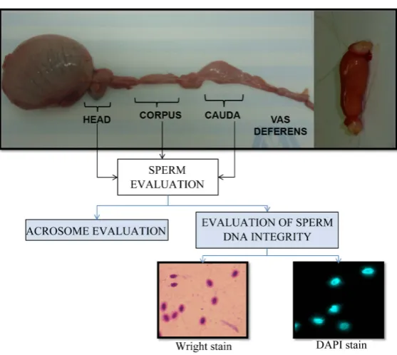
Elective castrations involving Azteca breed horses (n = 46) of 1 - 2 years of age, were performed to obtain each individual’s testes and epididymides. The collected testes and epididymides were immersed in saline solution (NaCl 0.9%), penicillin-G (100 UI/mL) and streptomycin sulfate (100 mg/mL), kept at a temperature between 4 and 8˚C, and transported to the laboratory (Figure 1), a trip of approximately 30 min. Epididymal sperm was obtained using the technique described by Muradas et al. [17] and the tubules of the body and head were minced when necessary. More specifically, the head, body and cauda were separated with Nylon No.1 and each segment was washed with 40 mL of Dulbecco’s Phosphate Buffered Saline (DPBS) at a temperature of 37˚C ± 2˚C (Dulbecco’s Phosphate Buffered Saline D8662 [2], performing 3 washes for each testicle and epididymis ob-tained. All of the animals used in this research were treated in accordance with the Federation of Animal Science Societies (FASS) guide for use of farm animals in research and teaching [18].
2.2. Sperm Evaluation
Figure 1.Azteca breed horse testicle and epididymide showing sperm DNA fragmentation analysis using Wright stain and DAPI stain.
Ni-U, Nikon®, Japan) with 10 µL of sample, sperm diluted 1:1 with DPBS. The percentage of dead sperm was determined with smears stained with eosin-nigrosin and with Diff Quick (Kubus®) to determine the percentage of abnormal morphologies, based on [6] [13], using a 400× optical light microscope. A total of 300 cells were counted per sample (Figure 1).
2.3. Acrosome Evaluation
A total of 1.5 mL of DPBS containing collected semen was centrifuged at 50 g × 5 min to obtain a sperm pellet from which 10 µL were removed and diluted with 1000 µL of fixing solution (2% paraformaldehyde, 2% for-maldehyde buffered) and stored at 4˚C until analysis [19] [20]. For the acrosome structure assessment, five washes of the sample with DPBS were performed. Afterwards, the sample was incubated for 30 min (while pro-tected from light) with 10 uL of lectin Pisum sativum (PSA) (FITC-PSA, Sigma, St Louis, MO, USA) at a 1:10,000 dilution, plus an antifade solution (Vectashield, Burlingame, USA) in a 1:1 ratio and evaluated using a fluorescence microscope (FV10i, OYMPUS) with a 1000X magnification, similar to the procedure in Farlin et al. [21].
2.4. Evaluation of Sperm DNA Integrity
The Halomax kit for horses (Halotech SL, Madrid, Spain) was used for assessing DNA fragmentation. The Sperm-Halomax® methodology is based on the sperm chromatin dispersion test (SCDt) [22]-[24]. Details of this technique have previously been reported for human and other mammalian species [23] [25]-[27].
In the present study, a sample of 15 × 106 sperm/mL was diluted in agarose to give a final concentration of 1 × 10⁶ sperm/mL, placed in racks, lysed, and stained with DAPI (SIGMA, 4’-6-diamidino Obtained 2-phenylindole) and Wright stain (Accustain, Sigma) to obtain two slides for each epididymal segment sample. Kjelland et al.
3. Results
The highest total concentration of sperm (P < 0.05) was found in the head of the epididymis. The percentage of normal morphology was higher in the cauda epididymis, but cauda samples also had a lower motility (P < 0.05) compared to the samples from the head and corpus segments. The highest percentage of damaged acrosomes was obtained in sperm from the head of the epididymis while the lowest percentage came from the cauda (P < 0.05). The DNA fragmentation indices were higher in the head and corpus (P < 0.01) compared to the cauda (Table 1). Notably, the values obtained regarding the DNA fragmentation index were 12% higher (P < 0.01) using Wright stain in sperm from the head and corpus (Table 2) compared to those stained with DAPI.Figure 2
andFigure 3 show epididymal sperm stained with DAPI and Wright stain.
4. Discussion
[image:4.595.90.539.426.570.2]Sperm maturation occurs in the tract of the epididymis, with the sperm acquiring important functions essential
Table 1.Spermatobioscopy parameters and sperm DNA fragmentation index (DNA FI) in the head, corpus, and cauda of Azteca breed horse epididymides.
Epididymal Segment
Spermatobioscopy Parameters Head Corpus Cauda SEM P-Value
Total Concentration (×106) 3.34a 1.39b 1.34b 0.089 <0.0001
Motility (%) 69.30a 67.02a 54.91b 0.873 <0.0001
Normal Morphology (%) 74.37b 75.26b 80.78a 0.637 <0.0001
Mortality (%) 25.04a 17.17b 18.21b 0.479 <0.0001
Acrosomes Damaged (%) 23.76a 20.61b 16.09c 0.472 <0.0001
DNA FI DAPI 20.96a 20.40a 16.47b 0.559 <0.0001
DNA FI Wright 23.48a 23.22a 16.72b 0.672 <0.0001
Different letters indicate statistically different values, P < 0.0001.
Table 2.Comparison of staining methods to assess the DNA fragmentation index (DNA FI) of epididymal sperm obtained from Azteca breed horses.
Stain
Epididymal Segment DAPI (DNA FI) WRIGHT (DNA FI) P-Value (Student’s t-Test)
Head 20.91 (0.66) 23.42 (0.69) 0.009
Body 20.35 (0.52) 23.16 (0.82) 0.005
Cauda 16.42 (0.50) 16.66 (0.49) 0.729
[image:4.595.87.541.619.705.2]Figure 2.Azteca breed horse sperm staining with DAPI. (A) visualization of a sperm using phase contrast; (B) and (C) sperm exhibiting DNA fragmenta-tion (fluorescence microscopy); (C) and (D) measurements of halos (fluores-cence microscopy); (E) sperm without fragmentation (combination of phase contrast and fluorescence microscopy); (F) technical application of the sperm chromatin dispersion test.
Figure 3. Azteca breed horse sperm stained with Wright stain. (A) sperm
with-out fragmentation; (B) fragmented sperm.
for fertilization such as motility, differentiation of the midpiece and acrosome [11] [12] and changes and mod-ifications to the chromatin of the nucleus [5] [13] [31]. The differences detected in the sperm collected in dif-ferent segments of the epididymis reflect the biochemical changes that occur during sperm passage [32] [33]. When sperm reach the cauda they come in contact with various substances including immobiline which greatly reduces their metabolism to minimize the energy demand and minimize the occurrence of residues; primarily because the epithelium has not been developed for absorbing wastes, which are higher in the segment of the head and somewhat less in the corpus of the epididymis [33] [34].
The greater concentration of sperm was obtained from the head of the epididymis, and was higher than that reported by others, e.g., James et al. [35] and Guimarães et al. [3]. In other studies the unit of measure is given as the total sperm/mL [2] [36] but they do not report the total number of mLs used for conducting the evaluation; as such, it was reported as total concentration in the present study. The differences reported for epididymal sperm concentrations in these other studies can be attributed to the differences in the age of animals and/or en-vironmental temperatures utilized. The age of the animals and the individual effect should be considered as im-portant variables [37] [38].
The decrease in sperm motility in the cauda segment of the epididymis has been reported by Cary et al. [39]
[image:5.595.230.400.331.493.2]the aforementioned results, since the lowest percentage of damaged acrosomes were obtained from the cauda re-gion of the epididymis.
The increase in dead sperm in the head of the epididymis may be due to the biochemical regionalization of the epididymis. Fouchécourt et al. [34] identified the secretion of proteins during epididymal sperm transit, with the head segment secreting the most (73%), followed by the corpus (20.5%), and finally the cauda (6.5%). In the head of the epididymis, 44 different proteins were detected of which 22 were specific for this region, e.g., PGDS, GPX, and with the most abundant being the clusterines [44]-[46].
It is important to note that the horses used in this study were prepubertal, which likely affected the sperm DNA fragmentation index found in the present study. Monteiro et al. [40] reported a sperm DNA damage of 6.7% in cryopreserved washed ejaculates and 5.7% from epididymal sperm from adult horses. The aforementioned values are at least 10% below those found in the present study. López-Fernández et al. [22] performed sperm DNA fragmentation analysis using frozen-thawed straws of semen from stallions of various breeds and found that the average values at the time of thawing ranged between 10.3% and 25%, although in that study the Azteca breed was not included.
It is necessary to consider that the physiology of each segment of the epididymis may affect the extent to which sperm DNA becomes fragmented [14] [47], with associated influence on the quality of the embryo and its implantation [8] [48]. For instance, pregnancy rates in horses with the use of epididymal sperm have ranged from 8% - 92% [49] and this variation could be associated with sperm DNA fragmentation.
Statistically significant differences were found among the spermatobioscopy variables in different epididymal segments, meaning that there may be a higher risk of lower embryo production using sperm from one segment of the epididymis versus the other. The sperm tail must be analyzed and sperm separated by screening tests in which the largest number of immature or DNA fragmented sperm are excluded from being used in assisted re-productive techniques. Additionally, a full flushing of the epididymis, in terms of dissection of segments, is un-necessary as one would end up combining a high concentration of immature sperm with mature sperm, likely resulting in a less than ideal reproductive outcome. The epididymal sperm selection method must be conducted using such a segment selected method, most commonly the cauda, even though one sacrifices obtaining a greater volume of sperm. More advanced studies involving cultured sperm maturation should be evaluated for the head and body segments of the epididymis and the testis for Equine reproductive assisted techniques [42] [50] [51].
In a comparison of the DNA fragmentation index values using two different stains, Wright stain showed higher sperm DNA fragmentation index values than that evaluated with DAPI stain, with differences of around 12% (P < 0.05). Wright stain and the HALOMAX® kit have been used in several studies using bright field mi-croscopy [25] [52], since it contains Methylene Blue which has an affinity for proteins with a characteristic acidic property, binding to them via its positive charge thereby marking them a blue color. Given that the Wright Stain only requires bright field microscopy the sperm DNA fragmentation analysis can be much more economi-cal than using a fluorochrome, if it would require the purchase of a microscope with fluorescence capabilities. DAPI is widely used in various techniques to detect DNA damage by its affinity to join to the nitrogenous bases adenine and thymine [25] [53] [54], although its use has declined in DNA studies as it also binds to RNA, con-sidered as having low specificity relative to other intercalating, commercially available fluorochromes.
Studies will be needed to establish the correction for error with regard to the method of analysis of the sample, which must be conducted by individuals with other fluorochromes and different equipment characteristics found in each laboratory.
5. Conclusion
Some differences can be found in the characteristics of semen collected from the epididymal headcauda, and corpus; but less DNA fragmentation and less acrosome damage is observed in sperm from the cauda segment. Sperm DNA fragmentation analyses using Wright’s stain result in higher DNA fragmentation index values compared with analyses made with DAPI.
Conflicts of Interest
All authors declare that there are no conflicts of interest.
Acknowledgements
This work could not have been accomplished without the support of Grant No. 327973 of CONACYT and mounted police of the City of México Dr Manuel Morones and his collaborators.
References
[1] Braun, J., Sakai, M., Hochi, S. and Oguri, N. (1994) Preservation of Ejaculated and Epididymal Stallion Spermatozoa by Cooling and Freezing. Theriogenology, 41, 809-818. http://dx.doi.org/10.1016/0093-691X(94)90497-7
[2] Bruemmer, J.E. (2006) Collection and Freezing of Epididymal Stallion Sperm. Veterinary Clinics of North America:
Equine Practice, 22, 677-682. http://dx.doi.org/10.1016/j.cveq.2006.08.007
[3] Guimarães, T., Lopes, G., Ferreira, P., Leal, I. and Rocha, A. (2012) Characteristics of Stallion Epididymal Spermato-zoa at Collection and Effect of Two Refrigeration Protocols on the Quality of the Frozen/Thawed Sperm Cells. Animal Reproduction Science, 136, 85-89. http://dx.doi.org/10.1016/j.anireprosci.2012.10.028
[4] Samper, J.C. (2009) Equine Breeding Management and Artificial Insemination. Elsevier Health Sciences.
[5] Ball, B.A. (2008) Diagnostic Methods for Evaluation of Stallion Subfertility: A Review. Journal of Equine Veterinary Science, 28, 650-665. http://dx.doi.org/10.1016/j.jevs.2008.10.003
[6] Betancur, G.R., Suárez, A.Ú. and Rojano, B.A. (2014) Técnicas para el análisis de la fertilidad potencial del semen equino. (Techniques for Analyzing the Potential Fertility of Stallion Semen) (Métodos de avaliação do potencial de fertilidade do sêmen equino). Revista CES Medicina Veterinaria y Zootecnia, 8, 115-127.
[7] Papa, F.O., Melo, C. and Fioratti, E. (2008) Freezing of Stallion Epididymal Sperm. Animal Reproduction Science, 107, 293-301. http://dx.doi.org/10.1016/j.anireprosci.2008.05.003
[8] Monteiro, G.A., Papa, F.O., Zahn, F.S., Dellaqua Jr., J.A., Melo, C.M., Maziero, R.R.D., Avanzi, B.R., Alvarenga, M.A. and Guasti, P.N. (2011) Cryopreservation and Fertility of Ejaculated and Epididymal Stallion Sperm. Animal Re- production Science, 127, 197-201. http://dx.doi.org/10.1016/j.anireprosci.2011.08.002
[9] Heise, A., Thompson, P.N. and Gerber, D. (2011) Influence of Seminal Plasma on Fresh and Post-Thaw Parameters of Stallion Epididymal Spermatozoa. Animal Reproduction Science, 123, 192-201.
http://dx.doi.org/10.1016/j.anireprosci.2010.11.017
[10] Dias, G., Retamal, C., Tobella, L., Arnholdt, A. and Lopez, M. (2006) Nuclear Status of Immature and Mature Stallion Spermatozoa. Theriogenology, 66, 354-365. http://dx.doi.org/10.1016/j.theriogenology.2005.10.024
[11] Clemmons, A.J., Thompson, D. and Johnson, L. (1995) Local Initiation of Spermatogenesis in the Horse. Biology of Reproduction, 52, 1258-1267. http://dx.doi.org/10.1095/biolreprod52.6.1258
[12] Baumann, C., Daly, C.M., McDonnell, S.M., Viveiros, M.M. and De La Fuente, R. (2011) Chromatin Configuration and Epigenetic Landscape at the Sex Chromosome Bivalent during Equine Spermatogenesis. Chromosoma, 120, 227- 244. http://dx.doi.org/10.1007/s00412-010-0306-5
[13] Varner, D.D., Blanchard, T.L., Brinsko, S.P., Love, C.C., Taylor, T.S. and Johnson, L. (2000) Techniques for Evaluat-ing Selected Reproductive Disorders of Stallions. Animal Reproduction Science, 60-61, 493-509.
http://dx.doi.org/10.1016/S0378-4320(00)00115-9
matozoa. Journal of Reproduction and Fertility. Supplement, No. 56, 133-140.
[20] Meyers, S. (2001) Equine Sperm-Oocyte Interaction: The Role of Sperm Surface Hyaluronidase. Animal Reproduction Science, 68, 291-303. http://dx.doi.org/10.1016/S0378-4320(01)00166-X
[21] Farlin, M., Jasko, D., Graham, J. and Squires, E. (1992) Assessment of Pisum sativum Agglutinin in Identifying Acro-somal Damage in Stallion Spermatozoa. Molecular Reproduction and Development, 32, 23-27.
http://dx.doi.org/10.1002/mrd.1080320105
[22] López-Fernández, C., Crespo, F., Arroyo, F., Fernández, J.L., Arana, P., Johnston, S.D. and Gosálvez, J. (2007) Dy-namics of Sperm DNA Fragmentation in Domestic Animals: II. The Stallion. Theriogenology, 68, 1240-1250.
http://dx.doi.org/10.1016/j.theriogenology.2007.08.029
[23] Fernández, J., Gosálvez, J., Santiso, R., Goyanes, V. and López-Fernández, C. (2008) Adaptation of the Sperm Chro-matin Dispersion (SCD) Test to Determine DNA Fragmentation from Bull Sperm. New Cell Apoptosis Research. No-va Science Publishers, Inc., New York.
[24] González-Marín, C., Roy, R., López-Fernández, C., Diez, B., Carabaño, M., Fernández, J., Kjelland, M., Moreno, J. and Gosálvez, J. (2011) Bacteria in Bovine Semen Can Increase Sperm DNA Fragmentation Rates: A Kinetic Experi-mental Approach. Animal Reproduction Science, 123, 139-148. http://dx.doi.org/10.1016/j.anireprosci.2010.11.014
[25] Fernández, J.L., Muriel, L., Goyanes, V., Segrelles, E., Gosálvez, J., Enciso, M., LaFromboise, M. and De Jonge, C. (2005) Simple Determination of Human Sperm DNA Fragmentation with an Improved Sperm Chromatin Dispersion test. Fertility and Sterility, 84, 833-842. http://dx.doi.org/10.1016/j.fertnstert.2004.11.089
[26] Zee, Y.P., López-Fernández, C., Arroyo, F., Johnston, S.D., Holt, W.V. and Gosalvez, J. (2009) Evidence that Sin-gle-Stranded DNA Breaks Are a Normal Feature of Koala Sperm Chromatin, While Double-Stranded DNA Breaks Are Indicative of DNA Damage. Reproduction, 138, 267-278. http://dx.doi.org/10.1530/REP-09-0021
[27] Dogan, S., Vargovic, P., Oliveira, R., Belser, L.E., Kaya, A., Moura, A., Sutovsky, P., Parrish, J., Topper, E. and Me-mili, E. (2015) Sperm Protamine-Status Correlates to the Fertility of Breeding Bulls. Biology of Reproduction, 92, 92.
http://dx.doi.org/10.1095/biolreprod.114.124255
[28] Kjelland, M., González-Marín, C., Gosálvez, J., López-Fernández, C., Lenz, R., Evans, K. and Moreno, J. (2011) DNA Fragmentation Kinetics and Postthaw Motility of Flow Cytometric-Sorted White-Tailed Deer Sperm. Journal of Ani-mal Science, 89, 3996-4006. http://dx.doi.org/10.2527/jas.2011-4014
[29] Green, S.B. and Salkind, N.J. (2010) Using SPSS for Windows and Macintosh: Analyzing and Understanding Data. Prentice Hall Press, Upper Saddle River.
[30] Pallant, J. (2010) SPSS Survival Manual: A Step by Step Guide to Data Analysis Using SPSS. McGraw-Hill Interna-tional, New York.
[31] Vieira, L., Gadea, J., García-Vázquez, F., Avilés-López, K. and Matás, C. (2013) Equine Spermatozoa Stored in the Epididymis for up to 96 h at 4 ˚C Can Be Successfully Cryopreserved and Maintain Their Fertilization Capacity. Ani-mal Reproduction Science, 136, 280-288. http://dx.doi.org/10.1016/j.anireprosci.2012.10.027
[32] Neto, C.R., Monteiro, G.A., Soares, R.F., Pedrazzi, C., Dell’aqua Jr., J.A., Papa, F.O. and Alvarenga, M.A. (2013) Ef-fect of Removing Seminal Plasma Using a Sperm Filter on the Viability of Refrigerated Stallion Semen. Journal of Equine Veterinary Science, 33, 40-43. http://dx.doi.org/10.1016/j.jevs.2012.04.008
[33] Monteiro, G.A., Guasti, P.N., Rocha, A.S., Martin, I., Sancler-Silva, Y.F.R., Freitas Dell’Aqua, C.P., Dell’Aqua Jr., J.A. and Papa, F.O. (2013) Effect of Storage Time and Temperature of Equine Epididymis on the Viability, Motion Parameters, and Freezability of Epididymal Sperm. Journal of Equine Veterinary Science, 33, 169-173.
http://dx.doi.org/10.1016/j.jevs.2012.06.010
Repro-duction, 62, 1790-1803. http://dx.doi.org/10.1095/biolreprod62.6.1790
[35] James, A., Green, H., Hoffman, S., Landry, A., Paccamonti, D. and Godke, R. (2002) Preservation of Equine Sperm Stored in the Epididymis at 4 ˚C for 24, 48, 72 and 96 Hours. Theriogenology, 58, 401-404.
http://dx.doi.org/10.1016/S0093-691X(02)00883-X
[36] Monteiro, G.A., Freitas-Dell’Aqua, C.P., Guasti, P.N., Dell’Aqua Jr., J.A., Alvarenga, M.A., Landim, F.C. and Papa, F.O. (2013) Comparison of Apoptotic Cells between Cryopreserved Ejaculated Sperm and Epididymal Sperm in Stal-lions. Journal of Equine Veterinary Science, 33, 552-556. http://dx.doi.org/10.1016/j.jevs.2012.08.010
[37] Leme, D.P., Papa, F.O. and Roser, J.F. (2012) Reproductive Characteristics of Stallions during the Breeding and Non-Breeding Season in a Tropical Region. Tropical Animal Health and Production, 44, 1703-1707.
http://dx.doi.org/10.1007/s11250-012-0127-z
[38] Claes, A., Ball, B.A., Almeida, J., Corbin, C.J. and Conley, A.J. (2013) Serum Anti-Müllerian Hormone Concentra-tions in Stallions: Developmental Changes, Seasonal Variation, and Differences between Intact Stallions, Cryptorchid Stallions, and Geldings. Theriogenology, 79, 1229-1235. http://dx.doi.org/10.1016/j.theriogenology.2013.03.019
[39] Cary, J.A., Madill, S., Farnsworth, K., Hayna, J.T., Duoos, L. and Fahning, M.L. (2004) A Comparison of Electroeja-culation and Epididymal Sperm Collection Techniques in Stallions. The Canadian Veterinary Journal, 45, 35.
[40] Olaciregui, M., Gil, L., Monton, A., Luno, V., Jerez, R.A. and Marti, J.I. (2014) Cryopreservation of Epididymal Stal-lion Sperm. Cryobiology, 68, 91-95. http://dx.doi.org/10.1016/j.cryobiol.2013.12.009
[41] Johnson, L. (1995) Efficiency of Spermatogenesis. Microscopy Research and Technique, 32, 385-422.
http://dx.doi.org/10.1002/jemt.1070320504
[42] Parks, J., Lee, D., Huang, S. and Kaproth, M. (2003) Prospects for Spermatogenesis in Vitro. Theriogenology, 59, 73-
86. http://dx.doi.org/10.1016/S0093-691X(02)01275-X
[43] Hess, R.A. and de Franca, L.R. (2009) Spermatogenesis and Cycle of the Seminiferous Epithelium. In Molecular Me-chanisms in Spermatogenesis (pp. 1-15). Springer, New York.
http://dx.doi.org/10.1007/978-0-387-09597-4_1
[44] Dacheux, J.-L., Castella, S., Gatti, J.L. and Dacheux, F. (2005) Epididymal Cell Secretory Activities and the Role of Proteins in Boar Sperm Maturation. Theriogenology, 63, 319-341.
http://dx.doi.org/10.1016/j.theriogenology.2004.09.015
[45] Gatti, J.-L., Castella, S., Dacheux, F., Ecroyd, H., Metayer, S., Thimon, V. and Dacheux, J.-L. (2004) Post-Testicular Sperm Environment and Fertility. Animal Reproduction Science, 82, 321-339.
http://dx.doi.org/10.1016/j.anireprosci.2004.05.011
[46] Dacheux, J., Belghazi, M., Lanson, Y. and Dacheux, F. (2006) Human Epididymal Secretome and Proteome. Molecu-lar and CelluMolecu-lar Endocrinology, 250, 36-42. http://dx.doi.org/10.1016/j.mce.2005.12.022
[47] Sostaric, E., Aalberts, M., Gadella, B. and Stout, T.A. (2008) The Roles of the Epididymis and Prostasomes in the At-tainment of Fertilizing Capacity by Stallion Sperm. Animal Reproduction Science, 107, 237-248.
http://dx.doi.org/10.1016/j.anireprosci.2008.04.011
[48] Morris, L., Tiplady, C. and Allen, W. (2002) The in Vivo Fertility of Cauda Epididymal Spermatozoa in the Horse.
Theriogenology, 58, 643-646. http://dx.doi.org/10.1016/S0093-691X(02)00742-2
[49] Heise, A., Kähn, W., Volkmann, D.H., Thompson, P.N. and Gerber, D. (2010) Influence of Seminal Plasma on Fertili-ty of Fresh and Frozen-Thawed Stallion Epididymal Spermatozoa. Animal Reproduction Science, 118, 48-53.
http://dx.doi.org/10.1016/j.anireprosci.2009.06.009
[50] Honaramooz, A., Snedaker, A., Boiani, M., Schöler, H., Dobrinski, I. and Schlatt, S. (2002) Sperm from Neonatal Mammalian Testes Grafted in Mice. Nature, 418, 778-781. http://dx.doi.org/10.1038/nature00918
[51] Sato, T., Katagiri, K., Gohbara, A., Inoue, K., Ogonuki, N., Ogura, A., Kubota, Y. and Ogawa, T. (2011) In Vitro Pro-duction of Functional Sperm in Cultured Neonatal Mouse Testes. Nature, 471, 504-507.
http://dx.doi.org/10.1038/nature09850
[52] Enciso, M., López-Fernández, C., Fernández, J.L., García, P., Gosálbez, A. and Gosálvez, J. (2006) A New Method to Analyze Boar Sperm DNA Fragmentation under Bright-Field or Fluorescence Microscopy. Theriogenology, 65, 308- 316. http://dx.doi.org/10.1016/j.theriogenology.2005.05.044
[53] Sun, J.-G., Jurisicova, A. and Casper, R.F. (1997) Detection of Deoxyribonucleic Acid Fragmentation in Human Sperm: Correlation with Fertilization in Vitro. Biology of Reproduction, 56, 602-607.
http://dx.doi.org/10.1095/biolreprod56.3.602
[54] Chohan, K.R., Griffin, J.T., Lafromboise, M., Jonge, C.J. and Carrell, D.T. (2006) Comparison of Chromatin Assays for DNA Fragmentation Evaluation in Human Sperm. Journal of Andrology, 27, 53-59.