Synthesis, Spectroscopic and Crystal Structure Studies on
Ethyl 5,7-Dimethyl Coumarin-4-Acetate
Kiran K. Pujar1, G. N. Anilkumar2, Manohar V. Kulkarni1*
1Department of Chemistry, Karnatak University, Dharwad, India 2M S Ramaiah Institute of Technology, Department of Physics, Bangalore, India
Email: *[email protected]
Received November 1,2012; revised January 3, 2013; accepted January 11, 2013
Copyright © 2013 Kiran K. Pujar et al. This is an open access article distributed under the Creative Commons Attribution License, which permits unrestricted use, distribution, and reproduction in any medium, provided the original work is properly cited.
ABSTRACT
Ethyl ester of 5,7-dimethyl coumarin-4-acetic acid has been synthesized from 3,5-Xylenol in a two step sequence of reaction involving Pechmann cyclisation and acid catalyzed esterification. The title compound 2 crystallizes in Mono-clinic form, space group P 1 21/c 1, with a = 8.6248(4) Ǻ, b = 18.9103(8) Ǻ, c = 8.4204(4) Ǻ, β = 101.241(2), V = 1347.00(11) Ǻ, D cal = 1.283 Mg/cm3, Z = 4. The molecule is stabilized by intermolecular C-H … O bonds.
Keywords: Coumarin-4-Acetic acid Ethyl Ester; X-Ray Diffraction
1. Introduction
Coumarins are a group of naturally occurring lactones with wide ranging biological activities [1]. Varieties of 4-substituted coumarins possessing hydroxyl [2], ami-noalkyl [3] arylaminomethyl [4] sulphonamido [5] and aryloxymethyl [6] groups have exhibited anti coagulant, anti microbial and anti inflammatory activities. Coumarin 4-acetic acids have been found to exhibit good inflam-mation inhibiting activity in animal models [7]. They have been employed as key intermediates in the design and synthesis of polycyclic coumarins related to Protober-berine alkaloids [8]. The reactivity of C4-methylene
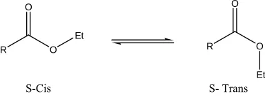
group has been employed for the construction of many 4-substituted bi-heterocyclic coumarins. Solid state con-formational studies on coumarin 4-acetic acid [9], ester [10,11] and dithionate [12] have been reported recently. It needs to be emphasized that the crystallographic study on esters is of considerable biological interest [13] since it determines the preferred orientation of the rotamer
(Figure 1), in turn the direction of the electron pair on
R O
O Et
R O
O
[image:1.595.76.268.639.709.2]Et S-Cis S- Trans
Figure 1. General structure for Rotamers of Esters.
alkyl oxygen. In view of the biological significance of 4-substituted coumarins and interest associated with the geometry of esters the title compound has been synthe-sized, charac terized and studied for its solid state con-formation by X-ray studies.
2. Experimental
2.1. Synthesis of the Title Compound
Compound 1 has been prepared by the reaction of
3,5-xylenol and citric acid by using sulphuric acid as condensing agent according to general procedure re-ported for coumarin 4-acetic acids [14]. Compound 1
was further converted into its ester (compound 2) by
re-fluxing in dry ethanol with catalytic quantity of sulphuric acid by Fischer esterification. The resulting solution was poured into crushed ice, the precipitate was filtered off, washed with 5% NaHCO3, and with water and
recrystal-lized from ethanol. (Figure 2) M.P. 123-125.C, yield
95%. The authenticity of the compound has been estab-lished by UV, IR, 1H, 13C NMR, and Mass spectra.
Crys-tals suitable for diffraction study were grown by a slow evaporation technique using 1:1 mixture of ethanol and dioxan at room temperature.
2.2. Characterization and Physical Measurements
The melting point was determined in open capillaries and is uncorrected. The IR absorption spectrum was obtained
by KBr pellet using a FT IR spectrometer. UV-vis spec-trum was recorded in the range of 200 nm to 800 nm using a Lambda 35 Perkin-Elmer spectrophotometer. The
1H and 13C NMR spectra were determined on a Bruker
Avance 400 model at 400 and 200 MHz respectively. Solvents were dried by refluxing with the appropriate drying agents and distilled before use.
2.3. X-Ray Crystallography
For the crystal structure determination, the single-crystal of the compound C15H16O4 was used for data collection.
The structure was solved and refined using the Bruker SHELXTL Software Package, using the space group P 1 21/c 1, with Z = 4 for the formula unit C15H16O4. The
final anisotropic full-matrix least-squares refinement on F2 with 176 variables converged at R1 = 4.03%, for the observed data and w R2 = 12.44% for all data. The goodness-of-fit was 1.008. The largest peak in the final difference electron density synthesis was 0.148 e−/Ǻ and
the largest hole was −0.136 e−/Ǻ with an RMS deviation
of 0.029 e−/Ǻ. On the basis of the final model, the
calcu-lated density was 1.283 g/cm3 and F(000), 552 e−/Ǻ.
Molecular structure of the compound showing the atomic numbering scheme is shown in Figure 3. The
crystallo-graphic parameters of the compound were presented in
Table 1. Selected bond distances and bond angles are
listed in Table 2 and selected Hydrogen-bonding
pa-rameters listed in Table 3.
3. Results and Discussion
Title compound crystallizes in the monoclinic centro-symmetric space group P 1 21/c 1with Z = 4. The struc-
ture of the compound consists of ethyl ester group. Se-lected bond lengths and bond distances for the title com ding parameters are listed in Table 3.
3.1 Molecular Skeleton
Coumarin ring is planar and carbethoxy group attached to the C4-CH2 is out of plane by an angle of 11.26. The
methyl group oriented at an angle of 90˚ to the mean plane of the bicyclic system. The ester carbonyl is ori-ented perpendicular to the C3-C4 double bond. The
molecule adopts the S-cis arrangement across the al-kyl-oxygen bond as revealed in the ORTEP diagram
(Figure 3). The exocyclic Oxygen is involved in
inter-molecular hydrogen bonding (O4····H-C1) with the CH3
group of ester. A weak hydrogen bonding (O2····.H-C4)
is also observed between the C4-CH2 and the carbonyl
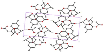
oxygen of ester. The molecular packing diagram (Figure 4) shows hydrogen bonding and the crystalline state is
stabilized by vander waals forces of attraction. The S-cis arrangement is also supported by the dihedral angle of 1.8 between C2-O1-C3-O2 indicating an eclipsed
ar-rangement (Table 3).
3.2. Bond Lengths
Bond lengths have been discussed in Table 2. The
aro-matic C-C bond lengths are in the range of 1.30 - 1.40 Å.
The ester C13-O4 bond shows a bond length of 1.20Å
which is similar to the lactone distance of 1.19Åbetween
O2-C3 .The alkyl oxygen O1-C3 bond is 1.32 Åwhereas
the corresponding bond of O3-C13 bond is slightly longer
by 1.35 Å.
H3C OH
CH3
COOH COOH
COOH HO
O O
H3C CH3
H2SO4 EtOH / H+
Compound 1 Compound 2
OH O
O O
H3C
CH3 O
O
CH2CH3
Figure 2. Chemical structure and synthetic pathway of the title compound.
H3C O O
CH3 O
O H3C H
H
H
H
Figure 4. Packing diagram dotted lines indicate C-H····O hydrogen bonds of the molecule viewed down a-axis.
Table 1. Crystallographic data and structure refinement parameters.
Empirical formula C15H16O4 Formula weight 260.28 g/mol
Crystal colour Temperature
Wavelength
Colourless 296 K 0.71073 Å Crystal system Monoclinic
Space group P21/c
Unit cell dimensions
a, b, c (Å) α, β, (˚)
8.6248 (4), 18.9103 (8), 8.4204 (4) 90, 101.241 (2), 90 Volume 1347.00 (11) (Å3)
Z, Calculated
density 4 Mg m−3 Absorption
coefficient 0.09 mm−1
F(000) 552 Crystal size 0.3 × 0.25 × 0.2 mm
Theta range for data
collection 2.41 to 26.070
Limiting indices −10 ≤ h ≤ 10, −23 ≤ k ≤ 22,10 ≤ l ≤ 10 Refinement method Full-matrix least-squares on F2
Absorption
correction Multi-scan
No. of measured, independent12192, 2675, 2113 and observed [I > 2(I)] reflections Rint0.021
(sin /)max (Å−1)max (Å−1) 0.618 Data/restraints/para
meters 2675/0/176 Goodness-of-
fit on F2 1.008 H-atom treatment H atoms constrained
∆ρmax, ∆ρmin (e Å−3) 0.15, −0.14 Final R indices R1 = 0.0403, wR2 = 0.1138 R indices(all data) R1 = 0.0533, wR2 = 0.1244
Large diff. peak
and hole 0.148 and −0.136e Å−3
3.3. Bond Angles
Bond angles have been discussed in Table 2. All the
aromatic C-C-C bond angles are in the expected range
Table 2. Selected bond lengths and bond angles (Å, ˚).
O1-C2 1.458 (2) C5-C14 1.346 (2) O1-C3 1.3240 (17) C6-C7 1.421 (2) O2-C3 1.1949 (18) C6-C12 1.402 (2) O3-C12 1.3777 (19) C7-C8 1.384 (2) O3-C13 1.359 (2) C7-C15 1.507 (2) O4-C13 1.207 (2) C8-C9 1.385 (2) C1-C2 1.482 (3) C9-C10 1.511 (3) C3-C4 1.504 (2) C9-C11 1.363 (3) C4-C5 1.504 (2) C11-C12 1.380 (2) C5-C6 1.4586 (19) C13-C14 1.437 (2) C2-O1-C3 117.29 (11) C6-C7-C15 124.66 (14) C12-O3-C13 122.35 (13) C8-C7-C15 116.36 (15) O1-C2-C1 110.46 (16) C7-C8-C9 123.46 (16) O1-C3-O2 123.71 (13) C8-C9-C10 120.61 (19) O1-C3-C4 110.87 (11) C8-C9-C11 117.94 (15) O2-C3-C4 125.41 (13) C10-C9-C11 121.45 (18) C3-C4-C5 112.83 (11) C9-C11-C12 120.23 (16) C4-C5-C6 124.09 (12) O3-C12-C6 122.36 (13) C4-C5-C14 116.56 (13) O3-C12-C11 114.29 (14) C6-C5-C14 119.33 (13) C6-C12-C11 123.34 (14) C5-C6-C7 127.90 (12) O3-C13-O4 117.41 (16) C5-C6-C12 116.07 (12) O3-C13-C14 116.33 (14) C7-C6-C12 116.02 (12) O4-C13-C14 126.25 (17) C6-C7-C8 118.97 (14) C5-C14-C13 123.52 (14) C3-O1-C2-C1 −86.04 (18) C5-C6-C7-C15 4.1 (2) C2-O1-C3-O2 −1.8 (2) C12-C6-C7-C8 1.8 (2) C2-O1-C3-C4 179.39 (12) C12-C6-C7-C15 −176.54 (15) C13-O3-C12-C6 −1.2 (2) C5-C6-C12-O3 0.02 (19) C13-O3-C12-C11 −180.00 (14) C5-C6-C12-C11 178.72 (14)
C12-O3-C13-O4 178.99 (15) C7-C6-C12-O3 −179.43 (13) C12-O3-C13-C14 0.5 (2) C7-C6-C12-C11 −0.7 (2)
O1-C3-C4-C5 −166.99 (12) C6-C7-C8-C9 −1.8 (2) O2-C3-C4-C5 14.3 (2) C15-C7-C8-C9 176.68 (16) C3-C4-C5-C6 76.53 (17) C7-C8-C9-C10 −179.68 (17) C3-C4-C5-C14 −101.80 (15) C7-C8-C9-C11 0.6 (2)
C4-C5-C6-C7 2.9 (2) C8-C9-C11-C12 0.6 (2) C4-C5-C6-C12 −176.50 (13) C10-C9-C11-C12 −179.14 (17) C14-C5-C6-C7 −178.84 (14) C9-C11-C12-O3 178.28 (14) C14-C5-C6-C12 1.79 (19) C9-C11-C12-C6 −0.5 (2) C4-C5-C14-C13 175.85 (14) O3-C13-C14-C5 1.4 (2) C6-C5-C14-C13 −2.6 (2) O4-C13-C14-C5 −176.91 (18)
C5-C6-C7-C8 −177.55 (14)
[image:3.595.308.533.101.735.2] [image:3.595.67.280.245.687.2]Table 3. Selected hydrogen-bond parameters.
D-H···A D-H (Å) H···A (Å) D···A (Å) D-H···A (˚)
C1-H1B···O4i 0.9600 2.5100 3.448 (3) 164.00
C4-H4B···O2ii 0.9700 2.4500 3.3963 (19) 166.00
Symmetry code(s): (i) −x + 1, y − 1/2, −z + 1/2; (ii) x, −y + 1/2, z − 1/2
carbon. The angles O2-C3-O1 and O2-C3-C4 are 123.7˚
and 125.4˚ whereas the O1-C3-C4 angle is significantly
reduced to 110.8˚.The values supporting the fractional (sp) hybridized orbitals are involved in bonding.
3.4. Spectral Studies
UV—Visible spectrum—The UV spectrum of the title
compound 2 in DMSO shows a strong band at 297 nm whereas fluorescence band appears at 387 nm which shows emission over a long range.
FT IR spectrum—The FT IR spectrum of the title
compound 2 shows the strong characteristic absorption band at 1725 cm−1 region due to lactone carbonyl
stretching frequency and band at 1615 cm−1 observed due
to C = C stretching.
1
H NMR spectrum—The structure of the title
com-pound 2 was further characterized by NMR absorption. Important structural information can be obtained from its NMR Spectra. 1H NMR(400 MHz, DMSO-d
6) δ ppm:
2.34 (s, 3H, C5-CH3 ), 2.54 (s, 3H, C7-CH3 ), 1.15 (t, 3H,
CH3-of ester, J = 7.2Hz), 4.15 (q, 4H, C4-CH2 and CH2
of ester J = 7.2Hz) 6.39 (s, 1H, C3-H), 7.0 (s, 1H, C6-H),
7.1 (s, 1H, C8-H) 13
C NMR spectrum: 13C NMR spectrum (100 MHz
DMSO-d6 ) (δ ppm) 14-CH3, 20 C7-CH3, 22 C5-CH3, 40
C4-CH2, 61 O-CH2, 115, 118, 130, 136, 142, 150, 155
these shifts correspond to aromatic carbons. Low inten-sity signals at 159, 170 are for carbonyl carbons of cou-marin and ester respectively.
3.5. Crystal Structure
A specimen of C15H16O4 was used for the X-ray
crystal-lographic analysis. The X-ray intensity data were meas-ured. The total exposure time was 2.50 hours. The frames were integrated with the Bruker SAINT software pack-age using a narrow-frame algorithm. The integration of the data using a monoclinic unit cell yielded a total of 12192 reflections to a maximum θ angle of 26.07˚ (0.81 Å resolution), of which 2675 were independent (average redundancy 4.558, completeness = 99.9%, Rint = 2.13%,
Rsig = 1.71%) and 2113 (78.99%) were greater than
2σ(F2). The final cell constants of a = 8.6248 (4) Å, b =
18.9103 (8) Å, c = 8.4204 (4) Å, β = 101.241 (2)˚, vol-ume = 1347.00 (11) Å3, are based upon the refinement of
the XYZ-centroids of 3786 reflections above 20 σ(I) with
5.382˚ < 2θ < 51.85˚. Data were corrected for absorption effects using the multi-scan method (SADABS). The ratio of minimum to maximum apparent transmission was 0.920. The structure was solved and refined using the Bruker SHELXTL Software Package, using the space group P 1 21/c 1, with Z = 4 for the formula unit, C15H16O4. The final anisotropic full-matrix least-squares
refinement on F2 with 176 variables converged at R1 =
4.03%, for the observed data and wR2 = 12.44% for all data. The goodness-of-fit was 1.008. The largest peak in the final difference electron density synthesis was 0.148 e−/Å3 and the largest hole was −0.136 e−/Å3 with an RMS
deviation of 0.029 e−/Å3. On the basis of the final model,
the calculated density was 1.283 g/cm3 and F(000), 552 e−.
4. Conclusion
It can be concluded that the newly synthesized 5, 7-dime- thyl coumarin-4-acetic ethyl ester exists mainly in the S-cis arrangement in the solid state. The packing diagram revealed the existence of only S-cis isomer which is sta-bilized by C-H····O bonds.
5. Acknowledgements
Fellowship from SC-ST cell Karnatak University, Dhar-wad is gratefully acknowledged.
REFERENCES
[1] M. V. Kulkarni, G. M. Kulkarni, C. H. Lin and C. M. Sun, “Coumarins and 1-Azacoumarins as Biodynamic Agents,” Current Medicinal Chemistry,Vol. 13, No. 23, 2006, pp. 2795-2818. doi:10.2174/092986706778521968
[2] S. Roseman, C. F. Heubuer, R. Pankratz and K. P. Link, “Studies on 4-Hydroxy Coumarins. XVI. The Metabolism of 4-Hydroxy Coumarins in the Dog,” Journal of the Ame- rican Chemical Society, Vol. 76, No. 6, 1954, pp. 1650- 1652. doi:10.1021/ja01635a055
[3] P. Laurin, D. Ferroud, L. Schiol, M. Klich, C. Dupuis- Hamelin, P. Mauvais, P. Lassaigne, A. Bonnefoy and B. Musicki, “Structure-Activity Relationship in Two Series of Aminoalkyl Substituted Coumarin Inhibitors of Gyrase B,” Bioorganic and Medicinal Chemistry Letters, Vol. 9, No. 19, 1999, pp. 2875-2880.
[4] M. V. Kulkarni and V. D. Patil, “Studies on Coumarins (Part 1),” Archiv der Pharmazie, Vol. 314, No. 8, 1981, pp. 708-711. doi:10.1002/ardp.19813140810
thesis and Biological Activity of Some 4-(Sulphonamido- Methyl)-Coumarins,” Indian Journal of Chemistry, Vol. 24B, No. 4, 1985, pp. 459-461.
[6] M. Ghate and M. V. Kulkarni, “Synthesis of Vanillin Ethers from 4-Bromomethyl Coumarins as Anti-Inflammatory Agents,” European Journal of Medicinal Chemistry, Vol. 38, No. 3, 2003, pp. 297-302.
doi:10.1016/S0223-5234(03)00016-3
[7] M. Basanagouda, V. B. Jadhav, M. V. Kulkarni and R. Nagendra Rao, “Computer Aided Prediction of Biological Activity Spectra: Study of Correlation between Predicted and Observed Activities for Coumarin-4-Acetic Acids,” Indian Journal of Pharmaceutical Sciences, Vol. 73, No. 1, 2011, pp. 88-92.
[8] V. B. Jadhav, S. K. Nayak, T. N. Row and M. V. Kul- karni, “Synthesis, Structure and DNA Cleavage Studies of Coumarin Analogues of Tetrahydroisoquinoline and Protoberberine Alkaloids,” European Journal of Medici- nal Chemistry, Vol. 45, No. 9, 2010, pp. 3575-3580. [9] K. Sivakumar, F. Jesurethinam, K. Subramanian and S.
Natarajan, “7-Hydroxy-4-Coumarin Acetic Acid Mono- hydrate,” Acta Crystallographica Section C,Vol.46, 1990, pp. 1663-1665. doi:10.1107/S0108270189011522
[10] S. Naveen, S. M. Anandwalkar, et al., “7,8-Dimethyl Cou- marin 4-Acetic Acid Ethyl Esters,” Analytical Sciences, Vol. 22, 2006, pp. X173-X174.
[11] I. Fujii, Y. Mano and N. Hirayama, “Crystal of 4-Ethoxy- coumarin” Analytical Science, Vol. 21, 2005, pp. x7-x8 [12] N. M. Mahabaleshwaraiah, K. M. Kumar, O. Kotresh, W.
F. A. Al-Eryani and H. C. Devarajgowda, “(6-Methoxy-2- Oxo-2H-Chromen-4-yl)Methylpyrrolidine-1-Carbodithiate,” Acta Crystallographica, Vol. E68, No. 5, 2012, p. 1566. [13] P. Deslongchamps, U. O. Cheriyan, J. P. Pradere, P. Soucy
and R. J. Taillefer, “Hydrolysis and Isomerization of syn Unsymmetrical N,N-Dialkylated Immidate Salts. Ex-perimental Evidence for Conformational Changes and for Stereo Electronically Controlled Cleaves in Hemi-Or- thoamide Tetrahedral Intermediates,” Nouveau Journal de Chimie, Vol. 3, No. 2, 1979, pp. 343-350.