0095-1137/11/$12.00 doi:10.1128/JCM.02243-10
Copyright © 2011, American Society for Microbiology. All Rights Reserved.
Evaluation of the Rapid MGIT TBc Identification Test for Culture
Confirmation of
Mycobacterium tuberculosis
Complex
Strain Detection
䌤
Ming-Chih Yu,
1† Huang-Yao Chen,
1† Mei-Hua Wu,
2Wei-Lun Huang,
2Yuh-Min Kuo,
2Fang-Lan Yu,
3and Ruwen Jou
3*
Division of Pulmonary Medicine, Department of Internal Medicine, Wan Fang Hospital, Taipei Medical University, 111 Hsin-Long Road, Section 3, Taipei, Taiwan1; Reference Laboratory of Mycobacteriology, Research and Diagnostic Center, Centers for
Disease Control, 161 Kun-Yang Street, Nan-Kang, Taipei, 115, Taiwan2; and Department of Laboratory Medicine, Taipei Medical University-Wan Fang Hospital, 111 Hsin-Long Road,
Section 3, Taipei, Taiwan3
Received 8 November 2010/Returned for modification 7 December 2010/Accepted 20 December 2010
A culture confirmation test for the detection of Mycobacterium tuberculosis complex strains that uses a
lateral-flow immunochromatographic assay to detect the MPB64 antigen, the MGIT TBc identification (TBc
ID) test, has been developed. We evaluated the performance of the TBc ID test in the detection of the M.
tuberculosiscomplex in 222 primary-positive liquid cultures. We compared these results to those of nucleic acid-based identification and conventional biochemical tests. The validity of the TBc ID test was determined,
and all of the nontuberculous mycobacteria (NTM) andNocardiaspecies tested were found to be negative. The
detection limit of the TBc ID test was 5ⴛ105
CFU/ml, and for IS6110real-time PCR it was 5 CFU/ml. All of
the M. tuberculosis andM. africanumcultures were found to be positive, while M. bovis and M. bovisBCG cultures were negative. With the exception of 1 contaminated culture, the 221 culture-positive isolates
con-tained 171 (77.5%)M. tuberculosisisolates, 39 (17.6%) NTM species, and 11 (5.0%) unidentified species. Two
culture-positive isolates harbored a 63-bp deletion at position 196 of thempb64gene. The sensitivity, specificity,
positive predictive values, and negative predictive values of the TBc ID test were 98.8, 100, 100, and 95.1%, respectively. Furthermore, the approximate turnaround time for real-time PCR was 4 h (including buffer and sample preparation), while for the TBc ID test it was less than 1 h. We suggest an algorithm for the primary
identification of M. tuberculosis in liquid culture using the TBc ID test as an alternative to conventional
subculture followed by identification using biochemical methods.
In 2007, the World Health Organization (WHO) adopted a policy that recommended the use of liquid culture methods for culture and drug susceptibility tests as a standard for tubercu-losis (TB) diagnosis and case management (28). The Taiwan Centers for Disease Control (CDC) recommended that liquid and solid media be used simultaneously for mycobacterial cul-ture (7), and approximately 90% of clinical mycobacteriology laboratories in Taiwan use a liquid culture system for the
isolation of theMycobacterium tuberculosiscomplex from
clin-ical specimens. Acid-fast bacillin (AFB) smear tests then are performed on positive cultures to dismiss contamination (4,
20). The turnaround time (TAT) for the recovery of the M.
tuberculosiscomplex thus is reduced to 10 to 14 days (8, 18). Although the recovery of mycobacteria can be accelerated by using liquid culture systems, this practice provides only partial benefits if it is not accompanied by a rapid species
identifica-tion test (16). DifferentiatingM. tuberculosis from
nontuber-culous mycobacteria (NTM) as soon as possible is important,
particularly in situations in which NTM strains represent a considerable share of the clinical isolates.
The identification ofM. tuberculosisis time-consuming using
conventional biochemical methods. The subculturing of iso-lated mycobacteria from liquid cultures onto solid media and their subsequent identification using conventional biochemical methods requires an additional 3 to 5 weeks (25). In addition, ambiguous biochemical reactions can confuse the test results.
Thus, the identification of M. tuberculosisusing biochemical
methods is a complex, labor-intensive, and time-consuming process. Nucleic acid amplification (NAA) methods, such as real-time PCR, are both rapid and specific but are technically challenging, and they require the use of sophisticated instru-ments. For this reason, the WHO also recommends the use of rapid and affordable methods for the identification to the
spe-cies level of theM. tuberculosiscomplex and NTM organisms
(http://www.who.int/tb/dots/laboratory/policy/en/index.html). The Taiwan CDC conducted a TB laboratory diagnosis sur-vey in 2009. Their results showed that there are 36 clinical mycobacteriology laboratories in the country that perform four conventional bacteriological tests (smear, culture, identifica-tion, and drug susceptibility testing) for TB diagnosis. Of these 36 laboratories, 19 (52.8%) use biochemical tests, 9 (25%) use NAA, and 8 (22.2%) use both methods for the culture
identi-fication ofM. tuberculosiscomplex strains. Among these same
laboratories, 13 use commercial molecular diagnosis assays,
* Corresponding author. Mailing address: Reference Laboratory of Mycobacteriology, Research and Diagnostic Center, Centers for Dis-ease Control, Department of Health, 161 Kun-Yang Street, Nan-Kang, Taipei, 115, Taiwan. E-mail: [email protected].
† Ming-Chih Yu and Huang-Yao Chen contributed equally to this study.
䌤Published ahead of print on 29 December 2010.
802
on May 16, 2020 by guest
http://jcm.asm.org/
and 10 perform in-house PCR using IS6110 (11) or other probes. Therefore, to strengthen TB laboratory services and facilitate the timely management of TB cases, the implemen-tation of a simple and reliable test is needed in Taiwan.
In August 2009, BD Diagnostics (a division of Becton, Dick-son, and Company) launched the BD MGIT TBc identification
(TBc ID) test for the identification of theM. tuberculosis
com-plex from liquid culture in Africa and the European Union. The TBc ID test is a lateral-flow immunochromatographic assay based on the detection of MPB64 in liquid cultures using an MPT64-specific monoclonal antibody. MPB64 is a
myco-bacterial protein that is secreted byM. tuberculosisand certain
strains ofM. bovis(1, 22, 29). However, some substrains ofM.
bovisBCG in theM. tuberculosiscomplex produce no MPT64 antigen (19). This qualitative test is rapid (readable in 15 min), easy to use, and requires no processing or additional instru-mentation. No clinical trial of the TBc ID test was ever re-ported. In this study, we evaluated the performance of the TBc
ID, NAAs (such as IS6110real-time PCR), and biochemical
tests for the identification of culture-positive mycobacteria in liquid media.
MATERIALS AND METHODS
Reference strains, clinical specimens, and mycobacterial culture.The
valida-tion of the TBc ID test was conducted using 24 NTM strains, 18 mixtures ofM.
tuberculosisand NTM strains, 2M. bovisstrains, 1M. africanumstrain, and 1
Nocardiastrain. For prospective analysis, clinical respiratory specimens were
digested and decontaminated usingN-acetyl-L-cysteine (Sigma Chemical
Com-pany, St. Louis, MO) and 2% sodium hydroxide (NaLC-NaOH). The detection limits of both the TBc ID test and real-time PCR were evaluated using serial
dilutions of anM. tuberculosisstock (5⫻107
CFU/ml). We performed 10-fold
serial dilutions from stocked H37Rv DNA (5⫻107CFU/ml) to 0.05 CFU/ml as
the lowest concentration, and we separately evaluated the limit of each assay. The procedures were repeated twice. The processed specimens were concen-trated using centrifugation, inoculated in Bactec MGIT 960 culture tubes, and incubated in the BD Bactec MGIT system (Becton Dickinson Microbiology Systems, Cockeysville, MD) (10). Once a positive signal was detected using the system, a smear microscopy test was performed to screen the deposit in the culture tubes for AFB using an auramine fluorescent stain. The results were confirmed after staining using the Ziehl-Neelsen method, and samples were
classified as either scanty, 1⫹, 2⫹, 3⫹, or 4⫹(3). From January to February
2010, consecutive culture-positive MGIT samples recovered from 3,214 clinical specimens were included in this study.
Assays for culture-positive MGIT media.Both TBc ID and conventional biochemical tests were performed in the TB Laboratory at the Taipei Medical University-Wan Fang Hospital, one of the contracted mycobacteriology labora-tories of the Taiwan CDC. NAA tests and sequencing were performed in the Reference Laboratory of Mycobacteriology at the Taiwan CDC.
(i) Conventional biochemical tests.Bactec cultures that were AFB positive
were subcultured onto solid Lo¨wenstein-Jensen (L-J) slants and incubated at
37°C to obtain colonies for identification. Conventional biochemical tests, in-cluding niacin production, nitrate reduction, 3-day acrylsulfatase, 3-day Tween 80 hydrolysis, urease, semiquantitative catalase, and tolerance to 5% NaCl, were
performed to identify theM. tuberculosiscomplex (6).
(ii) NAA tests.For the identification ofM. tuberculosiscomplex strains in AFB-positive Bactec cultures (2 to 3 days after a positive signal was detected),
IS6110real-time PCR was performed according to the method of Cleary et al.
(9). The primers used for this assay were IS6 (5⬘-GGCTGTGGGTAGCAGAC
C-3⬘) and IS7 (5⬘-CGGGTCCAGATGGCTTGC-3⬘), as well as an internal
oligonucleotide probe (5⬘-[6-carboxyfluorescein]-TGTCGACCTGGGCAGGG
TTCG-[6-carboxytetramethylrhodamine]-3⬘).
(iii) TBc ID.TBc ID devices were inoculated with 0.1 ml of Bactec cultures within 3 days of the detection of AFB-positive growth using an MGIT 960 instrument. Cultures were used directly for the TBc ID assay according to the manufacturer’s recommendations (5). All inoculated devices were incubated for 15 min at room temperature before the results were visually assessed for positive detection (i.e., a visible test line) and reagent function (i.e., a visible control line).
(iv) PCR-RFLP.To identify mycobacteria at the species level, we performed PCR-restriction fragment length polymorphism (PCR-RFLP) analysis of a 65-kDa protein according to the method described by Telenti et al. (26). We compared the obtained patterns to those in the database at http://app.chuv .ch/prasite/index.html (Prasite; Centre Hospitalier Universitaire Vaudois Lausanne).
Analysis of discrepant results.Conventional confirmation tests were consid-ered the gold standard for performance calculations. When discrepant results between the NAA, TBc ID assays, and conventional biochemical confirmation
tests were observed, the results frommpb64gene sequencing were used to
resolve the discrepancies. Mutations in thempb64gene were analyzed using PCR
amplification, and the resulting product was sequenced using the following oligo-nucleotide primers: AMS-50-F (5⬘-TCGATCTGCTAGCTTGAGTCTGGT-3⬘)
and AMS-51-R (5⬘-ACCACCGCACCAAGGCTGCTGTCTA-3⬘) (23). The
PCR products were sequenced using an ABI 3730 automated sequencer (Ap-plied Biosystems, Life Technologies Corporation, CA) under standardized con-ditions. The data were analyzed using Sequencing Analysis 5.2.0 software (Ap-plied Biosystems, Life Technologies Corporation, CA).
Performance analysis.Using the results of the conventional biochemical iden-tification methods as the gold standard, the performance of the TBc ID and
IS6110real-time PCR tests was analyzed. To assess the performance of these
identification tests, the sensitivity, specificity, positive predictive value (PPV), and negative predictive value (NPV) were calculated after discrepant analysis (2). We also evaluated the TAT of the tests based on the protocols suggested by the manufacturers and the Taiwan CDC.
RESULTS
Validity of the TBc ID test.The detection limits of the TBc
ID and IS6110real-time PCR tests were 5⫻105and 5 CFU/
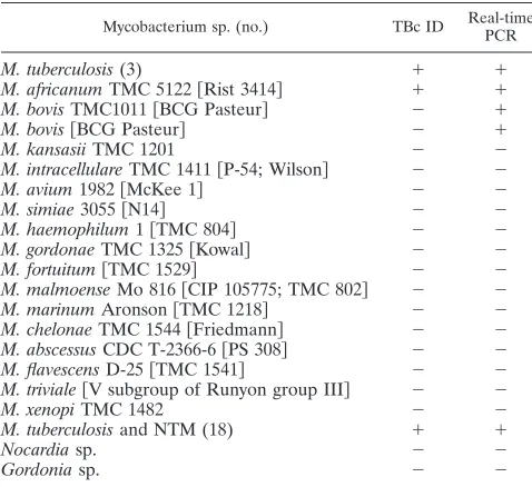
ml, respectively. The results of the identification of reference mycobacterial strains are summarized in Table 1. The analyt-ical specificity of the TBc ID test was assessed using reference
strains. Using the TBc ID test, 3M. tuberculosisstrains, 1 M.
africanumstrain, and 18 mixtures ofM. tuberculosisand NTM strains were identified as positive, while 18 NTM strains, 1
Nocardiastrain, 1 Gordonia sp., 1 M. bovis strain, and 1 M. bovisBCG strain were identified as negative.
Mycobacterial species identification. We obtained 221
(6.9%) AFB smear-positive Bactec cultures out of 3,214
dif-TABLE 1. Validation of the BD MGIT TBc identification test using reference mycobacterial strains
Mycobacterium sp. (no.) TBc ID Real-time
PCR
M. tuberculosis(3) ⫹ ⫹
M. africanumTMC 5122关Rist 3414兴 ⫹ ⫹
M. bovisTMC1011关BCG Pasteur兴 ⫺ ⫹
M. bovis关BCG Pasteur兴 ⫺ ⫹
M. kansasiiTMC 1201 ⫺ ⫺
M. intracellulareTMC 1411关P-54; Wilson兴 ⫺ ⫺
M. avium1982关McKee 1兴 ⫺ ⫺
M. simiae3055关N14兴 ⫺ ⫺
M. haemophilum1关TMC 804兴 ⫺ ⫺
M. gordonaeTMC 1325关Kowal兴 ⫺ ⫺
M. fortuitum关TMC 1529兴 ⫺ ⫺
M. malmoenseMo 816关CIP 105775; TMC 802兴 ⫺ ⫺
M. marinumAronson关TMC 1218兴 ⫺ ⫺
M. chelonaeTMC 1544关Friedmann兴 ⫺ ⫺
M. abscessusCDC T-2366-6关PS 308兴 ⫺ ⫺
M. flavescensD-25关TMC 1541兴 ⫺ ⫺
M. triviale关V subgroup of Runyon group III兴 ⫺ ⫺
M. xenopiTMC 1482 ⫺ ⫺
M. tuberculosisand NTM (18) ⫹ ⫹
Nocardiasp. ⫺ ⫺
Gordoniasp. ⫺ ⫺
on May 16, 2020 by guest
http://jcm.asm.org/
[image:2.585.300.539.90.309.2]ferent clinical specimens. However, 11 cultures (5.0%) yielded no colonies after being subcultured onto L-J slants; thus, no identification results were obtained for these cultures. We identified 210 L-J culture-positive samples using standard
bio-chemical tests: 171 (81.4%) were culture positive forM.
tuber-culosis, and 39 (18.6%) were culture positive for NTM. The most prominent NTM species identified using PCR-RFLP wereM. abscessus(48.7%),M. kansasii(15.4%),M. gordonae
(10.3%), andM. intracellulare(7.7%) (Table 2). None of the
210 culture-positive samples collected from patients were
iden-tified as containing mixed cultures ofM. tuberculosisand NTM.
TBc ID and NAA tests.The results of the NAA and TBc ID
tests were compared to the culture results, and the results are summarized in Table 2. The NAA test performed as well as gold-standard biochemical methods. In contrast, the TBc ID test successfully yielded 39 true-negative results for 39 NTM
cultures and 2 false-negative results for M. tuberculosis
cul-tures. The gene sequencing of the two false-negative samples
revealed a 63-bp deletion at position 196 in thempb64gene in
both. In addition, of the 11 AFB smear-positive Bactec cul-tures with no L-J growth, the results of all of the TBc ID tests were negative, while one true-positive NAA test result was observed. This false-negative TBc ID result also was seen in tests of two other specimens from the same patient that yielded
culture-positiveM. tuberculosis.Of the 39 samples that were
identified as negative by the TBc ID and NAA tests, each tested positive for NTM (Table 2).
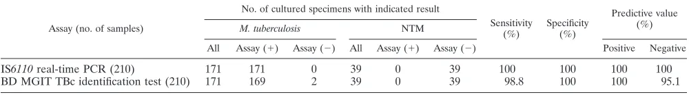
Correlation between tests. Compared to conventional
bio-chemical methods, the sensitivity of the TBc ID test was 98.8%, the specificity was 100%, the positive predictive value was 100%, and the negative predictive value was 95.1% (Table 3). In contrast, the same four performance indexes all were 100%
for the NAA method. For the primary identification of M.
tuberculosisfrom liquid culture, the TAT was 1 h using the TBc ID test and 1 to 3 days using the NAA test; in contrast, the average TAT for conventional biochemical tests was 24 days (range, 7 to 49 days).
DISCUSSION
In mycobacterial laboratories in Taiwan, the average
recov-ery rate of theM. tuberculosiscomplex in all clinical specimens
has decreased from approximately 70% in 2000 to 50% in 2009 (unpublished data). Therefore, a fast and cost-effective
method that allows for the qualitative detection of the M.
tuberculosis complex from acid-fast bacillus (AFB)-positive cultures is necessary. We conducted and reported the first clinical trial of the TBc ID test. We evaluated the use of a simple MPB64-based lateral-flow immunochromatographic as-say, the TBc ID test, to enhance TAT and facilitate the rapid
identification of theM. tuberculosiscomplex directly from
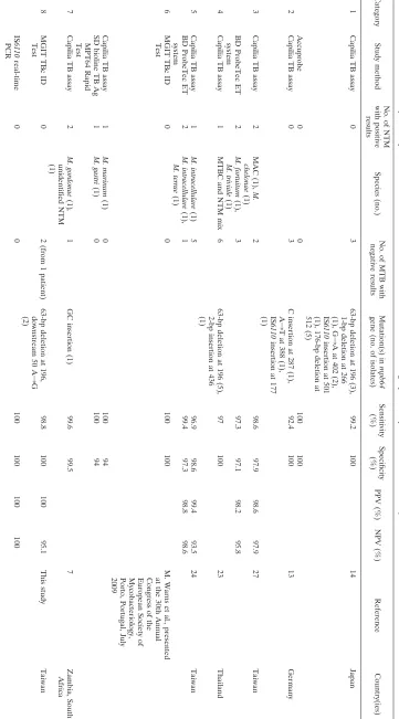
liq-uid culture. The lateral-flow immunochromatographic assay is useful because of its specificity and ease of use. Thus, several such tests are commercially available, including the Capilia TB assay (Tauns Laboratories, Inc., Numazu, Japan), the Tibilia rapid test (Hangzhou, China), the SD Bioline TB Ag MPT64 rapid test (Standard Diagnostics, South Korea), and the MGIT TBc ID test. Previous studies have demonstrated that these assays all had outstanding performance, exhibiting 92.4 to 100% sensitivity and 94 to 100% specificity in identifying the
M. tuberculosiscomplex (Table 4) (13, 14, 21, 23, 24, 27, and M. Warns et al., presented at the 30th Annual Congress of the European Society of Mycobacteriology, Porto, Portugal, July 2009). In this study, we found that the TBc ID test exhibited 98.8% sensitivity and 100% specificity compared to biochem-ical tests. However, to improve the accuracy of the identifica-tions and confirm negative TBc ID test results, a secondary NAA test must be employed after reculturing MGIT-positive cultures onto solid medium.
The specificity of the TBc ID test was shown to be 100% in
two studies. However, cross-reactivity withM. aviumcomplex
(MAC), M. chelonae, M. intracellulare, M. marinum, and M.
gordonaewas observed with the Capilia test, and
cross-reactiv-ity was observed withM. gastriwith the Bioline device (Table
[image:3.585.41.283.91.285.2]4). The sensitivity of the TBc ID test ranged from 98.8 to 100%, while the sensitivity of the Capilia test ranged from 92.4 to 99.6% in similar studies. In our study, the TBc ID test
TABLE 2. Comparison of the IS6110real-time PCR and BD MGIT TBc identification tests to culture confirmation tests
Mycobacterial species identified using biochemical
methods andhsp65
PCR-RFLPa
(no. of specimens)
IS6110real-time
PCR (no. of
specimens⫽210)
BD MGIT TBc identification
test (no. of
specimens⫽210)
Positive Negative Positive Negative
M. tuberculosis(171) 171 0 169 2
NTM (39) 0 39 0 39
M. abscessus(19) 19 19
M. kansasii(6) 6 6
M. gordonae(4) 4 4
M. intracellulare(3) 3 3
M. arupense(1) 1 1
M. avium(1) 1 1
M. chelonae(1) 1 1
M. fallax(1) 1 1
M. fortuitum(1) 1 1
M. holsaticum(1) 1 1
M. nonchromogenicum(1) 1 1
aA total of 222 specimens were primary positive in liquid culture, 11 were
culture negative on L-J medium, and 1 was contaminated.
TABLE 3. Correlation between culture confirmation assays and culture results
Assay (no. of samples)
No. of cultured specimens with indicated result
Sensitivity (%)
Specificity (%)
Predictive value (%)
M. tuberculosis NTM
All Assay (⫹) Assay (⫺) All Assay (⫹) Assay (⫺) Positive Negative
IS6110real-time PCR (210) 171 171 0 39 0 39 100 100 100 100
BD MGIT TBc identification test (210) 171 169 2 39 0 39 98.8 100 100 95.1
on May 16, 2020 by guest
http://jcm.asm.org/
[image:3.585.42.542.658.727.2]TABLE 4. Summary of the performance of MPB64-based lateral-flow immunochromatographic assays in the detection of Mycobacterium tuberculosis in dif ferent studies Category Study method No. of NTM with positive results Species (no.) No. of MTB with negative results Mutation(s) in mpb64 gene (no. of isolates) Sensitivity (%) Specificity (%) PPV (%) NPV (%) Reference Country(ies) 1 Capilia TB assay 0 3 63-bp deletion at 196 (3), 1-bp deletion at 266 (1), G 3 A at 402 (2), IS 6110 insertion at 501 (1), 176-bp deletion at 512 (5) 99.2 100 14 Japan Accuprobe 0 0 100 100 2 Capilia TB assay 0 3 C insertion at 287 (1), A 3 T at 388 (1), IS 6110 insertion at 177 (1) 92.4 100 13 Germany 3 Capilia TB assay 2 MAC (1), M. chelonae (1) 2 98.6 97.9 98.6 97.9 27 Taiwan BD ProbeTec ET system 2 M. fortuitum (1), M. triviale (1) 3 97.3 97.1 98.2 95.8 4 Capilia TB assay 1 MTBC and NTM mix 6 63-bp deletion at 196 (5), 2-bp insertion at 436 (1) 97 100 23 Thailand 5 Capilia TB assay 1 M. intracellulare (1) 5 96.9 98.6 99.4 93.5 24 Taiwan BD ProbeTec ET system 2 M. intracellulare (1), M. terrae (1) 1 99.4 97.3 98.8 98.6 6 MGIT TBc ID Test 0 0 100 100 M. Warns et al., presented at the 30th Annual Congress of the European Society of Mycobacteriology, Porto, Portugal, July 2009 Capilia TB assay 1 M. marinum (1) 0 100 94 SD bioline TB Ag MPT64 Rapid Test 1 M. gastri (1) 0 100 94 7 Capilia TB assay 2 M. gordonae (1) , unidentified NTM (1) 1 GC insertion (1) 99.6 99.5 7 Zambia, South Africa 8 MGIT TBc ID Test 0 2 (from 1 patient) 63-bp deletion at 196, downstream 50 A 3 G (2) 98.8 100 100 95.1 This study Taiwan IS 6110 real-time PCR 0 0 100 100 100 100
on May 16, 2020 by guest
http://jcm.asm.org/
[image:4.585.108.469.74.725.2]yielded two false-negative results for twoM. tuberculosis com-plex cultures from the same patient, using biochemical tests as a gold standard. These two isolates were confirmed as true
positives using IS6110real-time PCR and were found to
con-tain a 63-bp deletion at position 196 of thempb64gene.
Mu-tations (13, 14), deletions (14, 23), and IS6110or CG insertions
(13, 14, 21, 23) in the coding region (22, 29) of thempb64gene
that result in false-negative test results have been reported previously.
A recent clinical evaluation performed in South Africa re-vealed that the TBc ID test exhibited excellent performance compared to that of the niacin test (21) (Table 4). Similar results were obtained using the BD ProbeTec ET system (BD Diagnostic Systems, Sparks, MD) in two studies performed in Taiwan (24, 27). However, both the Gen Probe AccuProbe and
IS6110real-time PCR tests outperformed the TBc ID, which
occasionally was cross-reactive with NTM strains or yielded
false negatives forM. tuberculosiscomplex species with
muta-tions in thempb64gene (15). Furthermore, the
immunochro-matographic assay was less labor-intensive than other identifi-cation methods. In this study, the TAT for identifiidentifi-cation was 24 days in the clinical setting using standard biochemical tests, and it was 1 day for the TBc ID test. Thus, to avoid false-negative results and shorten the TAT of routine clinical prac-tices, we suggest a new algorithm for the primary identification ofM. tuberculosisstrains from liquid culture that uses the TBc ID test as an alternative to the currently available biochemical methods (Fig. 1).
Taiwan is considered a country with moderate TB preva-lence, with an incidence rate of 62 per 100,000 in 2008 (http:
//www.cdc.gov.tw/public/Data/9123117221971.pdf). However,
an increased incidence of NTM infections, from 32.3% in 2000 to 49.8% in 2008, was reported in a recent hospital-based
survey; this rise in NTM infections was due primarily to M.
aviumand M. abscessus infections (17). Therefore, to avoid inappropriate antituberculous treatment decisions, the
accu-rate differential diagnosis ofM. tuberculosisis imperative. The
increase in the number of NTM isolations has been attributed to the implementation of liquid culture systems (12) and the use of improved identification/differentiation techniques. In this study, we proved that the TBc ID test could
unambigu-ously differentiateM. tuberculosisfrom NTM strains and
mix-tures of both in a clinical setting.
In conclusion, the use of liquid culture systems together with a rapid, simple identification test has the potential to increase the speed of diagnosis and, most importantly, aid in the iden-tification of drug-resistant TB cases. Our findings suggest that lateral-flow immunochromatographic assays for the detection of MPB64, such as the TBc ID test, are crucial for the rapid and accurate diagnosis of TB to facilitate early treatment and reduce the spread of infections.
ACKNOWLEDGMENTS
We thank colleagues at the Laboratory of Mycobacteriology of the Taipei Medical University-Wan Fang Hospital for their technical as-sistance and BD Taiwan for providing the 250 MGIT TBc identifica-tion test kits to the hospital.
REFERENCES
1.Abe, C., K. Hirano, and T. Tomiyama.1999. Simple and rapid identification
of theMycobacterium tuberculosiscomplex by immunochromatographic
as-say using anti-MPB64 monoclonal antibodies. J. Clin. Microbiol.37:3693–
3697.
2.Altman, D. G., and J. M. Bland.1994. Statistics notes: diagnostic tests 2:
predictive values. BMJ309:102.
3.American Thoracic Society.1981. Diagnostic standards and classification of
tuberculosis and other mycobacterial disease. Am. Rev. Respir. Dis.123:
343–358.
4.Attorri, S., S. Dunbar, and J. E. I. Clarridge, Jr. 2000. Assessment of
morphology for rapid presumptive identification ofMycobacterium
tubercu-losisandMycobacterium kansasii. J. Clin. Microbiol.38:1426–1429. 5.BD Diagnostic Systems.2009. BD MGIT™ TBc ID identification test
package insert, BD document L8085917(01). BD Diagnostic Systems, Sparks, MD.
6.Centers for Disease Control.1981. Public health mycobacteriology. A guide for the level II laboratory. Centers for Disease Control, Atlanta, GA. 7.Centers for Disease Control, Taipei, Taiwan.2004. Laboratory manual of
mycobacteria. Centers for Disease Control, Taipei, Taiwan.
8.Chien, H. P., M. C. Yu, M. H. Wu, T. P. Lin, and K. T. Luh.2000. Com-parison of the Bactec MGIT 960 with Lowenstein-Jensen medium for re-covery of mycobacteria from clinical specimens. Int. J. Tuberc. Lung Dis.
4:866–870.
9.Cleary, T., G. Roudel, O. Casillas, and N. Miller.2003. Rapid and specific
detection ofMycobacterium tuberculosisby using the smart cycle instrument
and a specific fluorogenic probe. J. Clin. Microbiol.41:4783–4786.
10.Hanna, B. A., et al.1999. Multicenter evaluation of the Bactec MGIT 960
system for recovery of mycobacteria. J. Clin. Microbiol.37:748–752.
11.Hermans, P. W., et al.1990. Insertion element IS986 fromMycobacterium tuberculosis: a useful tool for diagnosis and epidemiology of tuberculosis.
J. Clin. Microbiol.28:2051–2058.
12.Herna´ndez-Gardun˜o, E., M. Rodrigues, and R. K. Elwood.2009. The inci-dence of pulmonary non-tuberculous mycobacteria in British Columbia,
Canada. Int. J. Tuberc. Lung Dis.13:1086–1093.
13.Hillemann, D., S. Ru¨sch-Gerdes, and E. Richter.2005. Application of the
Capilia TB assay for culture confirmation of Mycobacterium tuberculosis
complex isolates. Int. J. Tuberc. Lung Dis.9:1409–1411.
14.Hirano, K., A. Aono, M. Takahashi, and C. Abe.2004. Mutations including
IS6110insertion in the gene encoding the MPB64 protein of Capilia
TB-negativeMycobacterium tuberculosisisolates. J. Clin. Microbiol.42:390–392.
15.Ismail, N. A., K. Baba, D. Pombo, and A. A. Hoosen. 2009. Use of an
immunochromatographic kit for the rapid detection ofMycobacterium
tuber-culosisfrom broth cultures. Int. J. Tuberc. Lung Dis.13:1045–1047. 16.Katila, M. L., P. Katila, and R. Erkinjuntti-Pekkanen.2000. Accelerated
detection and identification of mycobacteria with MGIT 960 and COBAS
AMPLICOR Systems. J. Clin. Microbiol.38:960–964.
17.Lai, C. C., et al.2010. Increasing incidence of nontuberculous mycobacteria,
Taiwan, 2000–2008. Emerg. Infect. Dis.16:294–296.
FIG. 1. Suggested algorithm for the culture and identification of
theMycobacterium tuberculosiscomplex. The asterisk indicates that for
pending samples, a nucleic acid amplification (NAA) test should be performed when the morphology and growth rate suggest the presence of theM. tuberculosiscomplex.
on May 16, 2020 by guest
http://jcm.asm.org/
18.Lee, J. J., et al.2003. Comparative evaluation of the Bactec MGIT 960 system with solid medium for isolation of mycobacteria. Int. J. Tuberc. Lung
Dis.7:569–574.
19.Li, H., et al.1993. Evidence for absence of the MPB64 gene in some
substrains ofMycobacterium bovisBCG. Infect. Immun.61:1730–1734.
20.McCarter, Y. S., I. N. Ratkiewicz, and A. Robinson.1998. Cord formation in Bactec medium is a reliable, rapid method for presumptive identification of
Mycobacterium tuberculosiscomplex. J. Clin. Microbiol.36:2769–2771. 21.Muyoyeta, M., et al.2010. Evaluation of the Capilia TB assay for culture
confirmation ofMycobacterium tuberculosisin Zambia and South Africa.
J. Clin. Microbiol.48:3773–3775.
22.Nagai, S., H. G. Wiker, M. Harboe, and M. Kinomoto.1991. Isolation and partial characterization of major protein antigens in the culture fluid of
Mycobacterium tuberculosis. Infect. Immun.59:372–382.
23.Ngamlert, K., et al.2009. Diagnostic performance and costs of Capilia TB forMycobacterium tuberculosiscomplex identification from broth-based
cul-ture in Bangkok, Thailand. Trop. Med. Int. Health14:748–753.
24.Shen, G. H., et al.2009. Combining the Capilia TB assay with smear
mor-phology for the identification ofMycobacterium tuberculosiscomplex. Int. J.
Tuberc. Lung Dis.13:371–376.
25.Sun, J. R., S. Y. Lee, C. L. Perng, and J. J. Lu.2009. Detecting Mycobacte-rium tuberculosisin Bactec MGIT 960 cultures by inhouse IS6110-based PCR
assay in routine clinical practice. J. Formos. Med. Assoc.108:119–125.
26.Telenti, A., et al.1993. Rapid identification of mycobacteria to the species level by polymerase chain reaction and restriction enzyme analysis. J. Clin.
Microbiol.31:175–178.
27.Wang, J. Y., et al.2007. Performance assessment of the Capilia TB assay and
the BD ProbeTec ET system for rapid culture confirmation of
Mycobacte-rium tuberculosis.Diagn. Microbiol. Infect. Dis.59:395–399.
28.World Health Organization.2007. Use of liquid TB culture and drug sus-ceptibility testing (DST) in low and medium income settings. Summary report of the expert group meeting on the use of liquid culture media. WHO, Geneva, Switzerland.
29.Yamaguchi, R., et al.1989. Cloning and characterization of the gene for
immunogenic protein MPB64 ofMycobacterium bovisBCG. Infect. Immun.
57:283–288.