Associated Enterococci from
Enterococcus hirae
to Biofilm-Forming
Enterococcus faecalis
and Adherent
Escherichia coli
Anuradha Ghosh,cLuke Borst,b,eStephen H. Stauffer,aMitsu Suyemoto,bPeter Moisan,dLudek Zurek,cJody L. Gookina,e
Departments of Clinical Sciencesa
and Population Health and Pathobiology,b
College of Veterinary Medicine, North Carolina State University, Raleigh, North Carolina, USA; Department of Diagnostic Medicine and Pathobiology, Kansas State University, Manhattan, Kansas, USAc
; North Carolina Department of Agriculture and Consumer Services Veterinary Diagnostic Laboratory System, Raleigh, North Carolina, USAd
; Center for Comparative Medicine and Translational Research, College of Veterinary Medicine, North Carolina State University, Raleigh, North Carolina, USAe
Approximately 15% of foster kittens die before 8 weeks of age, with most of these kittens demonstrating clinical signs or
post-mortem evidence of enteritis. While a specific cause of enteritis is not determined in most cases, these kittens are often
empiri-cally administered probiotics that contain enterococci. The enterococci are members of the commensal intestinal microbiota but
also can function as opportunistic pathogens. Given the complicated role of enterococci in health and disease, it would be
valu-able to better understand what constitutes a “healthy” enterococcal community in these kittens and how this microbiota is
im-pacted by severe illness. In this study, we characterized the ileum mucosa-associated enterococcal community of 50 apparently
healthy and 50 terminally ill foster kittens. In healthy kittens,
Enterococcus hirae
was the most common species of ileum
muco-sa-associated enterococci and was often observed to adhere extensively to the small intestinal epithelium. These
E. hirae
isolates
generally lacked virulence traits. In contrast, non-
E. hirae
enterococci, notably
Enterococcus faecalis
, were more commonly
iso-lated from the ileum mucosa of kittens with terminal illness. Isolates of
E. faecalis
had numerous virulence traits and multiple
antimicrobial resistances. Moreover, the attachment of
Escherichia coli
to the intestinal epithelium was significantly associated
with terminal illness and was not observed in any kitten with adherent
E. hirae
. These findings identify a significant difference in
the species of enterococci cultured from the ileum mucosa of kittens with terminal illness compared to the species cultured from
healthy kittens. In contrast to prior case studies that associated enteroadherent
E. hirae
with diarrhea in young animals, these
controlled studies identified
E. hirae
as more often isolated from healthy kittens and adherence of
E. hirae
as more common and
extensive in healthy kittens than in sick kittens.
A
n estimated 74 million pet (
1
) and
⬃
70 million feral (National
Geographic News report [see
http://news.nationalgeographic
.com/news/2004/09/0907_040907_feralcats.html
]) cats currently
re-side in the United States. Population projections estimate that
these cats give birth to roughly 180 million kittens per year (Feral
Cat Spay/Neuter Project report [see
www.feralcatproject.org
]).
Each year inestimable numbers of kittens are abandoned,
or-phaned, or relinquished shortly after birth to be fostered by 4,000
to 6,000 U.S. animal shelters (Humane Society of the United States
report,
www.humanesociety.org
). While the exact statistics are
unknown,
⬃
15% of kittens fostered by these shelters die or are
euthanized because of illness before they reach 8 weeks of age
(
2–6
). An obvious cause of illness is unknown in as many as 20%
of these kittens (
3
); however, the majority of them are reported to
have clinical signs of diarrhea (
3
,
7
) or postmortem evidence of
enteritis at the time of death (
8
).
Many infectious agents are known or suspected causes of
gas-trointestinal morbidity in young kittens. Bacterial culprits,
how-ever, are difficult to decipher as they reside among a large and
diverse enteric microbiome. The Gram-positive enterococci are
an important part of the enteric microbiome and are generally
considered to be gastrointestinal commensals.
Enterococcus
fae-cium
in particular is commonly administered as a probiotic to
kittens having diarrhea (
9
,
10
). However, several other strains of
E.
faecium
and
Enterococcus faecalis
are recognized as serious
poten-tial pathogens. The clinical importance of these enterococci is
largely attributed to their ability to (i) acquire multiple
antimicro-bial resistance (
11
), (ii) opportunistically infect tissues outside the
gastrointestinal tract (
12–14
), and (iii) form resilient
environ-mental biofilms (
15
,
16
).
While the clinical significance of enterococci in
extragastroin-testinal and nosocomial infections is well recognized, little is
known about whether enterococci also play a significant role in
the pathogenesis of disease inside the gastrointestinal tract. In
re-cent years certain enterococci, most notably
Enterococcus hirae
and
Enterococcus durans
, have been observed in numerous
differ-ent species of neonatal animals to intimately and extensively
col-onize the mucosal surface of the small intestine in a manner
sim-ilar to that of enteropathogenic
Escherichia coli
(EPEC) (
17–25
).
While most of these reports describe animals having concurrent
signs of diarrhea, the association of enteroadherent enterococci
Received19 February 2013 Returned for modification23 March 2013
Accepted12 August 2013
Published ahead of print21 August 2013
Address correspondence to Jody L. Gookin, [email protected]. L.B., L.Z., and J.L.G. contributed equally to this work.
Supplemental material for this article may be found athttp://dx.doi.org/10.1128 /JCM.00481-13.
Copyright © 2013, American Society for Microbiology. All Rights Reserved. doi:10.1128/JCM.00481-13
on May 16, 2020 by guest
http://jcm.asm.org/
with clinical disease of the gastrointestinal tract in these young
animals remains unclear.
Given the conflicting roles of enterococci as both members of
the commensal microbiota and opportunistic pathogens, a better
understanding is needed of what constitutes the “healthy”
entero-coccal community in very young kittens and how this microbiota
is impacted by severe illness in this population. This is particularly
true with regard to the small intestine, where commensal
micro-bial-intestinal epithelial cell interactions can have a profound
im-pact on gastrointestinal function, colonization resistance, and
presumably neonatal survival. Consequently, the purpose of the
present study was to determine the prevalence, species diversity,
virulence traits, clonality, and antibiotic resistance of the
mucosa-associated enterococcal community of the small intestines of very
young kittens and their associations with disease mortality. In
addition, the prevalence and identity of enteroadherent bacteria
in the small intestines of these kittens were determined
in situ
.
MATERIALS AND METHODS
Study populations.One hundred unrelated kittensⱕ12 weeks of age were obtained from two collaborating facilities over a period of 18 months: fifty kittens in apparent good health and euthanized due to over-population at a local animal control facility (group A) and fifty kittens that had died or were euthanized due to severe illness while under foster care at a local county Society for Protection and Care of Animals (SPCA) (group B). No kittens were euthanized for the purpose of this study. Demo-graphic information was obtained for each kitten and premortem clinical signs were used to categorize group B kittens as having primarily gastro-intestinal, respiratory, or other/unknown underlying disease. Immedi-ately postmortem, kittens were kept refrigerated at 4°C and later trans-ported on ice packs from each collaborating facility to the North Carolina State University College of Veterinary Medicine for the procurement of study samples.
Light microscopic examination for enteroadherent bacteria and gastrointestinal pathology.Single full-thickness samples of the stomach, duodenum, ileum, and colon were obtained from each kitten and fixed in 10% neutral buffered formalin for a period ofⱖ24 h prior to processing into paraffin. Paraffin-embedded tissues were sectioned at 5-m thick-ness and stained with hematoxylin and eosin and Gram stains using rou-tine methods. All tissues from each kitten were examined independently by means of light microscopy by two study investigators (P.M. and J.L.G.). Each investigator, blinded to the origin and disease state of the kittens, assessed samples for the presence of colonies of bacteria that were inti-mately associated with the brush border of the intestinal epithelium, as has been described in all prior reports of enteroadherent enterococcal infections (17–25). This characteristic light microscopy finding has been shown in representative specimens to correlate with the observation of filamentous projections between the enterococci and epithelial microvilli using transmission electron microscopy (21–23,25). The final determi-nation of enteroadherent bacterial infection in each sample was reached by consensus. All tissues from each kitten were additionally examined by a board-certified veterinary pathologist (P.M.) for the presence of histo-pathological lesions consistent with gastrointestinal disease.
Fluorescencein situhybridization.Formalin-fixed, paraffin-embed-ded tissue samples from kittens with light microscopic evidence of en-teroadherent bacteria were sectioned at a thickness of 4m, mounted on poly-L-lysine-coated slides, and processed for fluorescencein situ hybrid-ization (FISH) as previously described in detail (25,26). Hybridizations were performed using the universal eubacterial probe Eub338 (26), la-beled at the 3=end with 6-carboxyfluorescein (6-FAM), and subsequently examined using a specific probe directed againstE. coli/Shigella(5= -Cy3-GCAAAGGTATTAACTTTACTCCC-3=) (26) or Enterococcus spp. (Enc221) (5=-Cy3-CACCGCGGGTCCATCCATCA-3=) (27) at working strengths of 5 ng/l (26) as previously described (25,26). Positive control
slides forE. coliincluded formalin-fixed, paraffin-embedded intestinal tissues from a pig and a dog that were diagnosed with enteropathogenicE. colibased on light microscopic observation of palisades of Gram-negative bacteria adhering to the intestinal epithelium, positive fecal culture forE. coli, and PCR amplification of the enterocyte attaching and effacing gene (eae). Positive control slides forEnterococcusspp. were formalin-fixed, paraffin-embedded intestinal tissue samples from a pig that was diag-nosed with enteroadherentEnterococcusinfection based on light micro-scopic observation of palisades of Gram-positive bacteria extensively col-onizing the mucosal surface as previously described in this species (19, 20). For each kitten identified with enteroadherent enterococci, the en-terococci were semiquantitatively described on the basis of the number of bacteria present (scant, mild, moderate, or severe) and extent of coloni-zation of the epithelium (focal or diffuse).
DNA extractions from formalin-fixed, paraffin-embedded intesti-nal tissue.In order to identify the species of adherent enterococci as observed by light microscopy and FISH, DNA was extracted from each corresponding paraffin-embedded tissue block. Fifteen 5-m serial sec-tions of each block were microwaved at 5-s intervals in 200l buffer ATL (Qiagen, Valencia, CA) until the paraffin liquefied. The solution was cen-trifuged (150⫻gfor 10 min), and the paraffin ring was removed using a sterile pipette tip. DNA extraction from the remaining solution was per-formed as previously described (25). Extractions perper-formed concurrently with the feline samples included paraffin wax spiked within vitrocultured E. hiraeATCC 8043 (positive control) and extraction reagents alone (neg-ative control). Between each tissue block, an equivalent amount of paraf-fin was excised from a block devoid of tissue to assess for the possibility of cross-contamination between samples.
PCR identification of enteroadherent enterococci and enteropatho-genicE. coli.Samples of DNA extracted from paraffin-embedded intes-tinal tissue as well as paraffin blocks devoid of tissue (negative controls) were first subjected to PCR amplification of an approximately 400-bp gene sequence of feline glyceraldehyde-3-phosphate dehydrogenase (GAPDH) DNA as previously described (28). Subsequent PCR was per-formed using species-specific primer pairs formur-2(E. hirae) (29),ddl (E. faeciumandE. faecalis) (30–32), andeae(EPEC) (33) using previously published reaction conditions (30–33). Amplicons were visualized by UV illumination after electrophoresis of 10l of the reaction solution in a 1.5% agarose gel containing ethidium bromide. The identity of each re-action product was confirmed by sequence analysis (Genewiz, Inc., Re-search Triangle Park, NC). Positive controls includedE. hirae(ATCC 8043),E. durans(ATCC 6056),E. faecalisandE. faecium(gifts from the Clinical Microbiology Laboratory at North Carolina State University), andeae-positiveE. coli(a gift from Karen Post, North Carolina Depart-ment of Agriculture and Consumer Services Veterinary Diagnostic Labo-ratory System).
Transmission electron microscopy.Intestinal tissue specimens (⬃80 mg), corresponding in location to the sites of microscopically observed enteroadherentE. hirae, were cut from the original paraffin blocks of specimens from two kittens and processed for transmission electron mi-croscopy as previously described (25).
Isolation and identification of enterococci.During necropsy of each kitten, the distal ileum was opened longitudinally and an area of the sur-face mucosa devoid of visual debris was rubbed with a sterile cotton-tipped applicator prior to streaking onto Columbia agar with colistin and nalidixic acid (CNA) containing 5% sheep blood (BD Diagnostic Systems, Sparks, MD). Individual catalase-negative colonies (range, 1 toⱖ10 per kitten) were subcultured onto Trypticase soy agar containing 5% sheep blood (BD Diagnostic Systems, Sparks, MD). Isolates were frozen in 2⫻ brain heart infusion broth plus 50% glycerol prior to shipment on dry ice to Kansas State University. Isolates were subsequently selected on m-En-terococcus agar (Difco, BD Diagnostic Systems, Sparks, MD) and con-firmed at the genus level by the esculin hydrolysis test using Enterococco-sel broth (Difco, BD Diagnostic Systems, Sparks, MD) incubated at 44.5°C for 24 h (34). Multiplex PCR was used to identify four common species,E.
on May 16, 2020 by guest
http://jcm.asm.org/
faecalis,E. faecium,Enterococcus casseliflavus, andEnterococcus gallinarum (30).Enterococcus faecalisATCC 19433,E. faeciumATCC 19434,E. casse-liflavusATCC 25788, andE. gallinarumATCC 49579 were used as positive controls. PCR amplification and sequencing of the manganese-dependent superoxide dismutase gene (sodA) were carried out for isolates that were not identified by multiplex PCR (35).
Screening for virulence traits by genotype and phenotype.Multiplex PCR was performed to screen the identified isolates for four putative virulence determinants (gelE, gelatinase;cylA, cytolysin;asa1, aggregation substance; andesp, enterococcal surface protein) (36). In addition,E. hirae-specific primer sets for the virulence genesgelE,cylA, andasa1were used to screen a subset of 60E. hiraeisolates from healthy kittens and 51E. hiraeisolates from sick kittens (37).E. faecalisMMH 594 andE. hirae AA-1c and Aa-3B were used as positive controls. Gelatinase activity was tested on Todd-Hewitt agar (Difco, BD Diagnostic Systems, Sparks, MD) supplemented with 1.5% skim milk (38). All identified isolates were spotted and after 16 to 20 h of incubation at 37°C were examined for clearance zones surrounding the colonies (39). Enterococcus faecalis OG1RF(pCF10) was used as a positive control. Polystyrene round-bot-tomed 96-well plates (Corning, Inc., Corning, NY) were used to detectin vitrobiofilm formation from identified isolates. The test strains were cul-tivated overnight in M17 broth (Difco, BD Diagnostic Systems, Sparks, MD) at 37°C. A ratio of 1:100 of the overnight culture was diluted in fresh M17 broth. Microtiter plates were incubated at 37°C without agitation for 24 h to allow for bacterial growth and biofilm formation (39). Biofilm formation was quantified using the crystal violet staining method as de-scribed by Hancock and Perego (40). All experiments included blank wells (medium without any inoculum),E. faecalisV583 (positive control for gelE and sprE expression and biofilm formation), and E. faecalis V583⌬gelE(negative control with isogenic deletion ofgelEthat does not form biofilm) and were replicated five times. Control strains (MMH 594, V583, OG1RF, and V583⌬gelE) were obtained from Lynn Hancock (Kan-sas State University). The deletion mutant V583⌬gelEstrain was described by Thomas et al. (41). Control strains ofE. hiraeAA-1c and Aa-3B were obtained from our previous study by Ahmad et al. (42).
Pulsed-field gel electrophoresis.Selected isolates ofE. hiraeandE. faecalis were genotyped by means of pulsed-field gel electrophoresis (PFGE) performed using SmaI according to a previously published pro-tocol (43) that was modified by replacing lysostaphin with mutanolysin (final concentration, 400 U/ml) in the bacterial digest. For accurate com-parison of gel images, the H9812 strain ofSalmonella entericaserotype Braenderup (ATCC BAA-664) digested with XbaI was included in 3 lanes of each gel. A position tolerance of 1% was used for band match-ing. Using BioNumerics 4.6 software (Applied Maths, Austin, TX), we created dendrograms using a similarity matrix of Dice coefficients and the unweighted pair group method with arithmetic mean (UPGMA) algo-rithm.
Antimicrobial susceptibility testing.MICs for selected isolates ofE. hiraeandE. faecaliswere determined by the broth dilution method using Mueller-Hinton broth, overnight incubation at 37°C in 5% CO2, and a commercially available MIC plate according to the manufacturer’s in-structions (Gram-positive National Antimicrobial Resistance Monitoring System plate [CMV3AGPF]; Trek Diagnostic Systems, Cleveland, OH). MICs were interpreted as either susceptible or nonsusceptible using Clin-ical and Laboratory Standards Institute (CLSI) guidelines (44) when avail-able; otherwise National Antimicrobial Resistance Monitoring System for Enteric Bacteria (NARMS) (45) breakpoints were used. Antimicrobials included on the plate were tigecycline (TGC), tetracycline (TET), chlor-amphenicol (CHL), daptomycin (DAP), streptomycin (STR), tylosin tar-trate (TYLT), quinupristin-dalfopristin (Q-D), linezolid (LZD), nitrofu-rantoin (NIT), penicillin (PEN), kanamycin (KAN), erythromycin (ERY), ciprofloxacin (CIP), vancomycin (VAN), lincomycin (LIN), and genta-micin (GEN).
Statistical analysis.Data were tested for significant differences in dis-tributions of observations between groups or culture isolates using
chi-square and Fisher exact tests. Differences in the mean values of continu-ous data between groups of kittens were analyzed using Student’sttests. Analyses were conducted using commercial software (SigmaPlot 12; Sys-tat Software, Inc., San Jose, CA) and an assignedPvalue of⬍0.05.
RESULTS
Kitten population demographics.
One hundred kittens were
in-cluded in this study (
Table 1
). Kittens that died or were euthanized
because of severe illness (group B) had a significantly lower
aver-age body weight than apparently healthy kittens (group A). Due to
the untimely nature of death or euthanasia of kittens with severe
illness, a significant difference in the duration of time between
death and necropsy was observed between the two groups.
Prevalence of clinical signs and light microscopic evidence of
gastrointestinal pathology in healthy versus sick kittens.
The
most commonly reported premortem clinical signs in group B
kittens were referable to gastrointestinal illness, mainly diarrhea.
Clinical signs of illness were not reported premortem for any
group A kittens (
Table 2
). Light microscopic evidence of
gastro-intestinal tract pathology and/or infectious agents were identified
in both groups of kittens (
Table 3
). Lesions observed via light
microscopy in the gastrointestinal tract of kittens were largely
nonspecific as to etiology and characterized in many cases as
con-sisting of mild inflammatory infiltrates and crypt abscesses (see
Table S1 in the supplemental material).
Adherence of
Enterococcus
spp. to the small intestinal
epithe-lium is more common and extensive in healthy versus sick
kit-tens.
A total of 9 group A and 7 group B kittens were observed by
means of light microscopy to have Gram-positive bacteria
adher-ent to the intestinal epithelium (
Table 4
). In each case, the
Gram-positive bacteria were identified as enterococci by means of
posi-tive hybridization to the
Enterococcus
-specific probe Enc221 (
Fig.
1
). For species identification of adherent enterococci, DNA was
extracted from each paraffin-embedded intestinal specimen in
which the adherent enterococci were observed. In 8/9 group A
kittens,
Enterococcus
species-specific PCR performed on the
ex-tracted DNA identified
E. hirae
and not
E. faecalis
or
E. faecium
as
the adherent enterococci. In contrast,
E. hirae
was identified as the
adherent
Enterococcus
sp. in only 2/7 group B kittens. The
remain-TABLE 1Demographic description of 100 kittens included in this studyaPopulation description Group A Group B
Age (avg⫾SD [range]) (wk)
6.2⫾3.6 (0.5–12) 5.8⫾2.4 (0.4–10)
Sex (no.)
Male 22 25
Female 26 23
Undetermined 2 2
Body wt (avg⫾SD [range]) (g)
529⫾250 (122–1,013) 360⫾162b(132–922)
Time from death to necropsy (avg⫾SD [range]) (h)
2.9⫾3.7 (0.75–20) 8.6⫾6.0b(1–18.75)
a
Group A consisted of 50 kittens that were apparently healthy and euthanized by a local animal control facility because of overpopulation. Group B consisted of 50 kittens that died (n⫽28) or were euthanized (n⫽22) due to severe illness while under foster care at a local SPCA.
b
Pisⱕ0.001 (Student’sttest).
on May 16, 2020 by guest
http://jcm.asm.org/
[image:3.585.298.545.86.232.2]ing group B kittens were PCR negative for
E. hirae
,
E. faecalis
, and
E. faecium
(
Table 4
). Feline GAPDH gene sequences were
ampli-fied from each extracted DNA sample.
The anatomic location, number, and extent of enteroadherent
enterococci present throughout the intestinal tract differed
de-scriptively between group A and group B kittens (
Table 3
). In
group A kittens, adherence of enterococci was restricted to the
small intestine and the bacteria were more often observed in large
numbers that extensively colonized the surface epithelium. In
group B kittens, adherence of enterococci was also demonstrated
in the colon and the bacteria were observed in scant numbers to
focally colonize the epithelium (
Table 5
and
Fig. 2
). Using two
representative specimens that were identified by light
micros-copy as having scant versus severe adherence of enterococci,
transmission electron microscopy (TEM) was utilized to
con-firm the ultrastructural presence of direct interaction between
the enterococci and the intestinal epithelial microvilli (
Fig. 3
).
There were no associations between the presence of
enteroad-herent enterococci and light microscopic evidence of
gastroin-testinal inflammation, duration of time between death and
necropsy, presence or absence of autolysis, or whether the
kit-ten had died or was euthanized.
[image:4.585.41.547.87.234.2]Adherence of
E. coli
to the small intestinal epithelium is
as-sociated with kitten mortality.
No kittens in group A and 9
kit-tens in group B were observed to have palisades of Gram-negative
bacteria adherent to the small intestinal (
n
⫽
6) or colonic
epithe-lium (
n
⫽
3) by means of light microscopy (
Table 4
). In each
kitten the Gram-negative bacteria were identified as
E. coli
by
pos-itive hybridization to the
E. coli/Shigella
-specific oligonucleotide
probe. In 3/9 group B kittens,
eae
gene sequences were amplified
TABLE 2Categorization of clinical signs reported premortem for 100 kittens with or without enteroadherentEnterococcusspp. or enteroadherentE. coliinfectionPremortem clinical sign
Group A Group B
No. (%) of kittens
No. of kittens with enteroadherent bacteria and respective clinical signs
No. (%) of kittens
No. of kittens with enteroadherent bacteria and respective clinical signs
Enterococcus E. coli Enterococcus E. coli
Gastrointestinal 0 (0) 25 (50) 2 5
Upper respiratory/ocular 0 (0) 20 (40) 1 3
Wounded or disabled 0 (0) 7 (14) 0 2
Failure to thrive 0 (0) 4 (8) 2 0
Unexpected death 0 (0) 3 (6) 1 0
None recorded 50 (100) 9 0 2 (4) 1 1
Total 50 9 0 50a 7 9
a
Eleven kittens in group B had more than one category of clinical signs reported premortem.
TABLE 3Numbers of kittens having histopathological lesions and/or infectious agents identified by light microscopy or FISH by anatomical location
Anatomical locationa
Group A (n⫽50) Group Bb(n⫽50)
No. (%) of kittens with abnormal histopathology
Infectious agents identified by light microscopy (no. of kittens)
No. of kittens with enteroadherent bacteria confirmed by
FISH No. (%) kittens
with abnormal histopathology
Infectious agents identified by light microscopy (no. of kittens)
No. of kittens with enteroadherent bacteria confirmed by FISH
Enterococcus E. coli Enterococcus E. coli
Stomach 2 (4) Helicobacter(2) 0 0 0 (0) Sarcina(1) 0 0
Duodenum 4 (8) 3 0 1 (2) 1 0
Ileum 3 (6) Coccidia (2),
spirochetes (2)
7 0 13 (26) Panleukopenia susp.
(3), coccidia (2), spirochetes (1), trichomonads (1)
6 6c
Colon 3 (6) Spirochetes (17),
trichomonads (1), ascarid eggs (1)
0 0 10 (20) Spirochetes (8),
trichomonads (1)
4 3
Total 10/50 (20) 21/50 (42) 9/50 (18) 0/50 (0) 19/50 (38) 13/50 (26) 7/50 (14) 9/50 (18)
Lung 1 (2) 7 (14) FHV-1 susp. (1),
Toxoplasmasusp. (2)
NE NE
Hepatobiliary 4 (8) 4 (8) Toxoplasmasusp. (1) NE NE
Pancreas 0 1 (2) Toxocara(1) NE NE
Total 5/50 (10) 0/50 (0) 11/50 (22) 4/50 (8)
aKittens may have had abnormal histopathology and/or infectious agents identified in more than one anatomical location. b
susp., suspected; FHV-1, feline herpesvirus 1; NE, tissues not examined by means of FISH.
cIn two kittens, the small intestinal location of enteroadherent bacteria could not be ascribed to duodenum versus ileum.
on May 16, 2020 by guest
http://jcm.asm.org/
[image:4.585.42.549.469.698.2]from DNA extracted from the corresponding paraffin-embedded
tissue specimen. Notably, enteroadherent enterococci were not
observed in any group B kittens diagnosed with adherent
E. coli
infection.
Ileum mucosa-associated enterococci are represented by
E.
hirae
in apparently healthy kittens and
E. faecalis
in sick kittens.
Selective culture of the ileum mucosa-associated bacteria for
en-terococci generated a total of 331
Enterococcus
isolates from 31
group A and 24 group B kittens. Six different species of
entero-cocci were represented.
E. hirae
was the most common species
isolated from the kittens and was identified significantly more
often in kittens from group A. In contrast,
E. faecalis
was isolated
significantly more often from kittens in group B (
Table 6
). There
were no significant differences in isolation of
E. hirae
in either
group of kittens based on the duration of time between death and
plating of intestinal swab samples. Sixteen kittens in group B
re-ceived oral antibiotics for an unspecified duration prior to death,
including amoxicillin (
n
⫽
9), cefazolin (
n
⫽
5), doxycycline (
n
⫽
2), amoxicillin-clavulanate (
n
⫽
1), and azithromycin (
n
⫽
1).
However, a history of antibiotic administration was not
signifi-cantly associated with isolation of either
E. faecalis
or
E. hirae
.
Enterococcus faecium
was isolated from 3 kittens in group B, one of
which had a history of receiving an
E. faecium
-containing
probi-otic (FortiFlora; Nestlé Purina, Vevey, Switzerland). The same
probiotic was historically given to 6 additional group B kittens,
none of which had
E. faecium
isolated from the ileum mucosa. An
unbiased representation of the diversity of isolates obtained from
individual group A and B kittens each having
ⱖ
8 isolates obtained
is shown in
Fig. 4
.
[image:5.585.41.285.86.208.2]Ileum mucosa-associated
E. hirae
lack phenotypic and
geno-typic determinants of enterococcal virulence.
To further
exam-ine the ileum mucosa-associated isolates of
E. hirae
for phenotypic
differences that may account for enteroadherence
in vivo
, isolates
were selected for determination of gelatinase activity and biofilm
formation
in vitro
. A total of 89 isolates from 6 group A and 3
group B kittens each having
ⱖ
9
E. hirae
isolates showed no
evi-dence of enteroadherent enterococcal infection
in vivo
. These
iso-lates were compared to 83 isoiso-lates from 6 kittens from group A
and 4 from group B for which
in vivo
enteroadherent enterococcal
or
E. coli
infections were observed. Irrespective of the presence of
adherent enterococci or
E. coli in vivo
, all tested isolates of
E. hirae
cultured from the ileum mucosa of group A and group B kittens
displayed similar weak gelatinase activity and failure to form
bio-film
in vitro
(
Fig. 5
). Putative genetic determinants of virulence
(
gelE
,
cylA
,
asa1
, or
esp
) were not identified in any isolates of
E.
hirae
(
n
⫽
172) obtained from the ileum mucosa of any group A or
group B kittens.
TABLE 4Kittens identified with enteroadherent bacteria and subsequent bacterium species identification
In situidentity of enteroadherent bacteria
No. of kittens with
enteroadherent bacteria/total no. tested (%)ain group:
A B
Enterococcusspp. 9/50 (18%) 7/50 (14%)
E. hiraeconfirmed by PCR 8/9 (89%) 2/7 (29%)b
Species undetermined by PCR 1/9 (11%) 5/7 (71%)b
Escherichia coli 0/50 (0%) 9/50 (18%)c
eaepositive by PCR 3/9 (33.3%)
eaeundetermined by PCR 6/9 (66.7%)
a
Enteroadherent bacteria were detected by light microscopy and identified to the genus level by fluorescencein situhybridization and to the species level by PCR performed on the same formalin-fixed, paraffin-embedded tissue specimens in which enteroadherent bacteria were observed. Kittens were apparently healthy (group A;n⫽50) or had died or were euthanized due to severe illness (group B;n⫽50).
bPisⱕ0.05 (Fisher’s exact test). c
Pisⱕ0.01 (Fisher’s exact test).
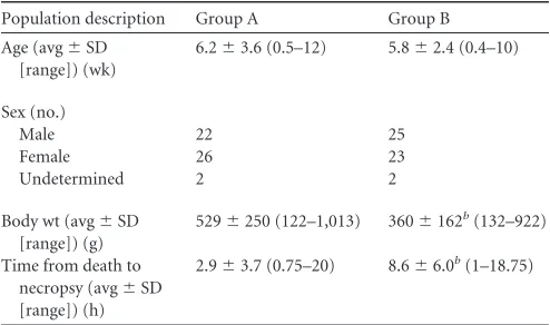
FIG 1Demonstration of enterococci adhering to the small intestinal epithe-lium of an apparently healthy kitten. Extensive colonies of bacteria are shown along the apical epithelium by means of Gram stain and fluorescencein situ hybridization using an oligonucleotide probe specific for eubacteria (Eub-338-FAM) orEnterococcusspp. (Enc-221-Cy3). Sections were nuclear counter-stained with DAPI (4=,6-diamidino-2-phenylindole). Bar⫽20m.
TABLE 5Semiquantitative description of the number and distribution of adherent enterococci in the intestinal tracts of kittens
No. of adherent enterococcia
Group A (no.) Group B (no.)
Kittens
Bacterial distribution
Kittens
Bacterial distribution
Focal Diffuse Focal Diffuse
Scant 1 1 4 4
Mild 3 2 1 2 2
Moderate 2 1 1 1 1
Severe 3 3 0
Total 9 4 5 7 6 1
aAdherent enterococci were identified by means of fluorescencein situhybridization.
Kittens were apparently healthy (group A;n⫽9) or had died or were euthanized due to severe illness (group B;n⫽7). Representative images of each description can be seen in
Fig. 2.
on May 16, 2020 by guest
http://jcm.asm.org/
[image:5.585.300.544.87.197.2] [image:5.585.62.265.367.667.2]Ileum mucosa-associated
E. faecalis
has gelatinase activity,
strong biofilm formation
in vitro
, and carries virulence traits.
Having demonstrated that non-
E. hirae
enterococci were isolated
almost exclusively from diseased kittens, we tested 81 non-
E. hirae
isolates that were cultured from the ileum mucosa of 12 group B
kittens for gelatinase activity and biofilm formation
in vitro
.
Iso-lates of
E. faecalis
(41/54) obtained from 6/8 group B kittens
dem-onstrated strong biofilm formation and/or gelatinase activity.
Ad-ditionally, all
E. faecalis
isolates carried one or more putative
virulence traits (
Fig. 5
).
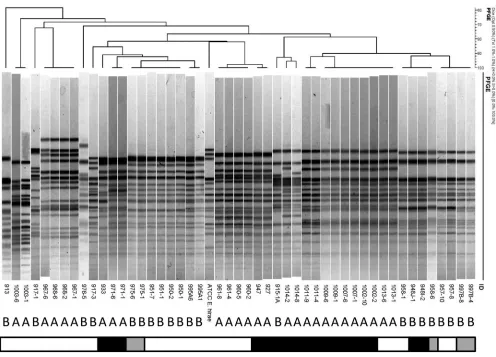
Pulsed-field gel electrophoresis supports genomic similarity
among
E. hirae
isolates cultured from the ileum mucosa of
kit-tens with enteroadherent enterococci.
To examine the genomic
similarities among ileum mucosa-associated
E. hirae
isolates and
their relationships to health versus illness of the kittens or the
presence of enteroadherent enterococci or
E. coli in vivo
, PFGE
was performed. One (if only 1 isolate) or 2 (if
ⱖ
2) isolates were
examined from each kitten that had
E. hirae
cultured from ileum
mucosa and either enteroadherent enterococci or
E. coli
identified
by FISH. Two isolates were examined from each kitten that had
ⱖ
10 isolates of
E. hirae
cultured from ileum mucosa and for which
neither enteroadherent enterococci nor
E. coli
isolates were
iden-tified (
Fig. 6
). There were no distinct differences in PFGE patterns
observed between isolates from group A and group B kittens.
However, kittens within each group frequently shared similar
ge-notypes of
E. hirae
but none of these genotypes was shared by both
groups. Accordingly, the
E. hirae
population appeared to be
geno-typically distinguishable among the healthy versus sick kittens.
There were no distinct differences in PFGE patterns observed
be-tween
E. hirae
isolates based on whether or not they were obtained
from kittens that had enteroadherent enterococci observed
in vivo
(
Fig. 6
).
Isolates of
E. faecalis
from the ileum mucosa of kittens are
genomically diverse.
To determine the genomic similarity among
E. faecalis
isolates, as predominantly obtained from kittens in
group B, PFGE was performed on 1 or 2 representative isolates
from each kitten (
Fig. 7
). No clusters could be assigned to the
typed
E. faecalis
isolates. With the use of a similarity index cutoff
value of
⬎
85%, the isolates appeared quite diverse among
differ-ent kittens. However, multiple isolates from the same kitten were
genotypically indistinguishable, indicating unique pulsotypes for
each kitten.
Multiple and specific antimicrobial resistance is more
com-mon acom-mong isolates of
E. faecalis
compared to those of
E. hirae
.
Antimicrobial susceptibility testing was performed on isolates of
E. faecalis
(
n
⫽
18) and
E. hirae
(
n
⫽
27), selected on the basis of
different PFGE profiles, from 12 and 26 kittens, respectively.
Re-sistance to multiple (
ⱖ
3) antimicrobial drugs was significantly
more common among the isolates of
E. faecalis
(17/18) than those
of
E. hirae
(16/27) (
P
⫽
0.014, Fisher’s exact test). The prevalences
of resistance to specific antimicrobial drugs among kittens
har-boring
E. hirae
or
E. faecalis
are shown in
Fig. 8
. Compared to
E.
hirae
,
E. faecalis
isolates were significantly more likely to be
resis-tant to chloramphenicol, quinupristin-dalfopristin,
erythromy-cin, lincomyerythromy-cin, and ciprofloxacin (see Table S2 in the
supple-mental material). There were no significant relationships between
multiple antimicrobial resistance and kitten group (A or B), PFGE
pulsotype, or history of antibiotic administration.
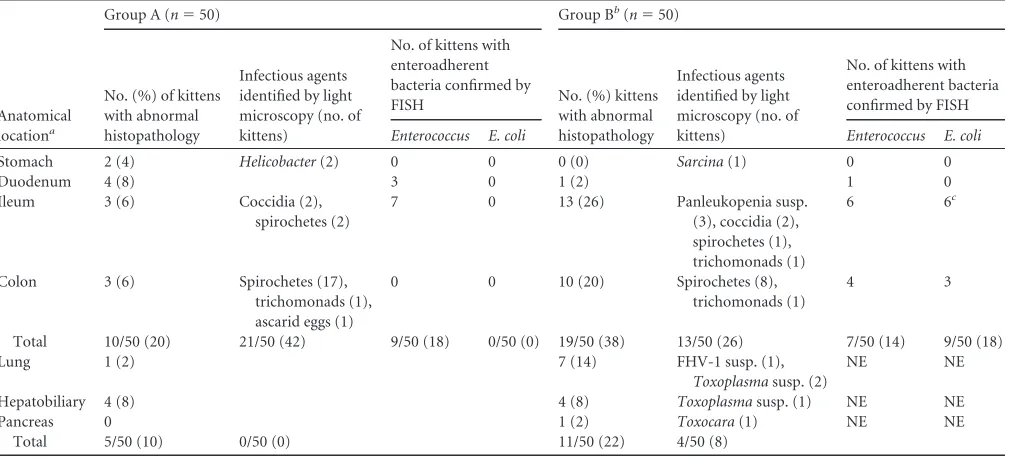
FIG 2Representative results of semiquantitative scoring of the number and extent of enteroadherent enterococci in 4 healthy group A kittens. (A) Scant, focal; (B) mild, focal; (C) moderate, diffuse; (D) severe, diffuse. Fluorescence in situhybridization was performed using an oligonucleotide probe specific for eubacteria (Eub-338-FAM) orEnterococcusspp. (Enc-221-Cy3). Specimens were nuclear counterstained with DAPI.
on May 16, 2020 by guest
http://jcm.asm.org/
[image:6.585.62.268.61.620.2]DISCUSSION
This study is the first to characterize the enterococcal community
of the distal small intestine of cats. We specifically focused on the
mucosa-associated enterococci in the ileum of very young kittens.
This decision was based on a strong association of small intestinal
disease with mortality in this population, recognition that overall
numbers of bacteria are highest in the ileum compared to other
regions of the small intestine, the importance of the ileum as a
portal of entry for invasive bacterial pathogens, and the likelihood
of also observing concurrent adherent enterococci in this region.
Moreover, bacteria that associate with the mucosal surface have a
unique composition and greater influence on intestinal epithelial
function than those generally residing in the lumen (
46
). In fact,
60% of the terminally ill kittens in this study were euthanized or
died with clinical signs and/or histopathological evidence of
gas-trointestinal tract disease; the majority of which was attributed to
the small intestine. It was not the purpose of this study to establish
the cause of death in these kittens, and light microscopic
exami-nation of the intestinal tract was generally unrewarding in
identi-fying any specific diagnoses.
[image:7.585.103.487.66.249.2]In this study we identified
E. hirae
as the dominant species of
enterococci to colonize the ileal mucosa in apparently healthy
FIG 3Transmission electron micrographs of enterococci interacting directly with the intestinal epithelial microvilli. The micrographs in the left panel were taken of a specimen with light microscopic evidence of scant, focal adherence of enterococci. The micrograph in the right panel was taken of a specimen with light microscopic evidence of severe diffuse adherence of enterococci. These specimens share the same origins as those shown inFig. 2AandD, respectively.TABLE 6Species identification of enterococci isolated from the ileum mucosa of apparently healthy kittens
Enterococcus spp. identified
Group A (n⫽153 isolates) Group B (n⫽178 isolates)
Total no. (%) of kittens
Total no. (%) of isolates
Total no. (%) of kittens
Total no. (%) of isolates
E. hirae 29 (58) 148 (97) 14 (28)c 75 (42)
E. faecalis 2 (4) 3 (2) 10 (20)b 64 (36)
E. faecium 1 (2) 1 (0.5) 3 (6) 12 (7)
E. avium 0 (0) 0 (0) 2 (4) 18 (10)
E. gallinarum 0 (0) 0 (0) 1 (2) 8 (4)
E. seriolicida 1 (2) 1 (0.5) 1 (2) 1 (1)
Total 31/50 (62)a 153/153 (100) 24/50 (48)a 178/178 (100)
aIn several kittens, multiple species of enterococci were isolated. Enterococcal species
were identified by either multiplex PCR (30) or sequencing of thesodAgene (35). The number (%) of total isolates of each species depicted is biased by variation between kittens in the number of isolates chosen for purification. An unbiased representation of the distribution of isolates between group A and B kittens is shown inFig. 3.
b
Pis⬍0.05.
cPis⬍0.01 (Fisher’s exact test).
FIG 4Population diversity of enterococcal species from 12 group A (n⫽120 isolates) and 13 group B (n⫽159 isolates) kittens, each of which hadⱖ8 individual isolates identified as to species. Four common species,E. faecalis,E. faecium,E. casseliflavus, andE. gallinarum, were identified using multiplex PCR (30). PCR amplification and sequencing of thesodAgene were carried out for isolates not identified by multiplex PCR (35).
on May 16, 2020 by guest
http://jcm.asm.org/
[image:7.585.40.287.530.662.2]young kittens. The only prior studies to examine the microbiota of
the feline small intestine focused on the lumen-dwelling bacteria
and were performed in older cats. In these studies, enterococci
were either not identified in the small intestine (
47
) or were
iden-tified as represented predominantly by
E. faecalis
(
48–50
).
Entero-coccus faecalis
(
51–54
), followed by
E. faecium
(
55
) and
E. hirae
(
11
), are the most commonly reported species of enterococci to
dominate the fecal flora of cats, where the numbers of enterococci
can average 10
6CFU per gram of feces (
11
).
In kittens that died or were euthanized due to severe illness,
there was a major difference in the species of enterococci
associ-ated with the ileum mucosa. Significantly greater numbers of these
kittens were colonized by
E. faecalis
, suggesting that the ileum
mucosa-associated microbiota in sick kittens reflects a more adult
fecal-like composition (
51–54
) of enterococci. Moreover, the
E.
faecalis
isolates obtained from these kittens were characterized as
carrying multiple genotypic and phenotypic attributes of
viru-lence. This is in contrast to
E. faecalis
isolates from healthy cats, in
which the carriage of virulence genes appears to be uncommon
(
56
), albeit there are a few reports of examinations of genotypic
virulence of
E. faecalis
in healthy cats (
11
,
57
). Most notable
among the virulence traits of
E. faecalis
from sick kittens was a high
prevalence of
gelE
, a gene encoding the zinc metalloproteinase
gelatinase, and concurrent documentation of gelatinase activity.
Gelatinase is implicated in enhancing enterococcal virulence by
conferring an ability to form solid-surface environmental biofilms
(
40
), and in the present study
E. faecalis
gelatinase activity was
associated with strong biofilm formation on polystyrene. There
was additionally a high prevalence of
asa1
, a gene encoding
aggre-gation substance, among
E. faecalis
isolates from sick kittens.
Ag-gregation substance is a surface bacterial adhesin that has been
demonstrated to increase attachment of
E. faecalis
to intestinal
epithelial cells
in vitro
(
58
). Whether
gelE
or
asa1
conferred to
E.
faecalis
an ability to outcompete
E. hirae
for representation within
the ileum mucosa-associated microbiota in these sick kittens is
unknown.
In addition to carrying multiple genetic virulence factors,
E.
faecalis
isolates were frequently resistant to tetracycline,
erythro-mycin, lincoerythro-mycin, quinupristin-dalfopristin, chloramphenicol,
linezolid, and ciprofloxacin. None of the
E. faecalis
isolates
dem-onstrated resistance to penicillin, aminoglycosides, or
vancomy-cin. Intrinsic or acquired resistance to quinupristin-dalfopristin,
tetracyclines, and macrolides is commonly reported among feline
isolates of
E. faecalis
(
11
,
52–54
). In contrast, resistance to
newer-generation antimicrobial drugs (linezolid) or those commonly
re-served for treatment of multidrug-resistant bacterial infections in
FIG 5Correlations among biofilm formation, gelatinase phenotype, and presence of virulence genes (gelE,asa1,esp, andcylA) in enterococci isolated from 12 group A kittens (n⫽120 isolates) (A) and 13 group B kittens (n⫽133 isolates) (B). The dotted lines indicate biofilm formation activity levels (⬍0.2, no biofilm; 0.2 to 0.7, biofilm;⬎0.7, strong biofilm). Kitten numbers are presented on thexaxis followed by the total numbers of characterized isolates in parentheses. Letters following the kitten numbers indicate kittens with adherent enterococci (X), no adherent bacteria (Y), or adherentE. coli(Z).E. faecalisV583 was used as a positive control. Bars correspond to the means⫾standard errors of the means (SEM) of 5 replicate experiments. OD550, optical density at 550 nm.on May 16, 2020 by guest
http://jcm.asm.org/
[image:8.585.44.541.68.392.2]cats (ciprofloxacin) was somewhat surprising (
11
,
52
). None of
the kittens had a history of treatment with fluoroquinolones.
Compared to isolates of
E. faecalis
, isolates of
E. hirae
obtained
from apparently healthy or sick kittens were significantly less often
resistant to multiple (
ⱖ
3) antimicrobial drugs. Less frequent
an-timicrobial resistance among
E. hirae
than
E. faecalis
and
E.
fae-cium
of fecal origin in cats has been previously documented (
11
).
Whether or not the virulent, antimicrobial-resistant isolates of
E. faecalis
colonizing the ileum mucosa of sick kittens in this study
originated from commensal
E. faecalis
or were acquired from the
environment is not known. Based on the PFGE genotypes of
rep-resentative
E. faecalis
isolates from each kitten,
multidrug-resis-tant strains did not share colonization of the ileum mucosa with
other strains. Sick kittens possibly had a greater susceptibility and
opportunity for colonization by resistant strains of
E. faecalis
, as
they spent time in a foster care or hospital environment in contrast
to the apparently healthy kittens that were frequently euthanized
shortly after their receipt to the animal control facility. An
oppor-tunistic infection of the sick kittens by virulent
E. faecalis
is
sup-ported by studies demonstrating frequent antibiotic resistance in
fecal enterococci in cats from catteries, hospitalized cats, and
res-ident cats in veterinary clinics (
11
,
53
,
54
). However, whether the
colonization of ileum mucosa-associated microbiota by
E. faecalis
was a contributing cause or consequence of gastrointestinal
dis-ease and terminal illness in the sick kittens reported here is
un-known. What appears more certain is that the intestinal
entero-cocci of sick kittens may serve as a reservoir for potential
transmission of antimicrobial resistance genes to the environment
and/or to other hosts.
Apart from being identified as the dominant cultivable species
of enterococci in the ileum mucosa-associated microbiota in
ap-parently healthy young kittens, overt and extensive adhesion of
E.
hirae
to the small intestinal epithelium was observed in 16% of the
kittens in this population. Given that detection of this event
re-quires light microscopy and only two biopsy specimens of the
small intestine from each kitten were examined, it is likely that the
prevalence of this phenomenon was grossly underestimated.
While adherent enterococci were also detected in the sick kittens,
considerably fewer bacteria were observed and they were
infre-quently identified to the species level by means of PCR. We
attri-bute our inability to identify the adherent enterococci in sick
kit-tens as due to the presence of fewer bacteria (and less enterococcal
DNA) and as less likely due to adhesion by non-
E. hirae
entero-cocci. Our finding that enteroadherent
E. hirae
is common and
FIG 6Pulsed-field gel electrophoresis of 48E. hiraeisolates cultured from the ileum mucosa of apparently healthy kittens (group A;n⫽15) and kittens that died or were euthanized due to severe illness (group B;n⫽12). Bar denotes light microscopic findings in the kitten from which the isolate was obtained (white, no enteroadherent enterococci or EPEC observed; black, enteroadherent enterococci present; gray, EPEC present). Numerical values indicate the identities of the kittens and isolates. The type strain isE. hiraeATCC 8043.on May 16, 2020 by guest
http://jcm.asm.org/
[image:9.585.44.540.63.420.2]extensive in apparently healthy kittens provides a contrast to the
numerous uncontrolled case reports describing enteroadherent
enterococci in association with diarrhea in young animals (
17–
25
). Although the identity and virulence attributes of the
en-teroadherent enterococci in most of these reports was not
deter-mined, it should be considered that the adherent enterococci may
not have been the primary cause of diarrhea in these animals.
The mechanism(s) by which
E. hirae
adhere to the intestinal
epithelium in these young kittens is not clear. Based on a
compar-ative lack of virulence among the
E. hirae
isolates tested from the
kittens, it is evident that the genotypic (
gelE
and
asa1
) and
pheno-typic (gelatinase activity) attributes generally regarded as essential
for biofilm formation
in vitro
are not required for enteroadhesion
in vivo
. PFGE revealed that the
E. hirae
population is genotypically
very diverse and clustering did not correlate with enteroadherence
in vivo
. A notable feature of enteroadherent
E. hirae
is a light
microscopic appearance indistinguishable from that of EPEC that
requires a Gram stain for their differentiation (
25
). It is likewise
intriguing that adherence of
E. coli
was documented commonly
and exclusively in sick kittens in this study but was not observed in
any sick kitten with enteroadherent enterococci. This not only
identifies enteroadherent
E. coli
as a potentially important
intes-tinal pathogen in young kittens but also suggests that
enteroad-herence of
E. hirae
might competitively inhibit or otherwise deter
the attachment of
E. coli
.
Results of this study identify
E. hirae
as the most common
mucosa-associated species of enterococci to inhabit the small
in-testine in apparently healthy young kittens. Furthermore,
adher-ence of
E. hirae
to the small intestinal epithelium was common
and extensive in this population. Isolates of
E. hirae
generally
lacked phenotypic and genotypic determinants of virulence. In
contrast, kittens that died or were euthanized due to severe illness
were significantly more often identified as colonized by
E. faecalis
.
This population of
E. faecalis
was characterized by a high level of
gelatinase activity, strong biofilm formation on polystyrene, the
presence of virulence determinants, and multiple antimicrobial
resistances. Moreover, attachment of
E. coli
to the intestinal
epi-thelium was exclusively and significantly associated with terminal
illness and was not documented in any kitten for which
enteroad-herent
E. hirae
was observed. These findings identify a significant
difference in the species of enterococci colonizing the ileum
mu-cosa of healthy versus terminally ill young kittens and suggest that
E. hirae
represents an important commensal in this population.
Given the significance of small intestinal disease as a cause of
mor-tality in young kittens, these findings have important implications
toward identifying species of enterococci for their potential to
significantly impact the survival of very young kittens.
ACKNOWLEDGMENTS
This work was supported by grants W09-022 and W11-013 from the Winn Feline Foundation.
We thank Maria Stone, Megan Fauls, Mondy Lamb, Lisa Kroll, Ashley Williams, Hannah Preedy, Allison Baker, Wendy Savage, and Klara Zurek for valuable technical assistance. Transmission electron microscopy was performed in the Laboratory for Advance Electron and Light Optical Methods in the College of Veterinary Medicine, North Carolina State University.
REFERENCES
1.American Veterinary Medical Association. 2012. Overview: total pet ownership and pet population, p 31–32.InU.S. pet ownership and demo-FIG 7Pulsed-field gel electrophoresis of 19E. faecalisisolates cultured from
the ileum mucosa of apparently healthy kittens (group A;n⫽2) and kittens that died or were euthanized due to severe illness (group B;n⫽10). Numerical values indicate identities of the kittens and isolates. The type strain isE. faecalis ATCC 29212.
FIG 8Antimicrobial susceptibility test results for ileal mucosa culture isolates ofE. hirae(n⫽27) from 15 group A and 11 group B kittens andE. faecalis(n⫽ 18) from 2 group A and 10 group B kittens. *,P⬍0.05; **,P⬍0.01; ***,P⬍ 0.001 (Fisher’s exact test). TGC, tigecycline; TET, tetracycline; CHL, chloram-phenicol; DAP, daptomycin; STR, streptomycin; TYLT, tylosin tartrate; SYN, quinupristin-dalfopristin; LZD, linezolid; NIT, nitrofurantoin; PEN, penicil-lin; KAN, kanamycin; ERY, erythromycin; CIP, ciprofloxacin; VAN, vanco-mycin; LIN, lincovanco-mycin; GEN, gentamicin.
on May 16, 2020 by guest
http://jcm.asm.org/
[image:10.585.45.257.66.381.2] [image:10.585.283.543.70.232.2]graphics sourcebook, section 1. American Veterinary Medical Associa-tion, Schaumburg, IL.
2.Scott FW, Geissinger C. 1978. Kitten mortality survey. Fel. Pract. 8:31–34.
3.Nutter FB, Levine JF, Stoskopf MK. 2004. Reproductive capacity of free-roaming domestic cats and kitten survival rate. J. Am. Vet. Med. Assoc.225:1399 –1402.
4.Young C.1973. Preweaning mortality in specific pathogen-free kittens. J. Small Anim. Pract.14:391–397.
5. Sparkes AH, Rogers K, Henley WE, Gunn-Moore DA, May JM, Gruffydd-Jones TJ, Bessant C. 2006. A questionnaire-based study of gestation, parturition and neonatal mortality in pedigree breeding cats in the UK. J. Fel. Med. Surg.8:145–157.
6.Root MV, Johnston SD, Olson PN.1995. Estrous length, pregnancy rate, gestation and parturition lengths, litter size, and juvenile mortality in the domestic cat. J. Am. Anim. Hosp. Assoc.31:429 – 433.
7.Murray JK, Skillings E, Gruffydd-Jones TJ.2008. A study of risk factors for cat mortality in adoption centres of a UK cat charity. J. Feline Med. Surg.10:338 –345.
8.Cave TA, Thompson H, Reid SW, Hodgson DR, Addie DD. 2002. Kitten mortality in the United Kingdom: a retrospective analysis of 274 histopathological examinations (1986 to 2000). Vet. Rec.151:497–501. 9.Bybee SN, Scorza AV, Lappin MR.2011. Effect of the probiotic
Entero-coccus faeciumSF68 on presence of diarrhea in cats and dogs housed in an animal shelter. J. Vet. Intern. Med.25:856 – 860.
10. Hart ML, Suchodolski JS, Steiner JM, Webb CB.2012. Open-label trial of a multi-strain synbiotic in cats with chronic diarrhea. J. Feline Med. Surg.14:240 –245.
11. Ghosh A, Kukanich K, Brown CE, Zurek L.2012. Resident cats in small animal veterinary hospitals carry multi-drug resistant enterococci and are likely involved in cross-contamination of the hospital environment. Front. Microbiol.3:62.
12. Wagner KA, Hartmann FA, Trepanier LA.2007. Bacterial culture results from liver, gallbladder, or bile in 248 dogs and cats evaluated for hepato-biliary disease: 1998 –2003. J. Vet. Intern. Med.21:417– 424.
13. Litster A, Moss S, Platell J, Trott DJ.2009. Occult bacterial lower urinary tract infections in cats— urinalysis and culture findings. Vet. Microbiol. 136:130 –134.
14. Costello MF, Drobatz KJ, Aronson LR, King LG. 2004. Underlying cause, pathophysiologic abnormalities, and response to treatment in cats with septic peritonitis: 51 cases (1990 –2001). J. Am. Vet. Med. Assoc. 225:897–902.
15. KuKanich KS, Ghosh A, Skarbek JV, Lothamer KM, Zurek L.2012. Surveillance of bacterial contamination in small animal veterinary hospi-tals with special focus on antimicrobial resistance and virulence traits of enterococci. J. Am. Vet. Med. Assoc.240:437– 445.
16. Hamilton E, Kaneene JB, May KJ, Kruger JM, Schall W, Beal MW, Hauptman JG, DeCamp CE.2012. Prevalence and antimicrobial resis-tance ofEnterococcusspp. andStaphylococcusspp. isolated from surfaces in a veterinary teaching hospital. J. Am. Vet. Med. Assoc.240:1463–1473. 17. Etheridge ME, Vonderfecht SL.1992. Diarrhea caused by a slow-growing
Enterococcus-like agent in neonatal rats. Lab. Anim. Sci.42:548 –550. 18. Etheridge ME, Yolken RH, Vonderfecht SL.1988. Enterococcus hirae
implicated as a cause of diarrhea in suckling rats. J. Clin. Microbiol.26: 1741–1744.
19. Cheon DS, Chae C.1996. Outbreak of diarrhea associated with Entero-coccus duransin piglets. J. Vet. Diagn. Invest.8:123–124.
20. Vancanneyt M, Snauwaert C, Cleenwerck I, Baele M, Descheemaeker P, Goossens H, Pot B, Vandamme P, Swings J, Haesebrouck F, Devriese LA.2001.Enterococcus villorumsp. nov., an enteroadherent bacterium associated with diarrhoea in piglets. Int. J. Syst. Evol. Microbiol.51:393– 400.
21. Rogers DG, Zeman DH, Erickson ED.1992. Diarrhea associated with Enterococcus duransin calves. J. Vet. Diagn. Invest.4:471– 472. 22. Tzipori S, Hayes J, Sims L, Withers M.1984.Streptococcus durans: an
unexpected enteropathogen of foals. J. Infect. Dis.150:589 –593. 23. Collins JE, Bergeland ME, Lindeman CJ, Duimstra JR.1988.
Enterococ-cus(Streptococcus)duransadherence in the small intestine of a diarrheic pup. Vet. Pathol.25:396 –398.
24. Lapointe JM, Higgins R, Barrette N, Milette S.2000.Enterococcus hirae enteropathy with ascending cholangitis and pancreatitis in a kitten. Vet. Pathol.37:282–284.
25. Nicklas JL, Moisan P, Stone MR, Gookin JL.2010.In situmolecular
diagnosis and histopathological characterization of enteroadherent En-terococcus hiraeinfection in pre-weaning-age kittens. J. Clin. Microbiol. 48:2814 –2820.
26. Janeczko S, Atwater D, Bogel E, Greiter-Wilke A, Gerold A, Baumgart M, Bender H, McDonough PL, McDonough SP, Goldstein RE, Simpson KW.2008. The relationship of mucosal bacteria to duodenal histopathol-ogy, cytokine mRNA, and clinical disease activity in cats with inflamma-tory bowel disease. Vet. Microbiol.128:178 –193.
27. Wellinghausen N, Bartel M, Essig A, Poppert S.2007. Rapid identifica-tion of clinically relevantEnterococcusspecies by fluorescencein situ hy-bridization. J. Clin. Microbiol.45:3424 –3426.
28. Gray SG, Hunter SA, Stone MR, Gookin JL.Assessment of reproductive tract disease in cats at risk forTritrichomonas foetusinfection. Am. J. Vet. Res.71:76 – 81.
29. Arias CA, Robredo B, Singh KV, Torres C, Panesso D, Murray BE. 2006. Rapid identification ofEnterococcus hiraeandEnterococcus durans by PCR and detection of a homologue of theE. hiraemur-2 gene inE. durans. J. Clin. Microbiol.44:1567–1570.
30. Kariyama R, Mitsuhata R, Chow JW, Clewell DB, Kumon H. 2000. Simple and reliable multiplex PCR assay for surveillance isolates of van-comycin-resistant enterococci. J. Clin. Microbiol.38:3092–3095. 31. Dutka-Malen S, Evers S, Courvalin P.1995. Detection of glycopeptide
resistance genotypes and identification to the species level of clinically relevant enterococci by PCR. J. Clin. Microbiol.33:24 –27.
32. Cheng S, McCleskey FK, Gress MJ, Petroziello JM, Liu R, Namdari H, Beninga K, Salmen A, DelVecchio VG.1997. A PCR assay for identifi-cation ofEnterococcus faecium. J. Clin. Microbiol.35:1248 –1250. 33. Franck SM, Bosworth BT, Moon HW.1998. Multiplex PCR for
entero-toxigenic, attaching and effacing, and Shiga toxin-producingEscherichia colistrains from calves. J. Clin. Microbiol.36:1795–1797.
34. Macovei L, Zurek L.2007. Influx of enterococci and associated antibiotic resistance and virulence genes from ready-to-eat food to the human di-gestive tract. Appl. Environ. Microbiol.73:6740 – 6747.
35. Poyart C, Quesnes G, Trieu-Cuot P.2000. Sequencing the gene encoding manganese-dependent superoxide dismutase for rapid species identifica-tion of enterococci. J. Clin. Microbiol.38:415– 418.
36. Vankerckhoven V, Van Autgaerden T, Vael C, Lammens C, Chapelle S, Rossi R, Jabes D, Goossens H.2004. Development of a multiplex PCR for the detection ofasa1,gelE,cylA,esp, andhylgenes in enterococci and survey for virulence determinants among European hospital isolates of Enterococcus faecium. J. Clin. Microbiol.42:4473– 4479.
37. Ribeiro TC, Pinto V, Gasper F, Lopes MFS.2008.Enterococcus hirae causing wound infections in a hospital. J. Chin. Clin. Med.3:150 –152. 38. Gilmore MS, Coburn PS, Nallapareddy R, Murray BE.2002.
Entero-coccal virulence, p 301–354.InGilmore MS, Clewell DB, Courvalin P, Dunny GM, Murray BE, Rice LB (ed), The enterococci: pathogenesis, molecular biology and antibiotic resistance. ASM Press, Washington, DC. 39. Macovei L, Ghosh A, Thomas VC, Hancock LE, Mahmood S, Zurek L. 2009.Enterococcus faecaliswith the gelatinase phenotype regulated by the fsr operon and with biofilm-forming capacity are common in the agricul-tural environment. Environ. Microbiol.11:1540 –1547.
40. Hancock LE, Perego M. 2004. The Enterococcus faecalis fsr two-component system controls biofilm development through production of gelatinase. J. Bacteriol.186:5629 –5639.
41. Thomas VC, Thurlow LR, Boyle D, Hancock LE.2008. Regulation of autolysis-dependent extracellular DNA release byEnterococcus faecalis ex-tracellular proteases influences biofilm development. J. Bacteriol.190: 5690 –5698.
42. Ahmad A, Ghosh A, Schal C, Zurek L.2011. Insects in confined swine operations carry a large antibiotic resistant and potentially virulent en-terococcal community. BMC Microbiol. 11:23. doi:10.1186/1471-2180 -11-23.
43. Luey CK, Chu YW, Cheung TK, Law CC, Chu MY, Cheung DT, Kam KM.2007. Rapid pulsed-field gel electrophoresis protocol for subtyping of Streptococcus suisserotype 2. J. Microbiol. Methods68:648 – 650. 44. Clinical and Laboratory Standards Institute.2010. Performance
stan-dards for antimicrobial susceptibility testing, 20th informational supple-ment. CLSI M100-S20. Clinical and Laboratory Standards Institute, Wayne, PA.
45. US Department of Agriculture ARS.2012. 2010 NARMS animal arm annual report. Food and Drug Administration/Centers for Disease Con-trol and Prevention/U.S. Department of Agriculture, Athens, GA.http:
on May 16, 2020 by guest
http://jcm.asm.org/
//ars.usda.gov/SP2UserFiles/Place/66120508/NARMS/NARMS2010/NA RMS%20USDA%202010%20Report.pdf.
46. Eckburg PB, Bik EM, Bernstein CN, Purdom E, Dethlefsen L, Sargent M, Gill SR, Nelson KE, Relman DA.2005. Diversity of the human intestinal microbial flora. Science308:1635–1638.
47. Ritchie LE, Steiner JM, Suchodolski JS.2008. Assessment of microbial diversity along the feline intestinal tract using 16S rRNA gene analysis. FEMS Microbiol. Ecol.66:590 –598.
48. Osbaldiston GW, Stowe EC.1971. Microflora of alimentary tract of cats. Am. J. Vet. Res.32:1399 –1405.
49. Sparkes AH, Papasouliotis K, Sunvold G, Werrett G, Clarke C, Jones M, Gruffydd-Jones TJ, Reinhart G.1998. Bacterial flora in the duodenum of healthy cats, and effect of dietary supplementation with fructo-oligosaccharides. Am. J. Vet. Res.59:431– 435.
50. Papasouliotis K, Sparkes AH, Werrett G, Egan K, Gruffydd-Jones EA, Gruffydd-Jones TJ.1998. Assessment of the bacterial flora of the proximal part of the small intestine in healthy cats, and the effect of sample collec-tion method. Am. J. Vet. Res.59:48 –51.
51. Devriese LA, Cruz Colque JI, De Herdt P, Haesebrouck F. 1992. Identification and composition of the tonsillar and anal enterococcal and streptococcal flora of dogs and cats. J. Appl. Bacteriol.73:421– 425. 52. Jackson CR, Fedorka-Cray PJ, Davis JA, Barrett JB, Frye JG. 2009.
Prevalence, species distribution and antimicrobial resistance of entero-cocci isolated from dogs and cats in the United States. J. Appl. Microbiol. 107:1269 –1278.
53. Moyaert H, De Graef EM, Haesebrouck F, Decostere A.2006. Acquired antimicrobial resistance in the intestinal microbiota of diverse cat popu-lations. Res. Vet. Sci.81:1–7.
54. Leener ED, Decostere A, De Graef EM, Moyaert H, Haesebrouck F. 2005. Presence and mechanism of antimicrobial resistance among entero-cocci from cats and dogs. Microb. Drug Resist.11:395– 403.
55. Rodrigues J, Poeta P, Martins A, Costa D.2002. The importance of pets as reservoirs of resistantEnterococcusstrains, with special reference to vancomycin. J. Vet. Med. B Infect. Dis. Vet. Public Health49:278 –280. 56. Gulhan T, Aksakal A, Ekin IH, Savasan S, Boynukara B.2006. Virulence
factors ofEnterococcus faeciumandEnterococcus faecalisstrains isolated from humans and pets. Turk. J. Vet. Anim. Sci.30:477– 482.
57. Harada T, Tsuji N, Otsuki K, Murase T.2005. Detection of the esp gene in high-level gentamicin resistantEnterococcus faecalisstrains from pet animals in Japan. Vet. Microbiol.106:139 –143.
58. Sartingen S, Rozdzinski E, Muscholl-Silberhorn A, Marre R. 2000. Aggregation substance increases adherence and internalization, but not translocation, ofEnterococcus faecalisthrough different intestinal epithe-lial cellsin vitro. Infect. Immun.68:6044 – 6047.