0095-1137/10/$12.00
doi:10.1128/JCM.00582-10
Copyright © 2010, American Society for Microbiology. All Rights Reserved.
Fast Duplex One-Step Reverse Transcriptase PCR for Rapid Differential
Detection of West Nile and Japanese Encephalitis Viruses
䌤
Jung-Yong Yeh,
1†* Ji-Hye Lee,
1Hyun-Ji Seo,
2Jee-Yong Park,
1Jin-San Moon,
1†* In-Soo Cho,
1Joong-Bok Lee,
3Seung-Yong Park,
3Chang-Seon Song,
3and In-Soo Choi
3Foreign Animal Disease Division, National Veterinary Research and Quarantine Service, 480, Anyang-6-Dong, Manan-gu,
Anyang-city, Gyeonggi-do, 430-824, South Korea
1; College of Medicine, Konkuk University, Hwayang-1-dong,
Gwangjin-gu, Seoul, 143-701, South Korea
2; and College of Veterinary Medicine, Konkuk University,
Hwayang-1-dong, Gwangjin-gu, Seoul, 143-701, South Korea
3Received 19 March 2010/Returned for modification 6 July 2010/Accepted 7 September 2010
The aim of this study was to develop a highly sensitive and specific one-step duplex reverse transcriptase
PCR (RT-PCR) assay for the simultaneous and differential detection of West Nile (WNV) and Japanese
encephalitis (JEV) viruses. The bioinformatic analysis of published sequences of WNV and JEV revealed
conserved regions not targeted by previously reported primers. A total of 13 primers were designed based
on these regions to detect all of the WNV and JEV lineages and to discriminate between the two viruses
by the generation of 482- and 241-bp cDNA products, respectively. The results indicate that single-tube
duplex PCR using these primers is a useful technique for the detection and differentiation of WNV and
JEV in plasma or brain tissue. The novel duplex RT-PCR described in this study enables the early
diagnosis of these two encephalitic flaviviruses. In addition, this technique may be useful as part of a
testing regimen for human patients, horses, and other susceptible animal species, as it is rapid (less than
3.5 h from RNA extraction), sensitive, and specific, and it may enable the differential diagnosis of clinical
samples.
Species within the
Flavivirus
genus can cause public health
problems around the world. For example, the increasing
num-ber of Dengue and Japanese encephalitis virus (JEV)
infec-tions in Asia, frequent outbreaks of yellow fever in Africa and
South America, and the ongoing spread of West Nile virus
(WNV) throughout the Americas exemplify the geographical
burden of flavivirus diseases. Global flavivirus incidence has
grown in the past 25 years, and this increase is due primarily to
unprecedented population growth and migration, uncontrolled
urbanization, and the lack of effective mosquito control (9).
WNV and JEV are arthropod-borne flaviviruses (termed
arborviruses) that can emerge or reemerge in many regions
due to climate change and increased travel (1, 5, 11, 26, 35, 38).
WNV and JEV are members of the
Flaviviridae
family and are
arboviruses associated with both human and equine
encepha-litis worldwide. WNV circulates in Africa, west Asia, southern
Europe, and Australia. It emerged in North America in 1999,
leading to an extensive and ongoing outbreak (40). Similarly to
the emergence and rapid spread of WNV in North America, it
is possible that WNV could be introduced into far-east Asia
from continents in which it is endemic through infected birds,
travelers, or insect vectors. If a human pathogen like WNV
emerges or reemerges in a given country, very strict
epidemi-ological regulations need to be implemented immediately. To
monitor infection and prevent rapid viral spread in these cases,
methods are required for rapid viral detection in suspect cases
and potential vectors.
In areas where JEV is endemic, such as Korea and Japan,
distinguishing between WNV and JEV is critical for the
detec-tion of WNV invasion, because JEV is a mosquito-borne
fla-vivirus from the JEV serocomplex that causes encephalitis in
humans and horses, and it is widespread throughout most of
Asia (12, 27). However, Japanese encephalitis serocomplex
flaviviruses cross-react antigenically with WNV and thus are
not readily differentiated by serology (10). Molecular
diagnos-tic methods therefore are preferred, and reverse transcriptase
PCR (RT-PCR) methods have been used to develop sensitive
and specific assays for the identification of WNV (13, 24).
Recently, more-sensitive assays, such as fluorogenic real-time
(TaqMan) PCR, SYBR green-based real-time PCR, and
loop-mediated isothermal amplification (LAMP), have been
devel-oped for the diagnostic detection of WNV genes (16, 21–22).
However, these diagnostic methods were designed only to
de-tect strains of WNV isolated in the United States that are
closely related to lineage 1, and thus they may not detect
lineage 2 strains associated with Africa (2) and, more recently,
with Europe (8, 17). In areas where JEV is endemic and WNV
is absent, it is possible that other WNV lineages can emerge
(i.e., lineage 2). Therefore, it is necessary to design specific
assays that recognize all lineages of WNV and can distinguish
between JEV and WNV in order to make a definitive
diagno-sis. Two molecular diagnostic methods that simultaneously
dis-criminate between strains of WNV and JEV have been
re-ported previously, one that uses RT-PCR and restriction
fragment length polymorphism (RFLP) analysis and another
that uses fluorogenic real-time PCR (TaqMan) (32–33). In this
* Corresponding author. Mailing address for J.-Y. Yeh: Foreign
Animal Disease Division, National Veterinary Research and
Quaran-tine Service, Anyang 430-824, South Korea. Phone: 82-31-467-1860.
Fax: 82-31-449-5882. E-mail: [email protected]. E-mail address for
J.-S. Moon: [email protected].
† These authors contributed equally to the manuscript.
䌤
Published ahead of print on 15 September 2010.
4010
on May 16, 2020 by guest
http://jcm.asm.org/
study, our aim was to develop a more-rapid molecular
diag-nostic method that could detect and distinguish between WNV
and JEV using a conventional RT-PCR format in a single-tube
duplex platform with a primer mixture specific to JEV strains
and all of the lineages of WNV.
MATERIALS AND METHODS
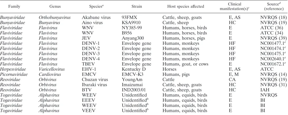
Viruses.WNV strains NY385-99 and B956 (American Type Culture Collec-tion [ATCC]) were used in this study. The NY385-99 strain (lineage 1) was isolated from a snowy owl in New York during the 1999 outbreak (36), and the B956 strain (lineage 2) was isolated from a woman in Uganda in 1937 (34). Anyang 300 (39), an attenuated JEV strain, also was used in this study. WNV and JEV also were used in specificity assays to evaluate primer sets for cross-ampli-fication. Because the envelope gene was the target gene selected for the differ-ential diagnosis of WNV and JEV in the multiplex RT-PCR, the envelope genes of dengue virus types 1 to 4 and tick-borne encephalitis viruses were synthesized to demonstrate the specificity of the assay among viruses belonging to the flavivirus family. The genes were cloned into the SacI/XhoI site in the pBlue-script II SK(⫹) vector from New England Biolabs (United Kingdom), as live viruses were not available due to their biosecurity status. The following related viruses, which cause neurological conditions in animals or humans, also were employed for specificity assays: Akabane virus, Aino virus, equine herpesvirus 1 (EHV-1), encephalomyocarditis virus (EMCV), bluetongue virus, and Western equine encephalitis virus. The RNAs of the following viruses also were included: RNAs extracted from the Cephalovac VEWT vaccine containing inactivated Eastern, Western, and Venezuelan equine encephalitis viruses (EEEV, WEEV, and VEEV). All of the viruses used in this study are listed in Table 1.
Virus cultures and quantitation of viral RNA.The flaviviruses Akabane virus, Aino virus, EMCV, and WEEV were grown in Vero cells (ATCC CCL-81) in alpha minimum essential medium (␣MEM) (GibcoBRL, Invitrogen Corpora-tion, Carlsbad, CA) containing 10% fetal bovine serum (FBS) (GibcoBRL, Invitrogen Corporation, Carlsbad, CA) and an antibiotic-antimycotic mixture (Invitrogen) at 37°C in a humidified 5% CO2environment. WNV manipulations were performed in a biosafety level 3 (BSL3) containment research laboratory at the National Veterinary Research and Quarantine Service (NVRQS) in accor-dance with the regulations of the South Korean government. EHV-1 was grown
in RK13 cells (ATCC CCL-37), and the bluetongue virus was grown in BHK cells. Noninfected Vero, RK13, and BHK cell line cultures were used as controls in the specificity assays. The detection limit of the assay was evaluated by both PFU and RNA copy numbers. For the titration of WNV and JEV infectivity, a plaque assay was performed according to the method described by Payne et al. (23). The viral titer was calculated and expressed as PFU per milliliter. One PFU represents a circumscribed area of cellular degeneration initially produced by one virion. To calculate RNA copy numbers, viral RNAs of WNV and JEV were quantified by the method of Shi et al. (30) and Santhosh et al. (29).
Primer design.The bioinformatic analysis of published sequences of WNV and JEV revealed conserved regions not targeted by previously reported primers. These conserved viral genome regions were chosen as the best candidates for the generation of specific primers. A total of 13 primers were designed within these regions for duplex RT-PCR to detect WNV and JEV based on the generation of 482- and 241-bp cDNA products, respectively (Table 2).
Optimization and performance evaluation.To optimize the reaction condi-tions, preliminary assays were performed to test different concentrations of each primer set in the duplex RT-PCR. Positive control plasma and brain homoge-nates were prepared by spiking samples with WNV and JEV cultured from cells, as positive brain and plasma samples were not available. Negative plasma and brain homogenate controls also were used. Plasma was harvested from live chickens and horses, and brain tissues were collected from carcasses of wild birds and horses. Each assay was performed in parallel with the corresponding uniplex RT-PCR for each viral and total RNA dilution. Finally, the combination of primer concentrations that yielded the best results for the target flaviviruses was selected and is shown in Table 2. Various amplification tests using the 13 primers were performed in several reactions, and incubation profile conditions were investigated to select the best type of DNA polymerase and to establish the optimal reaction protocol for the duplex RT-PCR assay. Experiments were performed in triplicate with known titers (or RNA copy numbers) to determine the detection threshold for each virus.
[image:2.585.43.542.81.281.2]RNA extraction and RT-PCR.Total nucleic acids were extracted from 400l of plasma and from 10% brain homogenate supernatants spiked with WNV and JEV. Automated extraction was performed using a BioRobot M48 workstation apparatus (Qiagen, GmBH, Hilden, Germany) with a MagAttract Virus Mini M48 kit (Qiagen, GmBH, Hilden, Germany). Nucleic acids were recovered in 50 l of elution buffer. Eluted RNA was stored at⫺70°C until use, and 10-fold serial dilutions were prepared with the same diluent. The duplex RT-PCR was
TABLE 1. Neurological viruses and flaviviruses used in this study
Family Genus Speciesa Strain Host species affected Clinical
manifestation(s)c
Sourced
(reference)
Bunyaviridae
Orthobunyavirus
Akabane virus
93FMX
Cattle, sheep, goats
E, AS
NVRQS (18)
Bunyaviridae
Bunyavirus
Aino virus
KSA9910
Cattle, sheep
HC
NVRQS (19)
Flaviviridae
Flavivirus
WNV
NY385-99
Humans, horses, birds
E
ATCC (36)
Flaviviridae
Flavivirus
WNV
B956
Humans, horses, birds
E
ATCC (34)
Flaviviridae
Flavivirus
JEV
Anyang300
Humans, horses, pigs
E
NVRQS (39)
Flaviviridae
Flavivirus
DENV-1
Envelope gene
Humans, monkeys
HF
NC001477.1
eFlaviviridae
Flavivirus
DENV-2
Envelope gene
Humans, monkeys
HF
NC001474.1
eFlaviviridae
Flavivirus
DENV-3
Envelope gene
Humans, monkeys
HF
NC001475.1
eFlaviviridae
Flavivirus
DENV-4
Envelope gene
Humans, monkeys
HF
NC002640.1
eFlaviviridae
Flavivirus
TBEV
Envelope gene
Humans, goat, or cows
E
NC001672.1
eHerpesviridae
Varicellovirus
EHV-1
Kentucky D
Horses
E, AS
ATCC
Picornaviridae
Cardiovirus
EMCV
EMCV-K3
Humans, pigs
E, M
NVRQS (14)
Reoviridae
Orbivirus
Chuzan virus
YoungAm
Cattle
CA
NVRQS (19)
Reoviridae
Orbivirus
Ibaraki virus
Imaizumai
Cattle, sheep, goats
HC
NVRQS (31)
Reoviridae
Orbivirus
BTV
IND2003/01
Cattle, sheep, goats
HC
IAH
Togaviridae
Alphavirus
WEEV
Unidentified
Humans, equids, birds
E
NVRQS
Togaviridae
Alphavirus
EEEV
Unidentified
bHumans, equids, birds
E
BI
Togaviridae
Alphavirus
WEEV
Unidentified
bHumans, equids, birds
E
BI
Togaviridae
Alphavirus
VEEV
Unidentified
bHumans, equids, birds
E
BI
a
WNV, West Nile virus; JEV, Japanese encephalitis virus; DENV, dengue virus; TBEV, tick-borne encephalitis virus; EHV-1, equine herpesvirus 1; EMCV, encephalomyocarditis virus; BTV, bluetongue virus; WEEV, Western equine encephalitis virus; EEEV, eastern equine encephalitis virus; VEEV, Venezuelan equine encephalitis virus.
b
RNAs extracted from the Cephalovac VEWT vaccine.
c
E, encephalitis (encephalomyelitis or meningoencephalitis); AS, abortion or still birth; M, myocarditis; CA, congenital abnormalities of the central nervous system; HC, hydranencephaly, cerebral cysts, or cerebellar hypoplasia; HF, hemorrhagic fever.
d
NVRQS, National Veterinary Research and Quarantine Service, Anyang, Republic of Korea; ATCC, American Type Culture Collection, Manassas, VA; IAH, Institute for Animal Health, Pirbright, United Kingdom; BI, Boehringer Ingelheim Vetmedica, MO.
e
GenBank accession number.
on May 16, 2020 by guest
http://jcm.asm.org/
performed using a one-step RT-PCR kit (Qiagen, GmBH, Hilden, Germany). The reaction mixtures were prepared in a volume of 25l containing 2l RNA, 1⫻buffer [Tris-Cl, KCl, (NH4)2SO4], 2.5 mM MgCl2, 0.2 mM deoxynucleoside triphosphates (dNTPs), 0.4M (each) the specific primers for uniplex RT-PCR or the primer mixture for duplex RT-PCR, 5 U RNase inhibitor (Intron Bio-technology, South Korea), and 1l enzyme mix (Omniscript and Sensiscript Reverse Transcriptases, HotStartTaq DNA polymerase; Qiagen, GmBH, Hilden, Germany). Reverse transcription amplification was accomplished in one step with the following optimized incubation program: 30 min at 50°C, 15 min at 95°C, 35 cycles of 94°C for 30 s, 55°C for 30 s, 72°C for 30 s, and 1 min at 72°C. RT-PCR amplifications were performed using an Eppendorf Mastercycler gra-dient thermal cycler (Eppendorf, Germany). RT-PCR amplification products (5 l) were analyzed by gel electrophoresis on a 3% agarose gel containing 0.5 g/ml of ethidium bromide.
Determination of intra- and interassay reproducibility.Spiked samples were prepared with different concentrations of the respective virus by diluting the virus-containing cell supernatant in plasma and 10% brain homogenate. The lowest viral titer (or RNA copy number) with a 100% detection rate was con-sidered the detection threshold. Samples were extracted and amplified three times in the same run to evaluate intraexperimental reproducibility and in eight different runs to evaluate interassay reproducibility.
RESULTS
Specificity.
The simplex and duplex RT-PCRS for WNV and
JEV were confirmed to be specific (Fig. 1 and 2). In addition,
the primer sequences used to detect WNV are common to all
WNV lineages, and all primers were designed using the most
recent WNV and JEV sequences published in GenBank. All
WNV- or JEV-positive samples tested using the duplex
RT-PCR assay amplified specifically according to the virus present
in the sample, indicating that the assay was 100% specific for
both viruses. The 13 selected primers amplified 482- and
241-bp PCR products from WNV and JEV, respectively, and
products were not amplified from negative-control virus
sam-ples. To test whether the amplified PCR fragment
corre-sponded to the expected virus, the PCR product was run on a
gel, and the band was excised and sequenced. Sequencing data
confirmed the amplification of the expected product. In
addi-tion, all control plasma and brain homogenate samples tested
negative, indicating that the assay was completely specific for
WNV and JEV.
Sensitivity.
To evaluate the sensitivity of this method, three
separate duplex RT-PCR experiments were performed on
se-rial 10-fold dilutions of plasma and 10% brain homogenate
suspensions containing a known titer of each target virus.
WNV isolates NY385-99 and B956 were detected at a
mini-TABLE 2. Sequences of oligonucleotide primers designed and used in this study
Virus
and direction Position Final sequence
a Primer
concn (M)
Melting
temp (°C) Name
Product size (bp)
WNV
Sense
1428
CTACTCCACACAGGYTGGAGCCACTC
0.834
64.3
WF1
482
CTACYCCACACAGATTGGGGCC
0.834
66.2
WF2
TTATTCAACACAGATAGGGGCCACCCAG
1.866
66.2
WF3
TTTGTCCGCCCAGGATGCAGC
1.866
65.8
WF4
Antisense
1910
GTTTGAGAATCTGAATGCCTTTGCACACAC
1.668
66.1
WR1
CAAKAAACTTGAARGCCTTTGAACAGAC
1.668
65.1
WR2
YCCAAGAAACTTRAAAGCCTTTGAACAGAC
1.252
68.9
WR3
AGTGTTTGAGAATCTGAATGCCTTTGCACATAC
1.252
66.4
WR4
AGTCCCAACAAATTTGAACGCTTTTGAACATAC
0.834
66.8
WR5
JEV
Sense
1439
TTACTCAGCGCAAGTAGGAGCGTCTCAAG
0.933
66.2
JF1
241
TTACTCAGCGCAAGTTGGGGCGTC
0.933
66.8
JF2
Antisense
1680
ATGCCGTGCTTGAGGGGGACG
0.626
66.8
JR1
CAYGCTGTGCTCGAAGGGGACG
0.626
67.0
JR2
[image:3.585.44.544.81.272.2]aAbbreviations are for a mixed-base code. Y⫽C, T (pyrimidine); K⫽G, T (keto); R⫽A, G (purine).
FIG. 1. Gel electrophoresis of the uniplex RT-PCR products. JEV
is indicated by the PCR product of 241 bp and WNV by the PCR
product of 482 bp. Lane M, 1-kb DNA molecular size marker (100-bp
DNA ladder; Bioneer); lane J, Anyang300 strain of JEV; lane WN,
NY385-99 strain of WNV; lane WB, B956 strain of WNV.
FIG. 2. Multiplex RT-PCR amplification of WNV and JEV. Lane
M, 1-kb DNA molecular size marker (100-bp DNA ladder; Bioneer);
lane JEV, Anyang300 strain of JEV; lane WNV, NY385-99 strain of
WNV; lanes WNV and JEV, WNV and JEV, respectively, in a single
tube.
on May 16, 2020 by guest
http://jcm.asm.org/
[image:3.585.360.483.572.678.2] [image:3.585.58.267.577.677.2]mum titer of 10
2.2PFU/ml (corresponding to 10
4.5copies of
RNA) and 10
1.8PFU/ml (corresponding to 10
3.7copies of
RNA), respectively, in brain homogenates and in plasma.
Ex-periments comparing the sensitivity of uniplex RT-PCR and
duplex RT-PCR indicated that the duplex assay was 10-fold
more sensitive for both lineage 1 and 2 WNV, while for JEV
the sensitivity was similar for both reactions (10
2.4PFU/ml
[corresponding to 10
4.1copies of RNA] in brain homogenate
and plasma), as seen in Fig. 3.
Intra- and interassay reproducibility.
Different dilutions of
the reference solutions were used as controls to assess the
precision and reproducibility of the assay. The coefficient of
variation was determined based on the values obtained from 10
replicates (intra-assay variation) and between experiments
(in-terassay variation). The intra-assay coefficient of variation
ranged from 7 to 9% for WNV and from 3 to 7% for JEV. The
coefficient of interassay variation ranged from 5 to 7% for
WNV and from 2 to 11% for JEV in one-step single-tube
duplex RT-PCR. This analysis was conducted in triplicate in
eight independent experiments.
DISCUSSION
Molecular techniques are more rapid and sensitive than
culture-based techniques (particularly when
immunocom-plexes are formed) for detecting viruses and do not require a
BSL3 laboratory. To our knowledge, the RT-PCR assay
de-scribed in this study is the first one-step single-tube duplex
RT-PCR assay developed that allows the simultaneous
detec-tion of WNV and JEV. Under laboratory condidetec-tions,
auto-mated nucleic acid extraction (55 min) followed by RT-PCR
amplification and gel electrophoresis (150 min) provides a
diagnostic result in approximately 3.5 h. In addition to
diag-nosis, the method described here may be useful for
epidemi-ological surveillance and screening blood donors, and thus it
could be used during outbreak periods. This method also is
cost-effective, as two flaviviruses can be detected in a single
assay from a single extract. Duplex RT-PCR also might be
useful for identifying viruses in coinfected mosquitoes and
measuring their relative abundance in areas where targeted
arboviruses circulate.
This study shows that WNV and JEV can be detected using
the same plasma extract or brain homogenate through a novel
duplex RT-PCR assay, enabling the early diagnosis of these
flaviviruses. This is of particular interest because these viruses
can produce similar symptoms, and there is a risk of
overlook-ing exotic WNV cases in an area where JEV is endemic. To
date, a WNV outbreak has not been reported in far east Asia.
However, some reports have documented the risk of WNV
introduction into this region (20, 28, 37). Recent increases in
travel enhance the chance that arboviral diseases will emerge
or reemerge in tropical regions, as demonstrated by the recent
Chikungunya outbreak in India (25), the emergence of dengue
virus in Hawaii (6), and the extensive West Nile fever outbreak
in the United States (4). In addition, nontropical areas are
also at risk due to climate change (15), as shown by WNV
cases in Europe and the Mediterranean basin (40) and
spo-radic Chikungunya cases in Italy (7). Arboviral diseases also
are endemic in some regions, such as in Brazil (3), where
dengue virus outbreaks are recurrent. To prevent these
out-breaks, there is a need for methods to rapidly detect viruses in
suspect cases and determine the presence of viruses in vectors.
Epidemiological surveillance is essential for outbreak
monitor-ing and disease control, and ideally it should involve diagnostic
tools such as the duplex assays described herein.
The development of a rapid, specific, and sensitive duplex
one-step RT-PCR assay for the detection of all WNV lineages
and JEV described in this study allows for the detection and
differentiation of WNV and JEV in a single run from a single
extract. Thus, this assay has the potential for use in clinical
diagnosis and epidemiological surveillance. It also is
cost-ef-fective compared to corresponding simplex assays. This novel
single-tube duplex PCR also may be useful as part of the
testing regimen for horses and human patients with viral
en-cephalitis or for the surveillance of birds or mosquitoes.
ACKNOWLEDGMENTS
We thank Hyung-Seok Lee, Hee-Soo Park, Mi-Ran Choi, and
Jin-Hwa Lee for their help with the experimental work.
This investigation was financially supported by a grant from the
National Veterinary Research and Quarantine Service, Ministry for
Food, Agriculture, Forestry and Fisheries, Republic of Korea.
REFERENCES
1.Anonymous.2005. Japanese encephalitis in a U.S. traveler returning from Thailand, 2004. MMWR Morb. Mortal. Wkly. Rep.54:123–125.
2.Burt, F. J., A. A. Grobbelaar, P. A. Leman, F. S. Anthony, G. V. Gibson, and R. Swanepoel.2002. Phylogenetic relationships of southern African West Nile virus isolates. Emerg. Infect. Dis.8:820–826.
3.CDC.2008. Outbreak notice, update: Dengue, tropical and subtropical re-gions. Centers for Disease Control and Prevention, Atlanta, GA. 4.CDC.2008. West Nile Virus–statistics, surveillance, and control. Centers for
Disease Control and Prevention, Atlanta, GA.
5.Charles, P. E., H. Zeller, B. Bonnotte, A. L. Decasimacker, J. B. Bour, P. Chavanet, and B. Lorcerie. 2003. Imported West Nile virus infection in Europe. Emerg. Infect. Dis.9:750.
6.Effler, P. V., L. Pang, P. Kitsutani, V. Vorndam, M. Nakata, T. Ayers, J. Elm, T. Tom, P. Reiter, J. G. Rigau-Perez, J. M. Hayes, K. Mills, M. Napier, G. G. Clark, and D. J. Gubler.2005. Dengue fever, Hawaii, 2001–2002. Emerg. Infect. Dis.11:742–749.
7.Enserink, M.2007. Infectious diseases. Chikungunya: no longer a third world disease. Science318:1860–1861.
8.Erde´lyi, K., K. Ursu, E. Ferenczi, L. Szeredi, F. Ratz, J. Skare, and T. Bakonyi.2007. Clinical and pathologic features of lineage 2 West Nile virus infections in birds of prey in Hungary. Vector Borne Zoonotic Dis.7:181– 188.
9.Gubler, D. J.2002. The global emergence/resurgence of arboviral diseases as public health problems. Arch. Med. Res.33:330–342.
10.Hirota, J., H. Nishi, H. Matsuda, H. Tsunemitsu, and S. Shimiz.2010. Cross-reactivity of Japanese encephalitis virus-vaccinated horse sera in serodiagnosis of West Nile virus. J. Vet. Med. Sci.72:369–372.
11.Huba´lek, Z., L. Lukacova, J. Halouzka, P. Sirucek, J. Januska, J.
Precech-FIG. 3. Detection limit of multiplex RT-PCR assay for the
detec-tion of JEV (a) and WNV strains NY385-99 (b) and B956 (c) by using
primer mixtures designed in this study. Values are PFU per ml. WNV
isolates NY385-99 and B956 were detected at a minimum titer of 10
2.2PFU/ml (corresponding to 10
4.5copies of RNA) and 10
1.8PFU/ml
(corresponding to 10
3.7copies of RNA), respectively, while for JEV
the sensitivity was 10
2.4PFU/ml (corresponding to 10
4.1copies of
RNA) in brain homogenate and plasma.
on May 16, 2020 by guest
http://jcm.asm.org/
[image:4.585.43.285.68.143.2]telova, and P. Prochazka.2006. Import of West Nile virus infection in the Czech Republic. Eur. J. Epidemiol.21:323–324.
12.Igarashi, A.1992. Epidemiology and control of Japanese encephalitis. World Health Stat. Q.45:299–305.
13.Igarashi, A., M. Tanaka, K. Morita, T. Takasu, A. Ahmed, D. S. Akram, and M. A. Waqar.1994. Detection of West Nile and Japanese encephalitis viral genome sequences in cerebrospinal fluid from acute encephalitis cases in Karachi, Pakistan. Microbiol. Immunol.38:827–830.
14.Jeoung, H. Y., W. H. Lee, W. Jeong, Y. J. Ko, C. U. Choi, and D. J. An.2010. Immune responses and expression of the virus-like particle antigen of the porcine encephalomyocarditis virus. Res. Vet. Sci.89:295–300.
15.Kovats, R. S.2000. El Nino and human health. Bull. World Health Organ.
78:1127–1135.
16.Lanciotti, R. S., A. J. Kerst, R. S. Nasci, M. S. Godsey, C. J. Mitchell, H. M. Savage, N. Komar, N. A. Panella, B. C. Allen, K. E. Volpe, B. S. Davis, and J. T. Roehrig.2000. Rapid detection of West Nile virus from human clinical specimens, field-collected mosquitoes, and avian samples by a TaqMan re-verse transcriptase-PCR assay. J. Clin. Microbiol.38:4066–4071. 17.Lanciotti, R. S., J. T. Roehrig, V. Deubel, J. Smith, M. Parker, K. Steele, B.
Crise, K. E. Volpe, M. B. Crabtree, J. H. Scherret, R. A. Hall, J. S. MacK-enzie, C. B. Cropp, B. Panigrahy, E. Ostlund, B. Schmitt, M. Malkinson, C. Banet, J. Weissman, N. Komar, H. M. Savage, W. Stone, T. McNamara, and D. J. Gubler.1999. Origin of the West Nile virus responsible for an outbreak of encephalitis in the northeastern United States. Science286:2333–2337. 18.Lim, S. I., C. H. Kweon, D. S. Tark, S. H. Kim, and D. K. Yang.2007.
Sero-survey on Aino, Akabane, Chuzan, bovine ephemeral fever and Japa-nese encephalitis virus of cattle and swine in Korea. J. Vet. Sci.8:45–49. 19.Lim, S. I., C. H. Kweon, D. K. Yang, D. S. Tark, and J. H. Kweon.2005.
Apoptosis in Vero cells infected with Akabane, Aino and Chuzan virus. J. Vet. Sci.6:251–254.
20.Loktev, V. B.2004. West Nile virus, vulture–Russia (Vladivostok). ProMED-MAIL. http://www.promedmail.org/pls/apex/f?p⫽2400:1202:8037438595758357 ::NO::F2400_P1202_CHECK_DISPLAY,F2400_P1202_PUB_MAIL_ID:X,25848. 21.Papin, J. F., W. Vahrson, and D. P. Dittmer.2004. SYBR green-based real-time quantitative PCR assay for detection of West Nile Virus circum-vents false-negative results due to strain variability. J. Clin. Microbiol.42:
1511–1518.
22.Parida, M., G. Posadas, S. Inoue, F. Hasebe, and K. Morita.2004. Real-time reverse transcription loop-mediated isothermal amplification for rapid de-tection of West Nile virus. J. Clin. Microbiol.42:257–263.
23.Payne, A. F., I. Binduga-Gajewska, E. B. Kauffman, and L. D. Kramer.2006. Quantitation of flaviviruses by fluorescent focus assay. J. Virol. Methods
134:183–189.
24.Porter, K. R., P. L. Summers, D. Dubois, B. Puri, W. Nelson, E. Henchal, J. J. Oprandy, and C. G. Hayes.1993. Detection of West Nile virus by the polymerase chain reaction and analysis of nucleotide sequence variation. Am. J. Trop. Med. Hyg.48:440–446.
25.Ravi, V.2006. Re-emergence of chikungunya virus in India. Indian J. Med. Microbiol.24:83–84.
26.Rogers, B. A., L. Hueston, and I. Ratnam.2009. Imported West Nile virus encephalitis in an Israeli tourist. Med. J. Aust.191:232–234.
27.Rosen, L.1986. The natural history of Japanese encephalitis virus. Annu. Rev. Microbiol.40:395–414.
28.Saito, M., Y. Osa, and M. Asakawa.2009. Antibodies to flaviviruses in wild ducks captured in Hokkaido, Japan: risk assessment of invasive flaviviruses. Vector Borne Zoonotic Dis.9:253–258.
29.Santhosh, S. R., M. M. Parida, P. K. Dash, A. Pateriya, B. Pattnaik, H. K. Pradhan, N. K. Tripathi, S. Ambuj, N. Gupta, P. Saxena, and P. V. Laksh-mana Rao.2007. Development and evaluation of SYBR green I-based one-step real-time RT-PCR assay for detection and quantitation of Japanese encephalitis virus. J. Virol. Methods143:73–80.
30.Shi, P. Y., E. B. Kauffman, P. Ren, A. Felton, J. H. Tai, A. P. Dupuis, Jr., S. A. Jones, K. A. Ngo, D. C. Nicholas, J. Maffei, G. D. Ebel, K. A. Bernard, and L. D. Kramer.2001. High-throughput detection of West Nile virus RNA. J. Clin. Microbiol.39:1264–1271.
31.Shin, Y.-K., J.-K. Oem, S. Yoon, B.-H. Hyun, I.-S. Cho, S.-S. Yoon, and J.-Y. Song. 2009. Monitoring of five bovine arboviral diseases transmitted by arthropos vectors in Korea. J. Bacteriol. Virol.39:353–362.
32.Shirato, K., H. Miyoshi, H. Kariwa, and I. Takashima.2005. Detection of West Nile virus and Japanese encephalitis virus using real-time PCR with a probe common to both viruses. J. Virol. Methods126:119–125.
33.Shirato, K., T. Mizutani, H. Kariwa, and I. Takashima.2003. Discrimination of West Nile virus and Japanese encephalitis virus strains using RT-PCR RFLP analysis. Microbiol. Immunol.47:439–445.
34.Smithburn, K., T. Hughes, A. Burke, and J. Paul.1940. Neurotrophic virus isolated from blood of native of Uganda. Am. J. Trop. Med. Hyg.20:471– 492.
35.Soverow, J. E., G. A. Wellenius, D. N. Fisman, and M. A. Mittleman.2009. Infectious disease in a warming world: how weather influenced West Nile virus in the United States (2001–2005). Environ. Health Perspect.117:1049– 1052.
36.Steele, K. E., M. J. Linn, R. J. Schoepp, N. Komar, T. W. Geisbert, R. M. Manduca, P. P. Calle, B. L. Raphael, T. L. Clippinger, T. Larsen, J. Smith, R. S. Lanciotti, N. A. Panella, and T. S. McNamara.2000. Pathology of fatal West Nile virus infections in native and exotic birds during the 1999 outbreak in New York City, New York. Vet. Pathol.37:208–224.
37.Ternovoi, V. A., E. V. Protopopova, S. G. Surmach, M. V. Gazetdinov, S. I. Zolotykh, A. M. Shestopalov, E. V. Pavlenko, G. N. Leonova, and V. B. Loktev.2006. The genotyping of the West Nile virus in birds in the far eastern region of Russia in 2002–2004. Mol. Gen. Mikrobiol. Virusol.
4:30–35.
38.Weaver, S. C., and W. K. Reisen.2010. Present and future arboviral threats. Antiviral Res.85:328–345.
39.Yang, D. K., C. H. Kweon, B. H. Kim, S. I. Lim, J. H. Kwon, S. H. Kim, J. Y. Song, and H. R. Han.2005. Immunogenicity of baculovirus expressed re-combinant proteins of Japanese encephalitis virus in mice. J. Vet. Sci.6:125– 133.
40.Zeller, H. G., and I. Schuffenecker.2004. West Nile virus: an overview of its spread in Europe and the Mediterranean basin in contrast to its spread in the Americas. Eur. J. Clin. Microbiol. Infect. Dis.23:147–156.