Effect of vitamin D on ventricular
remodelling in heart failure: a
meta-analysis of randomised controlled trials
Jin-Dong Zhao, Jing-Jing Jia, Ping-Shuan Dong, Di Zhao, Xu-Ming yang, Dao-Lin Li, Hui-Feng Zhang
To cite: Zhao J-D, Jia J-J, Dong P-S, et al. Effect of vitamin D on ventricular remodelling in heart failure: a meta-analysis of randomised controlled trials. BMJ Open 2018;8:e020545. doi:10.1136/ bmjopen-2017-020545
►Prepublication history for this paper is available online. To view these files, please visit the journal online (http:// dx. doi. org/10.1136/ bmjopen-2017-020545).
Received 17 November 2017 Revised 13 June 2018 Accepted 25 July 2018
Department of Cardiology, The First Affiliated Hospital, and College of Clinical Medicine of Henan University of Science and Technology, Luoyang, China
Correspondence to Professor Ping-Shuan Dong; pingshuandong@ foxmail. com © Author(s) (or their employer(s)) 2018. Re-use permitted under CC BY-NC. No commercial re-use. See rights and permissions. Published by BMJ.
AbstrACt
Objectives The level of vitamin D is considered to be
associated with the development and progression of heart failure (HF). However, it is still unclear whether supplementation of vitamin D could improve ventricular remodelling in patients with HF. This study aimed to systematically evaluate the influence and safety of additional vitamin D supplementation on ventricular remodelling in patients with HF.
Design This study is a meta-analysis of randomised
controlled trials (RCTs).
setting The PubMed, EMBASE, CNKI, Cochrane library,
Web of Science databases and grey literature were searched for RCTs regarding the effect of vitamin D on ventricular remodelling in patients with HF (from database creation to October 2017). RevMan V.5.3 software was employed for data analysis.
Participants Seven RCTs with a total of 465 patients,
including 235 cases in the vitamin D group and 230 cases in the control group, were included.
Primary and secondary outcome measures Left
ventricular end-diastolic dimension (LVEDD), left ventricular ejection fraction (LVEF) and the incidence of adverse reactions.
results Compared with the control group, a decrease
in the LVEDD (mean difference (MD)=−2.31 mm, 95% CI −4.15 to −0.47, p=0.01) and an increase in the LVEF (MD=4.18%, 95% CI 0.36 to 7.99, p=0.03) were observed in the vitamin D group. Subgroup analysis also revealed a reduced LVEDD in adults (>18 years) and adolescents (<18 years) of the vitamin D group relative to that in those of the control group. High-dose vitamin D (>4000 IU/ day) was more effective at reducing the LVEDD than low-dose vitamin D (<4000 IU/day). Moreover, vitamin D supplementation was more effective at reducing the LVEDD and increasing the LVEF in patients with reduced ejection fraction than in patients without reduced ejection fraction.
Conclusion Vitamin D supplementation inhibits ventricular
remodelling and improves cardiac function in patients with HF.
trial registration number CRD42017073893.
IntrODuCtIOn
Heart failure (HF) is the main factor leading to economic loss due to poor prognosis and a high mortality rate.1 In the USA, there are
at least 5 000 000 patients with decreased contractile function; meanwhile, there is also the same number of patients with the same disease in Western Europe.2 3 In recent years,
the prognosis of HF has improved remark-ably, and the 5-year survival rate has increased from 43% to 52%.4 At present, the main
treatment methods for HF are still β-receptor blocking agents, ACEI/angiotensin receptor blocker and aldosterone receptor antago-nists; although these medications can reduce the incidence of adverse cardiac events and improve cardiac function,5 HF is still a main
cause of global mortality. Therefore, there is presently an urgent need for supplementary treatment methods and strategies. Lack of or insufficiency in vitamin D may result in cardio-vascular and cerebrocardio-vascular diseases.6–10
Many studies have discovered that11–14 there
is a remarkable association between lack of vitamin D and the progression of HF. Studies have shown that patients with HF generally lack vitamin D and have a poor prognosis; moreover, supplementation of vitamin D could reduce the mortality rate of patients with HF.15–17 Several studies have shown that
strengths and limitations of this study
► The results of this systematic review and
me-ta-analysis are highly dependent on the quality of the included primary research studies, and only ran-domised controlled trials were included in this study.
► Subgroup analyses were performed according
to clinical heterogeneity analysis of the included studies.
► This was the first review to systematically examine
the impact of vitamin D supplementation on ventric-ular remodelling in patients with heart failure.
► The results suggest that vitamin D may be used as
adjunctive heart failure medication in heart failure patients with an underlying lack of or insufficiency in vitamin D.
► The results need to be interpreted with caution as
there were few studies in each subgroup.
on September 22, 2020 by guest. Protected by copyright.
vitamin D acts as a negative regulator of the renin–angio-tensin–aldosterone system (RAAS)18–20 and modulates
myocardial extracellular matrix turnover. Consistently, vitamin D receptor (VDR) knockout mice show increased RAAS activity, which leads to hypertension, cardiac hyper-trophy, increased water intake and sodium retention,18
and VDR knockout mice show increased metalloprotease activity, which promotes the destruction of myocardial tissue, leading to ventricular remodelling.21 22 Therefore,
lack of vitamin D could result in deterioration of heart function and accelerate myocardial remodelling.
At present, different studies have reported controver-sial conclusions regarding the influence of vitamin D on ventricular remodelling in patients with HF. Therefore, the present study performed a meta-analysis to further clarify the influence of vitamin D on ventricular remodel-ling in patients with HF.
MAterIAls AnD MethODs search strategy
PubMed, EMBASE, Cochrane Central Register of Controlled Trials, CNKI and Web of Science were searched. Grey literature was also retrieved in Opengrey and ProQuest. The reference lists of identified articles and
the bibliographies of original articles were also reviewed. The medical subject headings used in the search were as follows: “heart failure”, “vitamin D”, “ventricular remodel-ling”, “heart function tests” and “randomized controlled trials”. The keywords used in the search were as follows: “cardiac failure”, “myocardial failure”, “heart decom-pensation”, “left ventricular remodelling”, “ventricular remodelling”, “ventricular myocardial remodelling”, “cholecalciferol”, “vitamin D3” and “controlled clinical trials”. The time frame for the retrieval was from the establishment of the database to 1 October 2017.
study selection
The inclusion criteria were as follows: (1) randomised controlled trials (RCTs) involving the effect of vitamin D on ventricular remodelling in patients with HF with or without blinding methods or allocation concealment methods; (2) parallel or crossover trials; (3) only the data before the washout period were used in a crossover test; (4) available baseline data and changes in the left ventricular end-diastolic dimension (LVEDD) and left ventricular ejection fraction (LVEF); (5) HF defined as New York Heart Association (NYHA) functional class ≥II or LVEF ≤40%; (6) participants of any gender, age or ethnicity; (7) participants without or changing
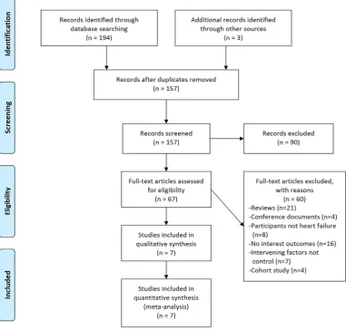
Figure 1 Flow diagram of study selection.
on September 22, 2020 by guest. Protected by copyright.
any micronutrient use except for vitamin D and (8) a minimum of 3 months of therapy was necessary for inclu-sion in the review to ensure that the intervention had sufficient time to produce a better effect. The exclusion criteria were as follows: (1) only the abstract was reported for reference; (2) studies with duplicated data, including the same group of patients or patients for whom there were updated results available; (3) the study had no outcomes of interest; (4) animal studies and reviews; (5) conference documents and (6) non-RCTs, cohort stud-iers and retrospective studies.
Data extraction and quality assessment
Data were independently extracted from each study by two authors (JDZ and JJJ) and entered into a struc-tured spreadsheet followed by a cross-check procedure. Disagreements were resolved by consensus or by a third investigator (PD). The following data were extracted from each trial: the first author’s surname, year of publication; demographic and methodological data; total number, mean age, gender distribution and race of enrolled patients; use of or change in drugs for HF; seated LVEDD
and LVEF at baseline, when available; number of patients randomly assigned to each intervention; duration of therapy; incidence and type of adverse events; number of dropouts or withdrawals because of adverse events; and change from baseline seated LVEDD and LVEF. Criteria for the RCT risk of bias evaluation listed in the Cochrane Handbook for Systematic Reviews of Interventions 5.1.0 were adopted, including the following: (1) random sequence generation, (2) allocation concealment, (3) blinding of patients and personnel, (4) blinding of outcome assess-ment, (5) incomplete outcome data and (6) selective reporting risk. Then, an evaluation system with ‘low risk’, ‘high risk’ and ‘not clear’ was established according to the six criteria described above.23
Outcomes assessed
The primary endpoint was LVEDD, and the secondary endpoints were LVEF and the incidence of adverse reactions.
Data analysis and synthesis
RevMan V.5.3 software was employed for data analysis. Continuous variables are reported as the mean differ-ence (MD) and 95% CI, and the test level was α=0.05. Following clinical heterogeneity analysis of the included studies, statistical heterogeneity was assessed using χ2
-based Cochran Q statistic and I2.24 For the Q statistic,
p≥0.1 indicates homogeneity among multiple similar studies, and the fixed-effects model was employed for the meta-analysis, while p<0.1 indicates statistically signif-icant heterogeneity, and the random-effects model was used for analysis. For the I2 statistic, I2 <25% indicates low
heterogeneity, while I2 >50% indicates moderate to high
heterogeneity.25
results
selection and description of studies
A total of 157 published papers were collected after the initial screening, and eventually, 7 RCTs26–32 with a total of
465 patients, including 235 cases in the vitamin D group and 230 cases in the control group, were included after reviewing the title, abstract and full text, as well as elim-inating duplicate documents, non-RCTs and studies that failed to meet the inclusion criteria. See figure 1 for the screening process.
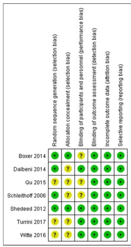
In the seven included studies,26–32 four reported an
appropriate randomisation method27–29 32; two adopted
allocation concealment29 32 and six used
double-blinding.26–30 32 See figure 2 for the evaluation of the
methodology of the studies.
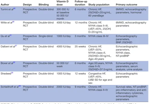
Data of the curative effect on the LVEDD were reported in five studies, and the LVEF was reported in all the included studies; two studies mentioned adverse reactions.26 30
Dropout or withdrawal from the research study was covered in all the included studies. The study characteristics are shown in table 1, and the basic information of the included population is shown in table 2.
Figure 2 Quality assessment.
on September 22, 2020 by guest. Protected by copyright.
The SD for changes from baseline on the LVEF were reported in four studies.27 28 30 32 According to
the Cochrane Handbook, the Corr were imputed by averaging the Corr of those four studies, and further imputed the SD for changes from baseline for other studies.26 29 31
The SD for changes from baseline on the LVEDD were reported in three studies.27 28 30 According to the Cochrane Handbook, the Corr were imputed by aver-aging the Corr of those three studies, and further imputed the SD for changes from baseline for other studies.26 29
effects of vitamin D on the lVeDD
Changes in the LVEDD of patients were reported in five studies,26–30 which showed high levels of
heteroge-neity among the results of the studies (heterogeheteroge-neity
χ2, p=0.07, I2=55%), thus supporting analysis using
the random-effects model. Compared with the control group, a decrease in the LVEDD was observed in the vitamin D group (MD=−2.31 mm, 95% CI −4.15 to −0.47, p=0.01) (figure 3).
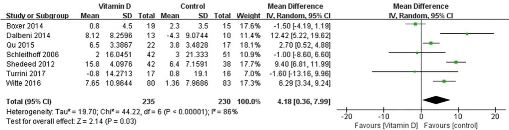
effects of vitamin D on the lVeF
Changes in the LVEF of patients were reported in all seven studies,26–32 which showed high levels of
hetero-geneity among the results of the studies (heteroge-neity χ2, p<0.001, I2=88%), thus supporting analysis
using the random-effects model. Compared with the control group, an increase in the LVEF was observed in the vitamin D group (MD=4.18%, 95% CI 0.36 to 7.99, p=0.03) (figure 4).
subgroup analysis
The analysis based on age stratification revealed that compared with the control group both the adults (aged ≥18 years) and non-adults (aged <18 years) with HF in the vitamin D group showed a decrease in the LVEDD (adults: heterogeneity χ2, p=0.65, I2=0%
;MD=−1.62 mm, 95% CI −2.83 to −0.42, p=0.008;
non-adults: MD=−5.51 mm, 95% CI −8.08 to −2.94, p<0.001) (figure 5). These results, however, need to be interpreted with caution as there was only one study in the subgroup of non-adults.
Table 1 Study characteristics
Author Design Blinding
Vitamin D dose
Follow-up
duration Study population Primary outcome
Turrini et al26 Prospective
RCT Double-blind 300 000 IU at baseline 50 000 IU/ month
6 months Chronic HF,
25(OH)D<20 ng/mL, 60 years<age
6MWD, echocardiography parameters, hormonal
Witte et al30 Prospective
RCT
Double-blind 4000 IU/day 12 months Chronic HF, NYHA class II–III, LVEF<45%, 25(OH) D<20 ng/mL
6MWD, echocardiography parameters
Qu et al31 Prospective
RCT Single-blind 1000 IU/day 3 months NYHA class III–IV Echocardiography parameters, BNP, 25(OH)D Dalbeni et al27 Prospective
RCT Double-blind 4000 IU/day 25 weeks Chronic HF, LVEF<55%, NYHA class>II, 25(OH)D<30 ng/mL, Age>40 years
Echocardiographic parameters, NYHA class, NT-proBNP
Boxer et al32 Prospective
RCT Double-blind 50 000 IU/week 6 months Age≥50 years, NYHA class II–IV, 25(OH)D<37.5 ng/mL
Echocardiographic parameters, serum analysis, urine analysis Shedeed29 Prospective
RCT Double-blind 1000 IU/day 12 weeks Congestive HF, LVEF<40%, LV<2 SD for age and sex
Echocardiographic parameters
Schleithoff et al28 Prospective
RCT Double-blind 2000 IU/day 9 months Chronic HF,NYHA class II–IV Survival rates, NT-proBNP,pro-inflammatory and anti-inflammatory cytokines, echocardiographic parameters
25(OH)D, 25-hydroxyvitamin D; 6MWD, 6-minute walk distance; HF, heart failure; IU, international units; LV, left ventricular; LVEF, left ventricular ejection fraction; NT, N-terminal pro-B-type natriuretic peptide; NYHA, New York Heart Association; RCT, randomised controlled trial.
on September 22, 2020 by guest. Protected by copyright.
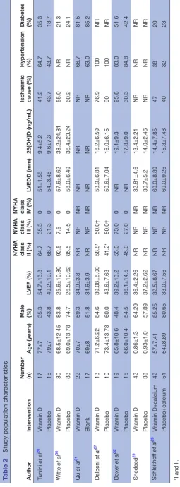
Table 2
Study population characteristics
Author
Intervention
Number (n)
Age (years)
Male (%)
LVEF (%)
NYHA class II (%) NYHA class III (%) NYHA class IV (%)
LVEDD (mm)
25(OH)D (ng/mL)
Ischaemic cause (%) Hypertension (%) Diabetes (%)
Turrini et al 26 Vitamin D 17 77±7 35.3 54.7±13.8 64.7 35.3 0 51±1.58 9.4±5.2 41.2 64.7 35.3 Placebo 16 79±7 43.8 49.2±19.1 68.7 21.3 0 54±3.48 9.6±7.3 43.7 43.7 18.7 Witte et al 30 Vitamin D 80 68.5±12.45 83.8 25.6±10.80 92.5 7.5 0 57.6±8.62 38.2±24.81 55.0 NR 21.3 Placebo 83 69.0±13.78 74.7 26.5±10.62 85.5 14.5 0 58.0±6.49 36.4±20.24 60.2 NR 24.1 Qu et al 31 Vitamin D 22 70±7 59.3 34.9±3.8 NR NR NR NR NR NR 66.7 81.5 Blank 17 69±8 51.8 34.6±3.9 NR NR NR NR NR NR 63.0 85.2 Dalbeni et al 27 Vitamin D 13 71.2±6.22 84.6 39.08±8.00 58.8* 50.0† 53.9±6.81 16.2±6.59 76.9 100 NR Placebo 10 73.4±13.78 60.0 43.6±7.63 41.2* 50.0† 50.6±7.04 16.0±6.15 90 100 NR Boxer et al 32 Vitamin D 19 65.8±10.6 48.4 39.2±13.2 55.0 73.0 0 NR 19.1±9.3 25.8 83.0 51.6 Placebo 15 66.0±10.4 54.5 36.1±14.5 45.0 27.0 0 NR 17.8±9.0 30.3 84.8 42.4 Shedeed 29 Vitamin D 42 0.86±1.3 64.29 36.4±2.26 NR NR NR 32.81±4.6 13.4±2.21 NR NR NR Placebo 38 0.93±1.0 57.89 37.2±2.62 NR NR NR 30.7±5.2 14.0±2.46 NR NR NR Schleithof f et al 28 Vitamin D+calcium 42 57±7.41 85.25 32.5±8.67 NR NR NR 69.0±8.89 14.4±7.85 47 38 20 Placebo+calcium 51 54±8.89 80.65 33.0±7.56 NR NR NR 69.0±9.26 15.3±7.48 40 32 23
*I and II. †III and IV
.
25(OH)D, 25-hydr
oxyvitamin D; L
VEDD, left ventricular end-diastolic dimension; L
VEF
, left ventricular ejection fraction; NR, not r
eported; NYHA, New Y
ork Heart Association.
on September 22, 2020 by guest. Protected by copyright.
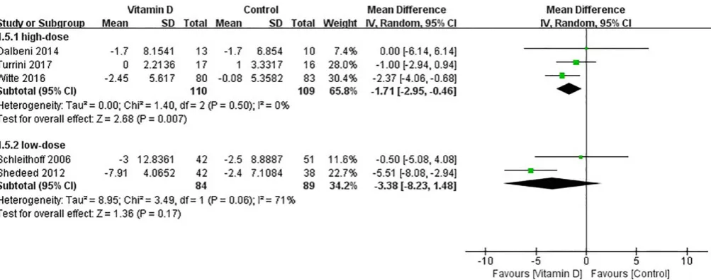
A subgroup analysis was performed according to the dosage of vitamin D. There was an effect from high-dose vitamin D on the reduction of the LVEDD (heteroge-neity χ2, p=0.50, I2=0%; MD=−1.71 mm, 95% CI −2.95 to
−0.46, p=0.007), but this effect was not seen with low-dose vitamin D treatment (heterogeneity χ2, p=0.06, I2=71%;
MD=−3.38 mm, 95% CI −8.23 to 1.48, p=0.17). (figure 6). According to patients with or without reduced ejection fraction, subgroup analyses were performed. Vitamin D supplementation was effective at reducing the LVEDD in patients with reduced ejection fraction (patients with reduced ejection fraction: heterogeneity χ2, p=0.07,
I2=62%;MD=−3.11 mm, 95% CI −5.67 to −0.55, p=0.02; patients without reduced ejection fraction: heterogeneity
χ2, p=0.76, I2=0%;MD=−0.91 mm, 95% CI −2.76 to 0.94,
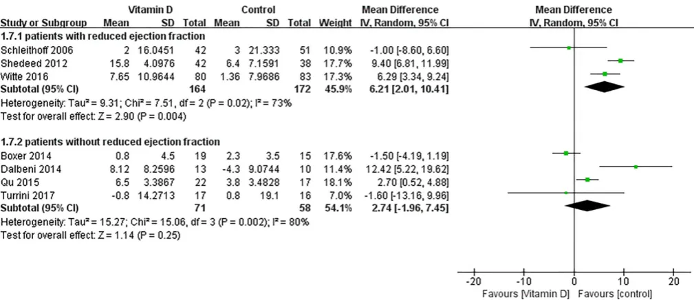
p=0.34) (figure 7). In addition, vitamin D supplementa-tion was effective at increasing the LVEF in patients with reduced ejection fraction (patients with reduced ejection fraction: heterogeneity χ2, p=0.02, I2=73%; MD=6.21%,
95% CI 2.01 to 10.41, p=0.004; patients without
reduced ejection fraction: heterogeneity χ2, p=0.002,
I2=80%; MD=2.74%, 95% CI −1.96 to 7.45, p=0.25)
(figure 8).
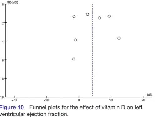
Publication bias
There was significant asymmetry in the funnel plot for the effect of vitamin D on the LVEDD, which may be due to publication bias and other causes (figure 9). On the other hand, no publication bias was found for the effect of vitamin D on the LVEF (figure 10).
Adverse event
Two studies26 30 reported adverse events. During the
follow-up, no significant adverse event was recorded. No data associated with the incidence of adverse events were recorded in the other studies.
DIsCussIOn
At present, different studies have reported controver-sial conclusions regarding the influence of vitamin D on ventricular remodelling in patients with HF. The results of this study show that compared with the control group supplementation of vitamin D could reduce the LVEDD (MD=−2.31 mm, 95% CI −4.15 to −0.47, p=0.01) and improve the LVEF (MD=4.18%, 95% CI 0.36 to 7.99, p=0.03) among patients with HF. In addition, a more pronounced effect is achieved using high-dose vitamin D and on patients with a reduced ejection fraction.
Most new treatment methods for chronic HF are expen-sive and require advanced technologies33; in addition, most
of these treatment methods have not passed strict phase III clinical trials. For patients with HF, vitamin D is not only cheap but also safe, and these patients may obtain more bene-fits from vitamin D therapy.34 Vitamin D toxicity is based not
only on the dosing but also on circulating 25-hydroxyvitamin D (25(OH)D) levels. The Institute of Medicine35 has set the
dosage for vitamin D at 4000 IU daily for healthy adults, and the Endocrine Society15 has set a dosage of 10 000 IU daily
for patients who are at risk of having circulating 25(OH) D levels<50 nmol/L. The Institute of Medicine35 considers
Figure 3 Forest plot showing the effect of vitamin D on left ventricular end-diastolic dimension.
Figure 4 Forest plot showing the effect of vitamin D on left ventricular ejection fraction.
on September 22, 2020 by guest. Protected by copyright.
circulating 25(OH)D levels below 30 nmol/L as deficient, levels between 30 and 49.99 nmol/L as inadequate, levels between 50 and 125 nmol/L as adequate and levels above 125 nmol/L as potentially harmful. In HF, cardiac contrac-tion and diastole are affected due to overload of Ca2+ ions
in myocardial cells. Lack of vitamin D may intervene with the functions of Ca2+ in myocardial cells, thus resulting
in cardiomyocyte hypertrophy and intra-organisational inflammatory reaction and fibrosis.36 37 Low vitamin D levels
can activate the renin–angiotensin system,38 give rise to
inflammatory reactions39 and result in endothelial
dysfunc-tion.40 The effects of vitamin D on the CV system are
addi-tionally mediated through elevated parathyroid hormone (PTH) levels, which are associated with the development of left ventricular (LV) hypertrophy.41 Although there is much
evidence showing that a lack of vitamin D could result in poor prognosis among patients with HF, different studies have reported controversial conclusions regarding whether
supplementation of vitamin D could benefit patients with HF.
In recent years, some relatively small RCTs have studied the influence of vitamin D on patients with HF. In the 2014 World Heart Failure Conference, Louise et al42 reported an
RCT that performed a 6-month study on 32 patients with HF, and the result showed that supplementation of vitamin D did not improve the LVEF or pro-BNP level. A recent meta-analysis43 also reported that vitamin D
supplementa-tion could not improve the LVEF (WMD: 4.11%, 95% CI −0.91 to 9.12, p=0.11) and 6-minute walk distance (6MWD) (WMD: 8.90 m, 95% CI: −48.47 to 66.26, p=0.76) in the treatment of chronic HF. In contrast, this study included three other studies, two of which showed a positive effect from vitamin D supplementation. The present studies have not shown that vitamin D supplementation can improve the 6MWD among patients26 30 44 45; thus, our
meta-anal-ysis did not evaluate this parameter. There are probably
Figure 5 Subgroup analysis of effect of vitamin D on left ventricular end-diastolic dimension according to patients’ age.
Figure 6 Subgroup analysis of effect of vitamin D on left ventricular end-diastolic dimension (LVEDD) according to dose of vitamin D.
on September 22, 2020 by guest. Protected by copyright.
many factors that affect exercise tolerance, such as phys-ical condition, obesity, habits and environment, and these confounding factors may obscure the weak force from the remodelled ventricle. However, other studies have drawn opposite conclusions. In 2015, Lowry et al46 reported in
the European Society of Cardiology a 12-month RCT, and the result showed that for patients with HF supplemen-tation of vitamin D could improve ventricular remodel-ling (LVEDD −4.46 mm; p=0.047); in addition, the LVEF showed an increasing trend. A non-RCT47 also showed
that supplementation of vitamin D could remarkably increase the LVEF value compared with baseline. Elidrissy
et al48 evaluated 61 cases of children with cardiomyopathy;
they concluded, based on the available evidence, that the most likely cause of cardiomyopathy is hypocalcaemia and suggested maternal supplementation of vitamin D during
pregnancy and lactation with up to 2000 units of vitamin D and 400 units for their infants for prevention, which was also similar to the results reported by Shedeed.29
Since there are disputes among different study results, we conducted a meta-analysis of relevant RCT studies to further clarify the influence of vitamin D supplemen-tation on ventricular remodelling in patients with HF. Results of this study show that supplementation of vitamin D inhibits myocardial remodelling and improves cardiac function in patients with HF.
Different studies have different results and conclu-sions, which may be attributed to the different recom-mended dosages of vitamin D; since there is no standard recommended dosage of vitamin D at present, there are differences in the recommended dosage of vitamin D in different trials, which may affect the study results.
Figure 7 Subgroup analysis of effect of vitamin D on left ventricular end-diastolic dimension according to patients with or without reduced ejection fraction.
Figure 8 Subgroup analysis of effect of vitamin D on left ventricular ejection fraction according to patients with or without reduced ejection fraction.
on September 22, 2020 by guest. Protected by copyright.
However, there has not been a report related to adverse effects from the dosage of vitamin D. Meanwhile, the study results are related to the selected group; for example, the study results of Schleithoff et al28 showed that there were
no remarkable changes in the LVEF and LVEDD, and the participants included in this study were predominantly those with NYHA level 3–4 and a high degree of HF, and there was a high rate of lost to follow-up visit (37%).
The data on changes in the LVEDD showed high levels of heterogeneity among studies. Shedeed29 studied the
cause of heterogeneity according to subgroup. There was a difference in the metabolism of vitamin D and cardiac recovery capacity between newborns and adults, which might be the cause of heterogeneity.
Although all trials included in this study are RCTs, there are still many limitations in this study (1) because the current studies found that vitamin D has a weak and uncertain effect on ventricular remodelling and cardiac function in patients with HF and cannot improve exercise tolerance or reduce cardiac mortality, additional large-scale clinical studies are needed. Future research needs to focus on whether different vitamin D dosages would be superior; in addition, different selection criteria need
to be defined (eg, ejection fraction, vitamin D threshold, PTH threshold, etc) and additional echocardiographic parameters, the 6MWD and cardiovascular mortality need to be evaluated; (2) this study exhibits heteroge-neity, and age stratification and whether there is reduced ejection fraction may be sources of clinical heterogeneity according to the subgroup analysis; (3) different recom-mended dosages of vitamin D are reported in different trials, which may affect study results; therefore, additional trials are required to explore the relationship between vitamin D dosage and effect; (4) the conclusions need to be interpreted with caution as the extent of detected improvement in the remodelling parameters is close to the range of the inter-observer or intra-observer vari-ability of the echocardiographic method itself. The base-line vitamin D level of patients and the follow-up duration may affect the study results, and except for Dalbeni et al,27 who have mentioned that no change in therapy was
made during follow-up, other studies have not reported adjustments in HF medication. Therefore, whether the weak improvement in the remodelling parameters from vitamin D are attributed to other HF drugs is unclear.
In conclusion, this study shows that supplementation of vitamin D inhibits myocardial remodelling in patients with HF and improves their cardiac function. Vitamin D may be used as adjunctive HF medication for patients with HF with an underlying lack of or insufficiency in vitamin D. This result is encouraging and of great clinical interest but still far from practical implications. The main implication is to encourage further research.
Contributors J-DZ: Makes a significant contribution to the concept or design of the work; or the acquisition, analysis or interpretation of work data; drafting work or critically modifying important knowledge content; final approval of the released version; and agreeing to all work Responsible for ensuring proper investigation and resolution of issues related to the accuracy or completeness of any part of the work. JJ J: Participate in writing. P-SD: Serving as a scientific consultant. DZ: Participated in the technical editing of the manuscript. X-MY: Critically reviewed the research proposal. D-LL: Data collected. H-FZ: Data collected.
Funding The authors have not declared a specific grant for this research from any funding agency in the public, commercial or not-for-profit sectors.
Competing interests None declared. Patient consent Not required.
ethics approval The study was conducted in accordance with the Helsinki Declaration and approved by the Ethics Committee of Henan University of Science and Technology.
Provenance and peer review Not commissioned; externally peer reviewed. Data sharing statement The authors declare that they are willing to share their data directly.
Open access This is an open access article distributed in accordance with the Creative Commons Attribution Non Commercial (CC BY-NC 4.0) license, which permits others to distribute, remix, adapt, build upon this work non-commercially, and license their derivative works on different terms, provided the original work is properly cited, appropriate credit is given, any changes made indicated, and the use is non-commercial. See: http:// creativecommons. org/ licenses/ by- nc/ 4. 0/.
reFerenCes
1. Jin M, Wei S, Gao R, et al. Predictors of Long-Term Mortality in
Patients With Acute Heart Failure. Int Heart J 2017;58:409–15.
Figure 9 Funnel plots for the effect of vitamin D on left ventricular end-diastolic dimension.
Figure 10 Funnel plots for the effect of vitamin D on left ventricular ejection fraction.
on September 22, 2020 by guest. Protected by copyright.
2. Heidenreich PA, Albert NM, Allen LA, et al. Forecasting the impact of heart failure in the United States: a policy statement from the
American Heart Association. Circ Heart Fail 2013;6:606–19.
3. Mosterd A, Hoes AW. Clinical epidemiology of heart failure. Heart
2007;93:1137–46.
4. Roger VL, Weston SA, Redfield MM, et al. Trends in heart failure
incidence and survival in a community-based population. JAMA
2004;292:344–50.
5. McMurray JJ, Adamopoulos S, Anker SD, et al. ESC guidelines
for the diagnosis and treatment of acute and chronic heart failure 2012: The Task Force for the Diagnosis and Treatment of Acute and Chronic Heart Failure 2012 of the European Society of Cardiology. Developed in collaboration with the Heart Failure Association (HFA)
of the ESC. Eur J Heart Fail 2012;14:803–69.
6. Sun Q, Shi L, Rimm EB, et al. Vitamin D intake and risk of
cardiovascular disease in US men and women. Am J Clin Nutr
2011;94:534–42.
7. Tamez H, Thadhani RI. Vitamin D and hypertension: an update and
review. Curr Opin Nephrol Hypertens 2012;21:492–9.
8. Karakas M, Thorand B, Zierer A, et al. Low levels of serum
25-hydroxyvitamin D are associated with increased risk of myocardial infarction, especially in women: results from the MONICA/
KORA Augsburg case-cohort study. J Clin Endocrinol Metab
2013;98:272–80.
9. Sun Q, Pan A, Hu FB, et al. 25-Hydroxyvitamin D levels and the
risk of stroke: a prospective study and meta-analysis. Stroke
2012;43:1470–7.
10. Desai CK, Huang J, Lokhandwala A, et al. The role of vitamin
supplementation in the prevention of cardiovascular disease events.
Clin Cardiol 2014;37:576–81.
11. Schierbeck LL, Jensen TS, Bang U, et al. Parathyroid hormone and
vitamin D–markers for cardiovascular and all cause mortality in heart failure. Eur J Heart Fail 2011;13:626–32.
12. Liu L, Chen M, Hankins SR, et al. Serum 25-hydroxyvitamin D
concentration and mortality from heart failure and cardiovascular disease, and premature mortality from all-cause in United States
adults. Am J Cardiol 2012;110:834–9.
13. Ford JA, MacLennan GS, Avenell A, et al. Cardiovascular disease
and vitamin D supplementation: trial analysis, systematic review, and
meta-analysis. Am J Clin Nutr 2014;100:746–55.
14. Wang L, Song Y, Manson JE, et al. Circulating 25-hydroxy-vitamin D
and risk of cardiovascular disease: a meta-analysis of prospective
studies. Circ Cardiovasc Qual Outcomes 2012;5:819–29.
15. Holick MF, Binkley NC, Bischoff-Ferrari HA, et al. Evaluation, treatment, and prevention of vitamin D deficiency: an Endocrine
Society clinical practice guideline. J Clin Endocrinol Metab
2011;96:1911–30.
16. Gotsman I, Shauer A, Zwas DR, et al. Vitamin D deficiency is
a predictor of reduced survival in patients with heart failure;
vitamin D supplementation improves outcome. Eur J Heart Fail
2012;14:357–66.
17. Pourdjabbar A, Dwivedi G, Haddad H. The role of vitamin D in
chronic heart failure. Curr Opin Cardiol 2013;28:216–22.
18. Li YC, Kong J, Wei M, et al. 1,25-Dihydroxyvitamin D(3) is a negative
endocrine regulator of the renin-angiotensin system. J Clin Invest
2002;110:e38.
19. Tomaschitz A, Pilz S, Ritz E, et al. Independent association
between 1,25-dihydroxyvitamin D, 25-hydroxyvitamin D and the renin-angiotensin system: the Ludwigshafen Risk
and Cardiovascular Health (LURIC) study. Clin Chim Acta
2010;411:e60.
20. Forman JP, Williams JS, Fisher ND. Plasma 25-hydroxyvitamin D and
regulation of the renin-angiotensin system in humans. Hypertension
2010;55:1283–8.
21. Weber KT, Weglicki WB, Simpson RU. Macro- and micronutrient dyshomeostasis in the adverse structural remodelling of
myocardium. Cardiovasc Res 2009;81:500e8.
22. Agarwal M, Phan A, Willix Jr R, et al. Is vitamin D deficiency associated with heart failure? A review of current evidence.
J Cardiovasc Pharmacol Ther 2011;16: 354:e63.
23. Higgins JPT, Green S, Cochrane Handbook for Systematic Reviews of Interventions Version 5.1.0, 2011. www. cochrane- hand- book. org (updated Mar 2011).
24. Higgins JP, Thompson SG, Deeks JJ, et al. Measuring inconsistency
in meta-analyses. BMJ 2003;327:557–60.
25. Higgins JP, Thompson SG. Quantifying heterogeneity in a
meta-analysis. Stat Med 2002;21:1539–58.
26. Turrini F, Scarlini S, Giovanardi P, et al. Effects of Cholecalciferol Supplementation in Patients with stable heart failure and LOw vITamin D levels (ECSPLOIT-D): a double-blind, randomized,
placebo-controlled pilot study. Minerva Cardioangiol 2017;65.
27. Dalbeni A, Scaturro G, Degan M, et al. Effects of six months
of vitamin D supplementation in patients with heart failure: a
randomized double-blind controlled trial. Nutr Metab Cardiovasc Dis
2014;24:861–8.
28. Schleithoff SS, Zittermann A, Tenderich G, et al. Vitamin D
supplementation improves cytokine profiles in patients with congestive heart failure: a double-blind, randomized,
placebo-controlled trial. Am J Clin Nutr 2006;83:754–9.
29. Shedeed SA. Vitamin D supplementation in infants with chronic
congestive heart failure. Pediatr Cardiol 2012;33:713–9.
30. Witte KK, Byrom R, Gierula J, et al. Effects of Vitamin D on Cardiac
Function in Patients With Chronic HF: The VINDICATE Study. J Am
Coll Cardiol 2016;67:2593–603.
31. Jun Q, Zhen W, hong-bo S, et al. The C linical Effect of V itam in D 3
Supplem entation on C ardiac Function in Patients w ith Ischem ic H
eart Failure. Chinese and Foreign M edical R esearch 2015;13:3–5.
32. Boxer RS, Hoit BD, Schmotzer BJ, et al. The effect of vitamin D on
aldosterone and health status in patients with heart failure. J Card
Fail 2014;20:334–42.
33. Amao R, Imamura T, Nakahara Y, et al. Reversible Motor Paralysis
and Early Cardiac Rehabilitation in Patients With Advanced Heart
Failure Receiving Left Ventricular Assist Device Therapy. Int Heart J
2016;57:766–8.
34. Witte KK, Byrom R. Micronutrients for chronic heart failure: end of
the road or path to enlightenment? JACC Heart Fail 2014;2:318–20.
35. Institute of Medicine. Dietary Reference Intakes: Calcium and Vitamin
D. Washington, DC: National Academies Press, 2011.
36. Weishaar RE, Simpson RU. Involvement of vitamin D3 with
cardiovascular function. II. Direct and indirect effects. Am J Physiol
1987;253:E675–83.
37. Chen S, Law CS, Grigsby CL, et al. Cardiomyocyte-specific deletion
of the vitamin D receptor gene results in cardiac hypertrophy.
Circulation 2011;124:1838–47.
38. Vaidya A, Williams JS. The relationship between vitamin D and the renin-angiotensin system in the pathophysiology of hypertension,
kidney disease, and diabetes. Metabolism 2012;61:450–8.
39. Zhang Y, Leung DY, Richers BN, et al. Vitamin D inhibits monocyte/
macrophage proinflammatory cytokine production by targeting
MAPK phosphatase-1. J Immunol 2012;188:2127–35.
40. Caprio M, Mammi C, Rosano GM. Vitamin D: a novel player in
endothelial function and dysfunction. Arch Med Sci 2012;8:4–5.
41. Saleh FN, Schirmer H, Sundsfjord J, et al. Parathyroid hormone and
left ventricular hypertrophy. Eur Heart J 2003;24:e60.
42. Schierbeck LLLouise, Madsen JL, Bang UC. The effect of vitamin D supplementation in vitamin D deficient men with heart failure. A
randomized controlled trial.:P1686. European journal of heart failure
2014;16:335.
43. Jiang WL, Gu HB, Zhang YF, et al. Vitamin D Supplementation in the
Treatment of Chronic Heart Failure: A Meta-analysis of Randomized
Controlled Trials. Clin Cardiol 2016;39:56–61.
44. Boxer RS, Kenny AM, Schmotzer BJ, et al. A randomized controlled
trial of high dose vitamin D3 in patients with heart failure. JACC Heart
Fail 2013;1:84–90.
45. Witham MD, Crighton LJ, Gillespie ND, et al. The effects of vitamin
D supplementation on physical function and quality of life in older
patients with heart failure: a randomized controlled trial. Circ Heart
Fail 2010;3:195–201.
46. Lowry J, Gierula J, Byrom R. Vitamin D supplementation improves the size and function of the left ventricle in patients with heart failure.
Eur Heart J 2015;36:665.
47. Zia AA, Komolafe BO, Moten M, et al. Supplemental vitamin D and
calcium in the management of African Americans with heart failure
having hypovitaminosis D. Am J Med Sci 2011;341:113–8.
48. Elidrissy AT, Munawarah M, Alharbi KM. Hypocalcemic rachitic
cardiomyopathy in infants. J Saudi Heart Assoc 2013;25:25–33.
on September 22, 2020 by guest. Protected by copyright.