DOI: 10.7324/JAPS.2018.8905 ISSN 2231-3354
© 2018 Gehan F. Balata. This is an open access article distributed under the terms of the Creative Commons Attribution License -NonCommercial- ShareAlikeUnported License (http://creativecommons.org/licenses/by-nc-sa/3.0/).
*Corresponding Author
Gehan F. Balata, Department of Pharmaceutics, Faculty of Pharmacy, Zagazig University, Zagazig, Egypt. E-mail: gf_balata @ yahoo.com
Formulation and Evaluation of Gliclazide in Vegetable Oil-Based Self
Emulsifying Delivery System
Gehan F. Balata
Department of Pharmaceutics, Faculty of Pharmacy, Zagazig University, Zagazig, Egypt.
ARTICLE INFO ABSTRACT
Article history: Received on: 05/05/2018 Accepted on: 17/07/2018 Available online: 30/09/2018
Objectives: To formulate a poorly water-soluble, antidiabetic drug, gliclazide, in the vegetable oil-based self-emulsifying delivery system (SEDDS), as a trial to improve its dissolution and hence its bioavailability. Material and methods: Solubility of gliclazide was investigated in different vegetable oils, surfactants and co-surfactants. The system was selected and the self-emulsification region was identified. Drug-loaded SEDDS was prepared and evaluated in terms of emulsification time, phase separation, droplet size and rate of drug dissolution. The formulation that showed good self-emulsifying properties, acceptable droplet size and high % drug release was further processed into solid- SEDDS by physical mixing with varying proportions of a mixture (2:1) of microcrystalline cellulose and lactose and characterized for crystalline, droplet size and in vitro drug dissolution properties. Results and Conclusions: Formulation of gliclazide in liquid and solid SEDDS using olive oil resulted in significant (P < 0.05) improvement in its dissolution properties. Both differential scanning calorimetry and x-ray diffraction studies demonstrated the presence of gliclazide in the molecular state in all the prepared solid formulations. Solid SEDDS of gliclazide containing a lower proportion of adsorbent (G1) had comparable dissolution properties with its liquid form. Thus formulation of gliclazide in the solid self-emulsifying system may be a promising strategy to improve its dissolution properties.
Key words:
gliclazide, solid-SEDDS, ternary phase diagram, dissolution, olive oil.
INTRODUCTION
Type 2 diabetes mellitus is a serious health problem in Egypt associated with high mortality rates due to macrovascular and microvascular complications (Hegazi et al., 2015).
Gliclazide, a second-generation hypoglycemic sulfonylurea, is the drug of choice in prolonged therapy for the control of Type 2 diabetes mellitus (Harrower, 1994). It stimulates insulin secretion from pancreatic beta cells (Jung et al., 1999) and increases the sensitivity of the peripheral tissue to insulin (Shavi et al., 2010). Gliclazide shows low aqueous solubility and consequently slow and variable gastrointestinal absorption with low bioavailability after oral administration (Palmer and Brogden, 1993). The solubility and dissolution rate of gliclazide had been improved by different methods including inclusion complexes with beta-cyclodextrin (Sapkal et al., 2007), solid dispersion with different carriers (Biswal et al., 2008; 2009) and surface solid
dispersion (Mahajan et al., 2012). Continuing that research, the self-emulsifying delivery system was suggested to improve the solubility and dissolution rate of gliclazide.
enzymatic or chemical degradation and activates lipoproteins promoting the lymphatic transport of lipophilic drugs (Craig, 1993). Moreover, emulsion droplets lead to a faster distribution of the drug in the gastrointestinal tract and hence minimizing the irritation due to the contact between the drug and the gut wall (Franceschinis et al., 2011). However, liquid SEDDS has many disadvantages that include: low drug loading capacity, drug leakage, low stability, incompatibility problems with the capsule shell and possibility of irreversible drugs/excipients precipitation (Kallakunta et al., 2012). Alternatively, solid- SEDDS has been already produced by loading liquid SEDDS on solid carriers using different technologies: extrusion/spheronisation (Iosio et al., 2008), wet granulation in high shear mixer (Franceschinis et al., 2005), spray drying (Yi et al., 2008) and physical mixing (Dixit and Nagarsenker, 2008). Solid SEDDS offer many advantages which include: stability, facility of manufacturing, can easily be packed in hard gelatin capsules and disperse freely in the gastrointestinal tract. In general, solid SEDDS combine the advantages of lipid-based drug delivery systems with those of solid dosage forms (Kallakunta et al., 2012).
Oils play a crucial role in the formulation of self-emulsifying formulations. Both long and medium chain triglyceride oils with different degrees of saturation are commonly used in the preparation of SEDDS (Khedekar and Mittal, 2013). Dietary vegetable oils with antioxidant activity such as olive oil and linseed oil are reported to be effective in cardiovascular diseases and associated metabolic disorders, including hyperglycemia and dyslipidemia (Berraaouan et al., 2013; Samarji and Balbaa, 2014; Kaithwas and Majumdar, 2012).
Previous literature reported the formulation of liquid and solid self-emulsifying system of gliclazide using Capryol 90 as the oil phase (Wankhade et al., 2012; Nipun and Ashraful Islam, 2014). The aim of this study is to incorporate gliclazide in vegetable oil-based solid-SEDDS in an attempt to improve its solubility and dissolution rate and hence its bioavailability. Liquid SEDDS was prepared using vegetable oil, Tween 80 as surfactant and propylene glycol as co-surfactant. Then the optimized formula was adsorbed on a mixture of microcrystalline cellulose and lactose by physical mixing and studied for its efficiency in improving drug dissolution properties.
MATERIAL AND METHODS
Gliclazide was obtained as a gift sample from Delta Pharma, Egypt. Tween 80, Tween 20 and Propylene glycol (PG) were purchased from Merck Specialities Pvt. Ltd., Mumbai. Polyethylene glycol 400 (PEG 400), Methanol,Soybean oil, Olive oil, Palm oil, Sunflower oil and Sesame oil were purchased from SD Fine Chemicals, Mumbai. Avicel PH 101 (Microcrystalline cellulose (MCC)) and Lactose were purchased from FMC
BioPolymer, USA.
Saturation solubility study
Vegetable oils are one class of lipid solvents that are safe as they are fully digested and absorbed after oral administration (Mohanvarma and Padavala, 2013). So, the solubility of gliclazide was determined in various vegetable oils, as well as, surfactants and co-surfactants. An excess amount of gliclazide (50 mg) was added to 5 ml of the selected vehicles in capped vials and the mixtures were sonicated (Ultrasonic model SS101H, Sonix IV, Huntington Beach, CA, USA) at 40-50°C to facilitate drug solubilization. Then the mixtures were transferred to a thermostatic shaker water bath (LSB-030S, Daihan Lab Tech Co., LTD, Indonesia) maintained at 25°C for 48 hr. After reaching equilibrium, vials were centrifuged (Nuve, NF 815, Ankara, Turkey) at 4000 rpm for 10 min and followed by filtration through a membrane filter (No 0.45 μm). The clear filtrate was diluted with methanol and measured spectrophotometrically at 226 nm (Apel Co., Ltd., Japan) (Palmer and Brogden, 1993). Triplicate measurements were taken and Mean ± SD was recorded.
Construction of ternary phase diagram & formulation optimization
Based on the results of solubility studies, oil, surfactant, and cosurfactant showing highest solubility of gliclazide were selected for construction of the phase diagram. Different blends were prepared using varying concentrations of oil (20, 30, 40 and 50%, w/w). Surfactant and cosurfactant (Smix) at each oil percentage were mixed by vortexing (JL6001-G/L, Mettler Toledo Co., Switzerland) in different weight ratios (1:1, 2:1, 3:1 and 4:1 w/w) (Table 1). About 0.2 ml of each blend was introduced into 100 ml of distilled water and the contents were mixed gently with a magnetic stirrer (magnetic stirrer, Thermolyne, Dubuque, IA, USA) at 100 rpm for 2 min at room temperature. The diluted products were inspected for clarity, phase separation and coalescence of droplets. Emulsions showing phase separation, cracking and coalescence of oil droplets were judged as unstable emulsions (Aboutaleb et al., 2016). Phase diagram was constructed to identify the self-emulsifying region using CHEMIX software. Preparation of gliclazide SEDDS
SEDDS formulations with gliclazide were prepared by adding the weighed amount of drug (Each 1 g formulation contained 40 mg of gliclazide) to surfactant followed by addition of co-surfactant and oil in a glass vials (Table 2). The resultant mixtures were stirred continuously by vortex mixing and heated at 40°C to obtain a homogeneous isotropic mixture. The SEDDS formulations were stored at ambient temperature until further use (Kallakunta et al., 2012).
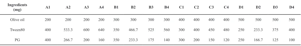
Table 1: Composition of different oil, surfactant and cosurfactant blends.
Ingredients
(mg) A1 A2 A3 A4 B1 B2 B3 B4 C1 C2 C3 C4 D1 D2 D3 D4
Olive oil 200 200 200 200 300 300 300 300 400 400 400 400 500 500 500 500
Tween80 400 533.3 600 640 350 466.7 525 560 300 400 450 480 250 233.3 375 400
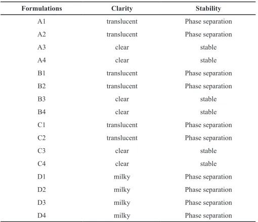
Table 2: Different formulations of gliclazide SEDDS.
Ingredients
(mg) A3 A4 B3 B4 C3 C4
Gliclazide 40 40 40 40 40 40
Olive oil 200 200 300 300 400 400
Tween80 600 640 525 560 450 480
PG 200 160 175 140 150 120
Characterization of SEDDS Assessment of emulsification time
The emulsification time for different formulations was determined using Dissolution Tester apparatus II, rotating paddle (SP6-400, G.B. CALEVA Ltd., Dorset, England). One gram of each formulation was added to 500 ml of distilled water with gentle agitation at 70 rpm and at a temperature of 37°C. The time to form homogenous nanoemulsion was determined (Abdalla et al., 2008).
Stability studies
(a) Phase separation & drug precipitation study
Since SEDDS are subjected to dilution in GIT after oral administration, so the ability of the prepared SEDDS to be diluted easily without any drug precipitation was determined. Different gliclazide SEDDS formulations were exposed to 100 and 500 folds dilution with distilled water. Examined formulations were stored at 25°C for 24 h and observed visually for phase separation and drug precipitation (Yin et al., 2009).
(b) Refrigeration thaw cycle
A single refrigeration thaw cycle was performed by taking 2 ml sample of each formulation which subjected to refrigeration at 2°C for 24 h then thawing at 40°C. The samples were then observed for phase separation and drug precipitation.
(c) Centrifugation
A 5 ml sample of each formulation was centrifuged (REMI,11, Cama Industrial Estate, Walbhat Road, Goregaon East, Mumbai – 400063, India) at 4,000 rpm for 5 min. Thereafter, the samples were observed for phase separation and drug precipitation. Determination of droplet size
The mean size of emulsion globules was determined by Zetasizer (Malvern Instruments, Malvern, UK) based on dynamic laser scattering. The dispersed formulations were measured after dilution (1:1000 v/v) with distilled water and mix for 1 minute with Cyclo mixer
In-vitro drug dissolution from liquid SEDDS
The in vitro dissolution test was performed in 900 ml distilled water maintained at 37 ± 0.5°C using USP Dissolution Tester apparatus II rotating at 70 rpm (SP6-400, G.B. CALEVA Ltd., Dorset, England). The SEDDS formulations (containing an amount equivalent to 40 mg gliclazide) were filled into hard gelatin capsules (size 00) and used for drug release studies compared with the plain drug. Samples (5 ml) were withdrawn and replaced with
fresh media after 5, 10, 15, 20, 30, 40, 50 and 60 min. Samples were filtered using a 0.45 μm filter and analyzed spectrophotometrically for gliclazide at 226 nm. Triple measurements were conducted for each formulation, and data presented as means ± SD. Percentage of cumulative drug dissolution at different time intervals was calculated and a graph plotted versus time. The dissolution data were analyzed in terms of initial dissolution rate over the first 10 min of dissolution (IDR = % dissolved/min) (Kallakunta et al., 2013) and dissolution efficiency after 60 min (DE60) which was calculated from the area under the dissolution curve at time‘t’ (measured using the trapezoidal rule) and expressed as percentage of the area of the rectangle described by 100% dissolution in the same time (Khan, 1975).
Formulation of solid SEDDS
The liquid SEDDS that exhibited acceptable emulsification efficiency and high dissolution parameters was selected for preparation of solid SEDDS, in three different ratios (liquid SEDDS:carrier mixture ratio was 1:1, 1:2 and 1:3 w/w). The formulations G1-G3 were prepared with a carrier mixture of MCC: lactose, 2:1 w/w. The liquid SEDDS was triturated with a mixture of MCC and lactose using a mortar and a pestle. The wet mass was passed through sieve No.120 and was dried at ambient temperature (Bhagwat and D’Souza, 2012).
Characterization of solid SEDDS
Differential scanning calorimetry (DSC)
The DSC thermograms of gliclazide, MCC, lactose and different solid SEDDS formulations were recorded using differential scanning calorimeter (Perkin Elmer, USA). The samples were heated in an open aluminum pan from 35 to 200°C at a scanning rate of 10°C/min under the stream of nitrogen.
X-ray diffraction studies (XRD)
X-ray diffraction measurements of gliclazide, MCC, lactose and different solid SEDDS formulations were carried out with X-ray diffractometer (X’Pert PRO PA Nalytical, USA). XRD patterns were recorded at room temperature using monochromatic CuKa-radiation over a range of 2θ angles from 3° to 50° with an angular increment of 0.02° per second.
Determination of droplet size
Droplet size of the dispersed solid SEDDS was determined using the same procedure as mentioned before for liquid SEDDS.
In-vitro drug dissolution
An amount of powder equivalent to 40 mg gliclazide was filled into hard gelatin capsules (size 00) and the dissolution of gliclazide from different solid SEDDS formulations were done under the same conditions as mentioned before for liquid SEDDS. RESULTS AND DISCUSSION
Saturation solubility study
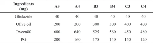
key factor for optimum drug loading and stability during storage (Miryala and Kurakula, 2013). The solubility of gliclazide in the screened vegetable oils, surfactants and co-surfactants is presented in Figure 1. Gliclazide exhibited high solubility in olive oil (1.09 mg/ml), tween 80 (2.01 mg/ml) and PG (1.94 mg/ml). Accordingly, olive oil, Tween 80 (surfactant) and PG (co-surfactant) were further used for the construction of ternary phase diagram.
Fig. 1: Solubility of gliclazide in different vegetable oils, surfactants and co-surfactants (Mean ± SD).
Superior gliclazide solubility in olive oil than the other investigated oils may be ascribed to its higher ester value (190.86). It was reported that the solvent capacity of oils for drugs is mainly dependent on the concentration of ester groups (Zambiazi et al., 2007). It is known that the ester value is a measure of the percentage of glycerol present in oil (Azeem et al., 2009).
Tween 80, a hydrophilic nonionic surfactant of HLB = 15 and has a high solubilizing capacity (Kallakunta et al., 2012). It was reported that nonionic surfactants with high HLB values (12– 15) facilitate the immediate formation of oil-in-water emulsions and rapid spreading of the formulation in aqueous media (Azeem et al., 2009). In addition, nonionic surfactants are most commonly used in oral formulations because of their lower irritation potential and toxicity, a wide range of compatibility, good physicochemical stability and being less affected by pH (Gershanik and Benita, 2000; Basalious et al., 2010). No remarkable difference of gliclazide solubility in either Tween 80 or Tween 20. However, Tween 80 was expected to produce better emulsification than Tween 20. Generally, surfactants are defined as molecules with polar heads and hydrophobic tails. Tween 20 is 20-mole ethoxylate of sorbitan monolaurate (12 C) while Tween 80 is 20-mole ethoxylate of sorbitan mono oleate (18 C). Thus, Tween 80 has a longer hydrophobic tail and expected to have bigger packing parameter and hence larger curvature of oil/water interfacial film with better emulsification (Mosca et al., 2013; Zeing et al., 2017).
For efficient emulsification, a single surfactant may not be able to produce the required reduction in O/W interfacial tension and fluid interfacial film, so the addition of co-surfactant is crucial. PG acts as a co-surfactant that is required to increase the flexibility of the interfacial film between oil and water and enable the dissolution of a large quantity of the drugs in the oil phase (Nepal et al., 2010). The flexibility of the surfactant film is
necessary to facilitate the existence of different structures including droplet-like shapes, aggregates and bicontinuous structures which enhance drug dissolution (Amemiya et al., 1999). In addition, PG increases the penetration of the oil phase into the surfactant hydrophobic tail thereby further decreases the interfacial tension (Zeing et al., 2017). Propylene glycol exhibited slightly higher solubilization capacity of gliclazide than polyethylene glycol that may be ascribed to a difference in surface tension of both alcohols i.e. 45.6 dyne/cm for PG compared to 48.4 dyne/cm for PEG (ACCU Dyne TestTM, www.accudynetest.com/visc_table.html; Talegaonkar et al., 2008).
Construction of ternary phase diagram & formulation optimization
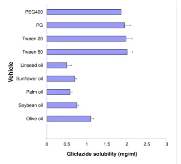
Construction of ternary phase diagrams gives an idea about the nature of the diluted dispersions and consequently, assists in selecting optimum proportions of oil, surfactant, and co-surfactant. The ternary phase diagram of olive oil - Tween80 - PG in different ratios was constructed (Figure 2) and the results of visual observations of the diluted blends are represented in Table 3. It is clear that blends containing 20-40% w/w of oil produced clear and stable emulsions with increasing Smix ratios (formulations A3, A4, B3, B4, C3, C4) whereas the resultant emulsions of blends with lower Smix ratios (formulations A1, A2, B1, B2, C1, C2) resulted in translucent emulsions followed by phase separation.
Fig. 2: Ternary phase diagram of SEDDS consisting of olive oil, Tween80 and PG.
Moreover, formulations containing higher oil content (50% w/w) produced milky emulsions with oil droplets on the surface when diluted with water.
According to the ternary phase diagram, formulations contained olive oil (20–40% w/w), Tween 80: PG ratio (3:1 & 4:1, w/w) were selected for the formulation of gliclazide loaded SEDDS.
also prevent coalescence of dispersed droplets resulting in stable nanoemulsions (Levy and Benita, 1990). Agubta et al., 2014, reported that efficient oil and water emulsification require the presence of a high concentration of surfactant to provide very low interfacial tension (≤103 mN/m). Gurram et al., 2015, reported that
large molecular volume oils including long-chain triglycerides such as olive oil require higher surfactant concentration than short chain mono/diglycerides.
Table 3: Formulation optimization.
Formulations Clarity Stability
A1 translucent Phase separation
A2 translucent Phase separation
A3 clear stable
A4 clear stable
B1 translucent Phase separation
B2 translucent Phase separation
B3 clear stable
B4 clear stable
C1 translucent Phase separation
C2 translucent Phase separation
C3 clear stable
C4 clear stable
D1 milky Phase separation
D2 milky Phase separation
D3 milky Phase separation
D4 milky Phase separation
Presence of co-surfactant is essential for the emulsification process. The flexibility of the interfacial film which is imparted by the presence of co-surfactant allows the exchange of co-surfactant from the interfacial film to continuous phase and dispersed phase and exchange of surfactant between the interfacial film and water and hence good emulsion formation (Bagwe et al., 2001). However, only small amounts of co-surfactant (Smix of 3:1 & 4:1) is required for the stability of the formed emulsion as large amounts lead to an expansion of the interfacial film with a subsequent increase in droplet size (Saifee et al., 2013). In addition, higher amounts of co-surfactant results in precipitation of drug upon dilution due to the partitioning of co-surfactant into the aqueous phase (Lawrence and Rees, 2009).
Characterization of SEDDS
Assessment of self-emulsification
The results of self-emulsification are given in Table 4. Emulsification time varied from 31s to 81s. The emulsification time was directly proportional to the amount of oil phase and inversely proportional to Smix ratio. This result may be due to the presence of higher concentration of tween80 that has high ability to reduce the interfacial tension between O/W interface which in turn shows a positive impact on spontaneous emulsification process (Bhikshapathi et al., 2013).Agubata and coworkers, 2014, reported that ease of emulsification is dependent on the ease with which water penetrates the oil-water interface with the formation of liquid crystalline phase resulting in rapid emulsification.
Furthermore, increasing oil concentration results in increased viscosity of the formulation and hence prolonged emulsification time. Similar results were reported by Harshal et al., 2011. Table 4: Emulsification time, droplet size and dissolution parameters of gli -clazide loaded in olive oil based SEDDS compared with the plain drug (mean ± SD, n = 3).
Formulation Emulsification time (Sec) Droplet size (nm) (% per min)IDR DE (%)
Plain drug NA NA 1.5 ± 0.05 27.9 ± 0.78
A3 40 ± 1.7 230 ± 5.6 5.1 ± 0.59 59.9 ± 1.64
A4 31 ± 1.2 215 ± 2.3 5.8 ± 0.57 66.8 ± 5.1
B3 53 ± 2.3 263 ± 3.5 7 ± 0.57 76.4 ± 5.5
B4 45 ± 1.5 245 ± 2.8 8.0 ± 0.75 86.3 ± 3.5
C3 81 ± 3.1 411 ± 5.2 5.1 ± 0.39 68 ± 2.82
C4 77 ± 3.5 360 ± 3.4 5.4 ± 0.55 66.1 ± 1.24
IDR: initial dissolution rate after 10 min.
DE%: dissolution efficiency % after 60 min.
Stability studies
All SEDDS samples which were subjected to appropriate dilutions and storage for 24 h, single refrigeration/thawing cycle or centrifugation were found to be stable with no evidence of phase separation or drug precipitation suggesting that diluted gliclazide SEDDS can remain stable in gastrointestinal fluids.
Determination of droplet size
Since the rate and extent of drug release, as well as its gastrointestinal absorption, mainly depend upon emulsion droplet size, the droplet size of the prepared formulations was determined and the results are represented in Table 4. The formulations had droplets size in the range of 215–411 nm.
It was clear that droplet size decreased with the increase in Smix which was consistent with previous studies. (Ostertag et al., 2012;Mehrnia et al., 2016) At high Smix, there will be a great reduction in interfacial tension with surfactant diffusion into the aqueous phase facilitating the formation of fine droplets. On the other hand, at low Smix, the surfactant concentration at the oil-water interface will not be enough to completely cover the surface of drops; coalescence of droplets occurs and hence increased droplets size (Zeing et al., 2017).
The droplet size was negatively affected by the oil concentration. This result may be explained on the basis of collision theory. As the concentration of oil increased, a larger number of emulsion droplets will be formed and consequently will collide with each other resulting in the formation of larger droplets (Shahavia et al., 2015).
Contour plot
The two-dimensional contour plots were drawn to understand the variation of emulsification time and droplet size with oil concentration and Smix (Figure 3A & B). At low oil concentration and high Smix, rapid emulsification (<40 sec) and small droplet size (<250 nm) were obtained.
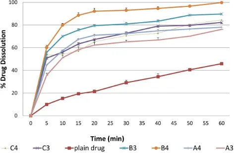
In-vitro drug dissolution from liquid SEDDS
and dissolution parameters of gliclazide from different SEDDS formulations, respectively. Table 5 summarizes the results of the ANOVA analysis. The dissolution parameters (IDR & DE%) of gliclazide from all SEDDS formulations were significantly (P = 0.000) higher than the plain drug. The maximal drug dissolution was obtained from the formulation B4 (containing oil of 30% w/w and surfactant/co-surfactant ratio of 4:1, w/w). The formulation B4 exhibited IDR of 8.0 ± 0.75 compared to only 1.5 ± 0.05 obtained by the plain drug, comprising a 5.3-folds increase in the initial dissolution rate within 10 min. Moreover, the dissolution
efficiency exhibited by the formulation B4 after 60 min was 86.3 ± 3.5% compared to only 27.9 ± 0.78% exhibited by the plain drug comprising a 3.1-folds increase in drug dissolution efficiency. This result may be ascribed to the presence of the drug in free molecular state or in emulsion form or in solubilized micellar solution when the formulation is subjected to aqueous medium compared to the crystalline form of the plain drug (Kallakunta et al., 2012). Moreover, the optimum dissolution properties (IDR & DE%) achieved by the formulation B4 may be due to the proper balance between oil percentage and a surfactant system.
Table 5: Analysis of variance for the different SEDDS formulations compared with the plain drug.
Value
Effect of the formulation of SEDDS
(Plain drug against different SEDDS)
Effect of oil % at Smix
3:1 (A3, B3, C3) Effect of oil % at Smix 4:1 (A4, B4, C4) Effect of Smix at 20% oil (A3, A4) Effect of Smix at 30% oil (B3, B4) Effect of Smix at 40% oil (C3, C4)
IDR DE% IDR DE% IDR DE% IDR DE% IDR DE% IDR DE%
P-value 0.000 0.000 0.007 0.004 0.005 0.000 0.227 0.089 0.14 0.039 0.453 0.346
R2 94.79% 97.6% 80.55% 83.64% 83.2% 92.54% 33.71% 55.55% 45.82% 69.63% 14.72% 22.19%
R2 adjusted 92.56% 96.58% 74.07% 78.19% 67.6% 90.05% 17.13% 44.44% 32.27% 62.04% 0.00% 2.74%
S-value 0.54 3.26 0.536 3.66 0.627 3.36 0.59 3.78 0.67 4.26 0.46 2.14
Fig. 3: Contour plots for the effect of oil concentration and Smix on (A)
emulsification time; (B) droplet size.
Fig. 4: In vitro dissolution profiles of gliclazide from liquid SEDDS formulations
compared with the plain drug (mean ± SD, n = 3).
The results revealed that both IDR and DE% were negatively affected by oil concentration, droplet size and emulsification time (nonsignificant effect) and positively affected
by Smix (nonsignificant effect), Table 5 & Figure 5. These results were in agreement with Nigade et al., 2012, who reported that surfactant concentration, oil/surfactant ratio, the polarity of the emulsion, droplet size and charge are critical parameters for oral drug absorption from SEEDS. Presence of excess surfactant in the formulation promotes the effective dispersion of the drug in the dissolution medium by the solubilization process, in addition to promoting drug spreading in fine oil droplets with larger surface area exposed to the dissolution medium (Suresh and Sharma, 2011). Moreover, the reduction in drug dissolution from formulations containing a higher percentage of oil (>30% w/w) may be due to the presence of insufficient proportion of surfactant and hence lesser reduction in interfacial tension between oil and aqueous phase followed by decreased drug partition into an aqueous medium (El-Laithy, 2008).
Values of correlation analysis are presented in Table 6 and Figure 6. Generally, the Pearson Correlation Coefficient (r) shows the nature and strength of correlation. The coefficient can vary between -1 and +1 while zero value describes no correlation. A positive sign of coefficient indicates a synergistic effect while a negative term indicates an antagonistic effect upon the response. It is clear that there was a nonsignificant correlation between dissolution parameters (IDR and DE%) and oil % and Smix. Alternatively, there was a strong positive significant correlation (r > 0.9, P < 0.05) between oil %, droplet size and emulsification time.
Formulation of solid SEDDS
Fig. 5: Correlation between IDR and DE% versus oil percentage, emulsification time, droplet size and Smix.
Fig. 6: Differential scanning calorimetric thermograms of plain gliclazide, MCC, lactose and different solid SEDDS.
The results showed that it was possible to convert the selected liquid SEDDS (B4) into solid forms with all liquid SEDDS:carrier mixture ratios used, while attempts to produce solid forms with higher proportions of liquid SEDDS (SEDDS:carrier mixture = 2:1) failed.
The selection of MCC was done on the basis of possessing high intraparticle porosity with good water swelling properties, as well as high adsorption capacity for lipid-based preparations (Parekh et al., 2017). Since MCC is a water-swellable polymer so it forms a network like structure in water which prevents nucleation, crystal growth and precipitation of drug upon dilution of SEDDS in GIT (Dokania and Joshi, 2015). In addition, MCC is a hydrogen bonding polymer which can form an intermolecular hydrogen bond with gliclazide resulting in retardation of drug
precipitation upon dilution in GIT (Chen et al., 2012).
Lactose is a water-soluble solid carrier with a small molecular weight which may provide good preservation of nanoemulsion droplet size after dilution (Li et al., 2013).
Table 6: Results of correlation analysis of IDR and DE% versus oil
percent-age, emulsification time, droplet size and Smix.
Factor Emulsification time (min) Droplet size (nm) (% per min)IDR Oil %
Droplet size (nm) r 0.978
P 0.001
IDR (% per min) r -0.334 -0.422
P 0.518 0.405
Oil % r 0.958 0.919 -0.076
P 0.003 0.01 0.887
Smix r -0.189 -0.193 0.309 0.00
P 0.72 0.714 0.552 1.00
DE% r -0.088 -0.153 0.953 0.177
p 0.886 0.773 0.003 0.773
IDR: initial dissolution rate after 10 min.
DE%: dissolution efficiency % after 60 min.
Characterization of solid SEDDS
Differential scanning calorimetry (DSC)
a broad peak at 85.24°C with ∆H = -22.96 mJ. Lactose showed a sharp endothermic peak at 153.4°C with ∆H = -327.11 mJ. No representative peaks for gliclazide were detected in all the prepared solid SEDDS with predominance of excipients peaks indicating the transformation of crystalline structure of gliclazide into molecularly dissolved state (Kallakunta et al., 2012).
Fig. 7: X-ray powder diffraction spectra of plain gliclazide, MCC, lactose and different solid SEDDS.
X-ray diffraction studies (XRD)
The XRD patterns of plain gliclazide, MCC, lactose and different solid SEDDS are shown in Figure 7. The diffraction pattern of gliclazide showed high-intensity peaks at 2θ of 10.59, 14.98, 17.2, 17.85, 18.15, 22.07 and 25.42° confirmed the crystalline nature of pure gliclazide (Biswal et al., 2009). Pure MCC showed three peaks at 2θ of 15, 16 and 23°. Lactose had distinct peaks at 2θ of 12.6, 19, 20 and 21°. The XRD patterns of all the prepared solid SEDDS showed the absence of drug peaks with a predominance of excipients peaks which may be due to the transformation of gliclazide into molecularly dissolved form in all the prepared solid SEDDS (Bhandari and Avachat, 2015). Determination of droplet size
Droplet size of the different solid SEDDS (G1-G3) after dispersion compared with the corresponding liquid formulation (B4) was presented in Table 7 and Figure 8. The droplet size of the resultant nanoemulsions from different solid preparations was in the range of 265–560 nm. The PDI values of the solid SEDDS (G1) and liquid SEDDS (B4) were below 0.3 indicating good uniformity in the globule size distribution after dilution with water (Jaiswal et al., 2014).
Fig. 8: Globule size distribution of (A) liquid SEDDS, B4, compared with (B) solid SEDDS, G1.
Comparing the droplet size of liquid SEDDS with those of the dispersed solid SEDDS, one can detect that they are significantly higher although still in the nano-size range which is required for absorption. In addition, the amount of adsorbent mixture had a positive significant effect on the droplet size. Jaiswal and coworkers reported that the presence of MCC as an adsorbent in the solid SEDDS leads to increase in globule size during dispersion of the formulations (Jaiswal et al., 2014). MCC hydrates and swells in water forming fibrils on which hydrophilic surfactant molecules adhere to its surface. So, MCC interferes with the complete reconstitution of the emulsion from the solid SEDDS results in a reduction in surfactant concentration at O/W interfaces with a subsequent increase in droplet size (Nikolakakis et al., 2015). This effect will increase with increasing proportions
of MCC. It is worth noting that the presence of lactose did not oppose the negative effect of MCC on the droplet size.
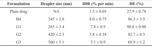
Table 7: Droplet size and dissolution parameters of gliclazide loaded in solid olive oil based SEDDS compared with its liquid form and plain drug (mean ± SD, n = 3).
Formulation Droplet size (nm) IDR (% per min) DE (%)
Plain drug NA 1.5 ± 0.05 27.9 ± 0.78
B4 245 ± 2.8 8.0 ± 0.75 86.3 ± 3.5
G1 265 ± 3.4 7.8 ± 0.9 90.6 ± 0.00
G2 420 ± 2.3 5.8 ± 0.34 82.7 ± 0.5
G3 560 ± 5.1 5.1 ± 0.9 69.9 ± 1.2
IDR: initial dissolution rate after 10 min.
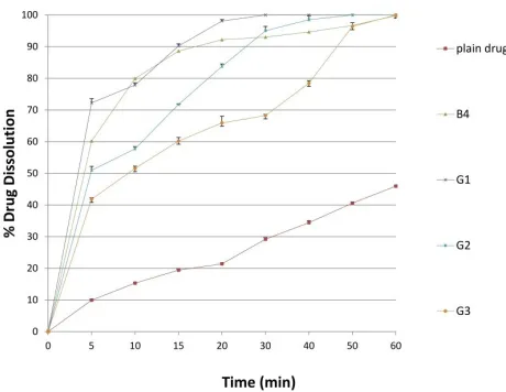
Fig. 9: In vitro dissolution profiles of gliclazide loaded in solid olive oil based
SEDDS compared with its liquid form and plain drug (mean ± SD, n = 3).
Fig. 10: Correlation analysis of IDR of gliclazide from liquid and solid SEDDS versus droplet size.
In-vitro drug dissolution
The calculated dissolution parameters (IDR & DE%) of the different solid SEDDS (G1-G3) compared with the corresponding liquid formulation (B4) and plain drug are presented in Table 7 and the release behavior is presented in Figure 9. It was clear that dissolution parameters of the different solid SEDDS were much superior (P = 0.00, R2 = 94.67% and R2 adjusted =
92.54%) when compared with the plain drug. About 4.2 and 2.9-folds increase in IDR and DE%, respectively of gliclazide from solid SEDDS formulations when compared with its plain form. Small droplet size of the formed nanoemulsion after the dispersion of the solid SEDDS results in a large surface area exposed for dissolution medium and hence rapid drug dissolution (Dixit and Nagarsenker, 2008; Cho et al., 2016).
Interestingly, for IDR and DE, there was no significant difference (P > 0.05) between liquid and solid SEDDS containing less adsorbent ratio (G1). However, gliclazide dissolution parameters from the other solid SEDDS were significantly (IDR: P = 0.003, R2 = 80.25% and R2 adjusted = 72.84%, DE%: P = 0.00,
R2 = 96.44% and R2 adjusted = 95.1%) reduced than the liquid
SEDDS. This result may be attributed to rapid emulsification and
small droplet size of the reconstituted dispersion. On the other hand, the reduced dissolution parameters of the preparations containing higher amounts of adsorbent (G2, G3) may be explained on the basis of increased droplet size of the solid-SEDDS after dispersion which is inversely proportional to the dissolution rate.
Correlation analysis revealed that there was a good correlation between the droplet size of the resultant nanoemulsions of the dispersed solid SEDDS and IDR values (Pearson correlation = -0.980, P = 0.020), Figure 10.
CONCLUSION
Olive oil based self-emulsifying delivery system containing 30% of oil and Tween 80: propylene glycol in 4:1 w/w can be successfully developed into solid form by mixing with a mixture of microcrystalline cellulose and lactose as an adsorbent in a weight ratio of 2:1 w/w. The solid self-emulsifying system of gliclazide (Formulation G1) showed improved dissolution properties which may be promising to overcome the problem of inter and intrasubject variability in bioavailability. In addition, the presence of olive oil in the formulation is expected to augment the antidiabetic effect of gliclazide which may confer an added value to the developed preparation. Future in vivo studies will be recommended.
CONFLICT OF INTEREST
The author discloses no possible conflicts of interest. REFERENCES
Abdalla A, Klein S, Mäder K. A new self-emulsifying drug delivery system (SEDDS) for poorly soluble drugs: Characterization, dissolution, in vitro digestion and incorporation into solid pellets. Eur J Pharm Sci, 2008; 3(5):457-464.
Aboutaleb HA, Kharshoum RM, Ismail HR. Formulation and optimization of solid self-nanoemulsifying system using porous carriers for oral delivery of cinnarizine. Int J Pharm Pharm Sci, 2016; 8(1):433-438.
ACCU Dyne TestTM. Available at: www.accudynetest.com/visc_ table.html.
Agubata CO, Nzekwe IT, Obitte NC, Ugwu CE, Attama AA, Onunkwo GC. Effect of oil, surfactant and co-surfactant concentrations on the phase behavior, physicochemical properties and drug release from self-emulsifying drug delivery systems. J Drug Discov Develop and Deliv, 2014; 1(1):1-7.
Amemiya T, Mizuno S, Yuasa H, Watanabe J. Emulsion type new vehicle for soft gelatin capsule available for preclinical and clinical trials: stabilization of new vehicle containing vitamin K2 with PEG 6000. Xenobiotics, 1999; 14(5):392-398.
Azeem A, Rizwan M, Ahmad FJ, Iqbal Z, Khar RK, Aqil M, Talegaonkar S. Nanoemulsion components screening and selection: A technical note. AAPS Pharm Sci Tech, 2009; 10:69-76.
Bagwe RP, Kanicky JR, Palla BJ, Patanjali PK, Shah DO. Improved drug delivery using microemulsions: Rationale, recent progress, and new horizons. Crit Rev Ther Drug Carrier Syst, 2001; 18:77-140.
Basalious EB, Shawky N, Badr-Eldin SM. SNEDDS containing bioenhancers for improvement of dissolution and oral absorption of lacidipine. I: development and optimization. Int J Pharm, 2010; 391:203-211.
Berraaouan A, Abid S, Bnouham M. Antidiabetic oils. Current Diabetes Reviews, 2013; 9(6):499-505.
Bhagwat DA, D’Souza JI. Development of solid self- microemulsifying drug delivery system with neusilin UF-2 for enhanced dissolution rate of telmisartan. Int J Drug Dev & Res, 2012; 4(4):398-407.
emulsifing pellets of carvedilol. Braz J Pharm Sci, 2015; 51(3):663-671. Bhikshapathi D, Madhukar P, Kumar BD, Kumar GA. Formulation and characterization of pioglitazone HCl self-emulsifying drug delivery system. Pharm Lett, 2013; 5(2):292-305.
Biswal S, Sahoo J, Murthy PN, Giradkar, RP, Avari JG. Enhancement of dissolution rate of gliclazide using solid dispersions with polyethylene glycol 6000. AAPS Pharma Sci Tech, 2008; 9(2):563-570.
Biswal S, Sahoo J, Murthy PN. Physicochemical properties of solid dispersions of gliclazide in polyvinyl pyrrolidone K-90. AAPS Pharm Sci Tech, 2009; 10(2):329-334.
Chen ZQ, Liu Y, Zhao JH, Wang L, Feng NP. Improved oral bioavailability of poorly water-soluble indirubin by a supersaturatable selfmicroemulsifying drug delivery system. Int J Nanomed, 2012; 7:1115-1125.
Cho H, Kang J, Ngo L, Tran P, Lee Y. Preparation and evaluation of solid-self-emulsifying drug delivery system containing paclitaxel for lymphatic delivery. J Nanomater, 2016; Article ID 3642418: 14 pages.
Craig DQM. The use of self-emulsifying systems as a means for improving drug delivery. Bull Tech Gattefossé, 1993; 86:21-31.
Czajkowska-Kośnik A, Szekalska M, Amelian A, Szymańska E, Winnicka K. Development and evaluation of liquid and solid self-emulsifying drug delivery systems for atorvastatin. Molecules, 2015; 20:21010-21022.
Dixit RP, Nagarsenker MS. Self-nanoemulsifying granules of ezetimibe: Design, optimization and evaluation. Eur J Pharm Sci, 2008; 35:183-192.
Dokania S, Joshi AK. Self-microemulsifying drug delivery system (SMEDDS) – challenges and road ahead. Drug Delivery, 2015; 22(6).
El-Laithy HM. Self-nanoemulsifying drug delivery system for enhanced bioavailability and improved hepatoprotective activity of biphenyl dimethyl dicarboxylate. Curr Drug Deliv, 2008; 5:170-176.
Fatouros DG, Deen GR, Arleth L, Bergenstahl B, Nielsen FS, Pedersen JS, Mullertz A. Structural development of self nano emulsifying drug delivery systems (SNEDDS) during in vitro lipid digestion monitored by small-angle X-ray scattering. Pharm Res, 2007; 24:1844-1853.
Franceschinis E, Bortoletto C, Perissutti B, Dal Zotto M, Voinovich D, Realdon N. Self-emulsifying pellets in a lab-scale high shear mixer: Formulation and production design. Powder Technology, 2011; 207:113-118.
Franceschinis E, Voinovich D, Grassi M, Perissutti B, Filipovic-Grcic J, Martinac A, Meriani-Merlo F. Self-emulsifying pellets prepared by wet granulation in high-shear mixer: influence of formulation variables and preliminary study on the in vitro absorption. Int J Pharm, 2005; 291:87-97.
Gershanik T, Benita S. Self-dispersing lipid formulations for improving oral absorption of lipophilic drugs. Eur J Pharm Biopharm; 2000; 50:179-188.
Gurram AK, Deshpande PB, Kar SS, Nayak YU, Udupa N, Reddy MS. Role of components in the formation of self-microemulsifying drug delivery systems. Indian J Pharm Sci, 2015; 77(3):249-257.
Harrower AD. Comparison of efficacy, secondary failure rate and complications of sulfonylurea. J Diabetes Complicat, 1994; 8:201-203.
Harshal M, Tanvir S, Dheera B, Rajendra WD. Design and development of solid self-micro-emulsifying drug delivery system (SMEDDS) of fenofibrate. Int J Pharm Pharm Sci, 2011; 3(4):163-166.
Hegazi R, El-Gamal M, Abdel-Hady N, Hamdy O. Epidemiology of and risk factors for type 2 diabetes in Egypt. Annals of Global Health, 81(6):814-820.
Iosio T, Voinovich D, Grassi M, Pinto JF, Perissutti B, Zacchigna M, Quintavalle U, Serdoz F. Bi-layered self-emulsifying pellets prepared by co-extrusion and spheronization: Influence of formulation variables and preliminary study on the in vivo absorption. Eur J Pharm Biopharm, 2008; 69:686-697.
Jaiswal P, Aggarwal G, Harikumar SL, Singh K. Development of self-microemulsifying drug delivery system and solid-self-microemulsifying drug delivery system of telmisartan. Int J Pharm Investig, 2014;
4(4):195-206.
Jung YJ, Dong YS, Lee SH, Kim KH, Yoon DS, Lee KH. Enhanced solubility and dissolution rate of itraconazole by a solid dispersion technique. Int J Pharm, 1999; 187:209-218.
Kaithwas G, Majumdar DK. In vitro antioxidant and in vivo antidiabetic, antihyperlipidemic activity of linseed oil against streptozotocin-induced toxicity in albino rats. Eur J Lipid Sci Technol, 2012; 114:1237-1245.
Kallakunta VR, Bandari S, Jukanti R, Veerareddy PR. Oral self-emulsifying powder of lercanidipine hydrochloride: Formulation and evaluation. Powder Technology, 2012; 221:375-382.
Kallakunta VR, Eedara BB, Jukanti R, Ajmeera RK, Suresh Bandari SA. Gelucire 44/14 and labrasol based solid self-emulsifying drug delivery system: formulation and evaluation. J Pharm Invest, 2013; 43:185-196.
Khan KA. The concept of dissolution efficiency. J Pharm Pharmacol, 1975; 27:48-49.
Khedekar K, Mittal S. Self emulsifying drug delivery system: A review. Int J Pharm Sci Res, 2013; 4(12):4494-4497.
Lawrence MJ, Rees GD. Microemulsion-based media as novel drug delivery systems. Adv Drug Deliv Rev, 2009; 45:89-121.
Levy MY, Benita S. Drug release from submicronized O/W emulsion: a new in vitro kinetic evaluation model. Int J Pharm, 1990; 66(1-3):29-37.
Li L, Yi T, Wai-Kei Lam C. Effects of spray-drying and choice of solid carriers on concentrations of labrasol® and transcutol® in solid self-microemulsifying drug delivery systems (SMEDDS). Molecules, 2013; 18:545-560.
Mahajan HS, Girnar GA, Nerkar P. Dissolution and bioavailability enhancement of gliclazide by surface solid dispersion using spray drying technique. Indian J Novel Drug Deliv, 2012; 4(2):115-124.
Mehrnia MA, Jafari SM, Zadeh BS, Maghsoudlou Y. Crocin loaded nano-emulsions: factors affecting emulsion properties in spontaneous emulsification. Int J Biol Macromol, 2016; 84:261-267.
Miryala V, Kurakula M. Self-nano emulsifying drug delivery system (SEDDS) for oral delivery of atorvastatin—Formulation and bioavailability studies. JDDT, 2013; 3:131-142.
Mohanvarma SK, Padavala MV. Oral lipid-based drug delivery systems – an overview. Acta Pharm Sin B, 2013; 3(6):361-372.
Mosca M, Cuomo F, Lopez, F, Ceglie A. Role of emulsifier layer, antioxidants and radical initiators in the oxidation of olive oil-in-water emulsions. Food Res Int, 2013; 50:377-383.
Nepal PR, Han HK, Choi HK. Preparation and in vitro-in vivo evaluation of Witepsol_ H35 based selfnanoemulsifying drug delivery systems (SNEDDS) of coenzyme Q10. European J Pharm Sci, 2010; 39(4), 224-232.
Nigade PM, Patil S, Shradha S. Self emulsifying drug delivery system (sedds): a review. IJPBS, 2012; 2(2):42-52.
Nikolakakis I, Panagopoulou A, Salis A, Malamataris S. Relationships between the properties of self-emulsifying pellets and of the emulsions used as massing liquids for their preparation. AAPS Pharm Sci Tech, 2015; 16(1):129-139.
Nipun TS, Ashraful Islam SM. SEDDS of gliclazide: Preparation and characterization by in-vitro, ex-vivo and in-vivo techniques. Saudi Pharm Journal, 2014; 22(4):343-348.
Ostertag F, Weiss J, McClements DJ. Low-energy formation of edible nanoemulsions: factors influencing droplet size produced by emulsion phase inversion. J Colloid Interface Sci, 2012; 388:95-102.
Palmer KJ, Brogden RN. Gliclazide, an update of its pharmacological properties and therapeutic efficacy. Drugs, 1993; 46:92-125.
Parekh VJ, Desai ND, Shaikh MS, Shinde UA. Self nanoemulsifying granules (SNEGs) of meloxicam: preparation, characterization, molecular modeling and evaluation of in vivo anti-inflammatory activity. Drug Dev Ind Pharm, 2017; 43(4).
drugs: non-emulsifying, self-emulsifying and ‘self-microemulsifying’ drug delivery systems. Eur J Pharm Sci, 2000; 11:S93-S98.
Pouton CW. Formulation of poorly water-soluble drugs for oral administration: physicochemical and physiological issues and the lipid formulation classification system. Eur J Pharm Sci, 2006; 29:278-287.
Saifee M, Zarekar S, Rao VU, Zaheer Z, Soni R, Burande S. Formulation and In vitro evaluation of solid-self- emulsifying drug delivery system (SEDDS) of Glibenclamide. AJADD, 2013; 1(3):323-340.
Samarji R, Balbaa M. Anti-diabetic activity of different oils through their effect on arylsulfatases. J Diabetes Metab Disord, 2014; 13:116.
Sapkal NP, Kilor VA, Bhusari KP, Daud AS. Evaluation of some methods for preparation of gliclazide-®-cyclodextrin inclusion complexes. Trop J Pharm Res, 2007; 6(4):833-840.
Seo YG, Kim DH, Ramasamy T, Kim JH, Marasini N, Oh YK, Kim DW, Kim JK, Yong CS, Kim JO, Choi HG. Development of docetaxel-loaded solid self-nanoemulsifying drug delivery system (SNEDDS) for enhanced chemotherapeutic effect. Int J Pharm, 2013; 452:412-420.
Shahavia MH, Hosseinia M, Jahanshahia M, Meyer RL, Najafpour G. Evaluation of critical parameters for preparation of stable clove oil nanoemulsion. Arabian Journal of Chemistry, 2015.
Shavi GV, Kumar AR, Usha YN, Armugam K, Ranjan O, Ginjupalli K, Pandey S, Udupa N. Enhanced dissolution and bioavailability of gliclazide using solid dispersion techniques. Int J Drug Deliv, 2010; 2:49-57.
Suresh PK, Sharma S. Formulation and in-vitro characterization of self-nanoemulsifying drug delivery system of cinnarizine. Pharmacie Globale (IJCP), 2011; 9(8):1-6.
Talegaonkar S, Azeem A, Ahmad FJ, Khar RK, Pathan SK, Khan
ZI. Microemulsions: a novel approach to enhanced drug delivery. Recent Pat Drug Deliv Formul, 2008; 2:238-257.
Tuleu C, Newton M, Rose MJ, Euler D, Saklatvala R, Clarke A, Booth S. Comparative bioavailability study in dogs of a self-emulsifying formulation of progesterone presented in a pellet and liquid form compared with an aqueous suspension of progesterone. J Pharm Sci, 2004; 93:1495-1502.
Yi T, Wan J, Xu H, Yang X. New solid self-microemulsifying formulation prepared by spray-drying to improve the oral bioavailability of poorly water soluble drugs. Eur J Pharm Biopharm, 2008; 70:439-444.
Wankhade VP, Atram SC, Nishan N Bobade NN, Pande SD, Tapar KK. Formulation and optimization of SNEDDS of gliclazide using response surface methodology. Asian J Pharm, 2012; 6(4):289.
Yin YM, Cui FD, Mu CF, Choi MK, Kim JS, Chung SJ, Shim CK, Kim DD. Docetaxel microemulsion for enhanced oral bioavailability: preparation and in vitro and in vivo evaluation. J Controlled Release, 2009; 140:86-94.
Zambiazi RC, Przybylski R, Zambiazi MW, Mendonca CB. Fatty acid composition of vegetable oils and fats. Curitiba, 2007; 25(1):111-120.
Zeing L, Xin X, Zhang Y. Development and characterization of promising Cremophor EL-stabilized o/w nanoemulsions containing short-chain alcohols as a cosurfactant. RSC Adv, 2017; 7:19815-19827.
How to cite this article: