Leszek Gromadziński
1, a–f, Ryszard Targoński
2, a, b, e, f, Piotr Pruszczyk
3, a, c, e, fAssessment of Right and Left Ventricular Diastolic
Functions with Tissue Doppler Echocardiography
in Congestive Heart Failure Patients
with Coexisting Acute Pulmonary Embolism
1 Department of Internal Medicine, Gastroenterology and Hepatology, University clinical Hospital,
University of Warmia and Mazury, Olsztyn, Poland
2 Department of Internal Medicine and cardiology, Municipal Hospital, Olsztyn, Poland 3 Department of Internal Medicine and cardiology, Medical University of Warsaw, Poland
A – research concept and design; B – collection and/or assembly of data; C – data analysis and interpretation;
D – writing the article; E – critical revision of the article; F – final approval of article; G – other
Abstract
Background. acute pulmonary embolism (aPe), despite improvements in diagnostic methods, often remains undiagnosed. Recently, systolic dysfunction has also been described as assessed by new echocardiographic tech-niques, such as tissue Doppler imaging (TDI).
Objectives. In our study we have attempted to assess diastolic function within the mitral and tricuspid annuli in congestive heart failure (cHf) patients with coexisting aPe.
Material and Methods. The study included 39 patients with cHf with sinus rhythm, 20 patients with confirmed aPe (Pe+), and 19 patients with excluded aPe (Pe–). aPe was confirmed or excluded on the result of spiral chest computed tomography. Tissue Doppler imaging (TDI) was performed to measure early diastolic velocity (em), late diastolic velocity (am) of both examined annuli, and em/amLV and em/amRV ratios.
Results. Pe+ subjects were found to have lower emRV than Pe– subjects [4.2 (2.0–14) vs. 6.5 (0.8–14) cm/s; p = 0.006]. The amLV was higher in the Pe+ vs. Pe– group [8.4 (3.0–15.2) vs. 3.0 (1.0–14.8), p = 0.0038]. em/amLV and em/amRV were significantly lower in the Pe + vs. Pe– group [0.55 (0.2–1.4) vs. 1.6 (0.16–5.4), p = 0.0089 and 0.41 (0.17–2.5) vs. 1.5 (0.05–5.5), p = 0.0069]. for the aPe diagnosis, the area under the ROc curve calculated for amLV and em/amLV was 0.771 (95% cI 0.509–0.890) and 0.742 (95% cI 0.577–0.868) respectively. for the aPe diagnosis, the sensitivity, specificity, positive and negative predictive values for amLV = 4.9 cm/s were: 95%, 68.4%, 76% and 92.9%, respectively and for em/amLV = 1.0 were: 95%, 63.2%, 73.1% and 92.3%, respectively.
Conclusions. TDI reveals changes in mitral and tricuspid annular velocities in cHf patients with confirmed aPe. These patients exhibit a reduced emRV and increased amLV (Adv Clin Exp Med 2014, 23, 3, 371–376).
Key words: tissue Doppler, diastolic function, acute pulmonary embolism.
adv clin exp Med 2014, 23, 3, 371–376 ISSN 1899–5276
ORIGINaL PaPeRS
© copyright by Wroclaw Medical University
a sudden obstruction of a section of the pulmo-nary vascular bed in patients with acute pulmopulmo-nary embolism (aPe) can lead to acute right ventricular (RV) pressure overload. In approximately 50% of aPe patients, echocardiographic signs of RV tolic dysfunction can be detected [1]. Recently, sys-tolic dysfunction has also been described as assessed by new echocardiographic techniques, such as tis-sue Doppler imaging (TDI) [2–13]. There are, how-ever, limited studies on the diastolic function of the heart muscle in aPe patients [14–17]. Patients with
coexisting congestive heart failure (cHf) constitute a specific group of patients exhibiting diastolic dys-function. In our study we have attempted to assess diastolic function within the mitral and tricuspid annuli in cHf patients with coexisting aPe.
Material and Methods
with coexisting confirmed aPe, and 19 patients with excluded aPe who served as the control group. cHf was diagnosed based on the clinical findings according to the framingham criteria and earlier medical records. Systolic heart failure (SHf) had occurred in 39% of patients, while heart failure with preserved ejection fraction (HfPef) had occurred in 61% of studied patients. aPe was confirmed or excluded on the result of spiral chest computed to-mography. all patients studied were admitted to the hospital due to cHf and clinical suspicion of aPe. Patients with the following conditions were ex-cluded from the study: detected non-sinus rhythm, acute coronary syndromes detected within the pre-vious 14 days, anemia with hemoglobin level be-low 11 g%, hyperthyroidism, significant valvular defects, condition after the replacement of the mi-tral or tricuspid valve, significant mimi-tral or tricus-pid annular calcification, shunt defects, disorders of the pericardium with fluid in the pericardial sac above 10 mm in diastole, chronic constrictive pericarditis, cardiac tamponade. The study proto-col was approved by the local bioethics commit-tee, and all patients provided informed consent.
Standard echocardiography was performed on admission in all patients using a 2.5 MHz transduc-er (System 5, Vingmed, Gentransduc-eral electric) and a si-multaneous ecG recording. In standard echocar-diography, left ventricular end-diastolic dimension (LVDD) and right ventricular end-diastolic di-mension (RVDD) were assessed in the parasternal long-axis view. In the apical view, right ventricular systolic pressure was measured by assessing tricus-pid regurgitation peak gradient (TRPG), and also left ventricular ejection fraction (LVef) using the simplified Simpson’s method [18].
color tissue Doppler echocardiography with computer image analysis (echopack 6.3, Ge Ving-med) was performed using a 2.5 MHz transducer for all patients. Doppler images were taken in the left lateral position and recordings were obtained during normal respiration. by placing the Doppler gate on the lateral mitral annulus at the posteri-or leaflet of the mitral valve, systolic and diastolic myocardial velocity profile was obtained. RV func-tion was assessed in the same way via the long-ax-is view, by measuring tricuspid lateral annular ve-locity at the anterior leaflet of the tricuspid valve. Systolic velocity was determined, discounting peak isovolumetric mitral annular systolic veloc-ity (SmLV) and peak isovolumetric tricuspid an-nular systolic velocity (SmRV). also, the following parameters were calculated: early diastolic velocity (em) and late diastolic velocity (am) of both ex-amined annuli, and the RV and LV Tei index. all parameters were calculated as the mean of mea-surements taken in 3 consecutive cardiac cycles.
The authors of this manuscript have certified that they have complied with the Principles of ethical Publishing present in the Declaration of Helsinki and that the study protocol was approved by a lo-cal ethics committee.
Statistical Analysis
Parameters with a normal distribution are pre-sented as a mean ± standard deviation, whereas val-ues with non-normal distributions are expressed as median and range. In order to compare both groups, a Student’s t-test and the Mann-Whitney test were used, depending on the parameter distri-bution. a χ2 test was used to compare qualitative
variables in contingency tables.
a value of p < 0.05 was considered to indicate sta-tistical significance. The stasta-tistical analysis was per-formed using Statistica 6.0 PL (Tulsa, OK, USa).
Receiver operating characteristic (ROc) curves served to determine the optimal cutoff points for identifying patients with aPe.
Results
Patients with cHf and acute pulmonary embo-lism (Pe+) comprised 20 patients; while the second group which included cHf patients with excluded aPe (Pe–) consisted of 19 patients. Table 1 pres-ents the clinical characteristics of these patipres-ents.
Table 2 presents the general characteristics of parameters assessed by tissue Doppler echocar-diography in both groups.
Mitral and tricuspid annular diastolic veloc-ities differed in both examined groups. Patients with confirmed aPe exhibited reduced early di-astolic tricuspid annular lateral velocity (emRV), whereas late diastolic tricuspid annular lateral ve-locities (amRV) were similar in both groups. The analysis of mitral annular diastolic velocities indi-cated that aPe patients were characterized by in-creased late diastolic mitral annular lateral veloc-ities (amLV) in comparison to patients without aPe. However, early diastolic mitral annular ve-locities (emLV) did not differ for both examined groups. The early to late LV diastole ratio (em/ /amLV), and early to late RV diastole ratio (em/ /amRV) were significantly lower in patients with confirmed aPe as compared to patients with no evidence of aPe.
ROC Analysis
amLV value differentiated aPe patients from pa-tients without aPe with a sensitivity of 95% and a specificity of 68.4%, positive and negative predic-tive values (PPV and NPV) were 76% and 92.9%, respectively.
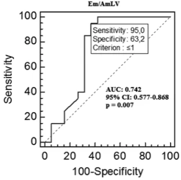
The optimal cutoff value in the ROc analysis for the parameter determining LV diastolic func-tion, i.e. em/amLV, was 1.0. The surface area un-der the ROc curve for the em/amLV parameter was 0.742 with a 95% cI (0.577–0.868), p = 0.007. at this value, the sensitivity of this parameter in
detecting aPe was 95% and the specificity was 63.2%; PPV and NPV amounted to 73.1% and 92.3%, respectively (fig. 1).
The optimal cutoff value in the ROc analysis for the emRV parameter was 5 cm/s. The surface area under the ROc curve for this value was 0.757 with a 95% cI (0.593–0.879). at this value, the sensitivity of this parameter in detecting aPe was 85% and the specificity was 68.8%; PPV and NPV amounted to 73.9% and 81.3%, respectively.
The optimal cutoff value in the ROc analysis
Table 1. General characteristics of the study population
Variable Whole group (n = 39) PE+ (n = 20) PE– (n = 19) p
Male gender, n (%) 14 (36) 6 (30) 8 (42) 0.19
age (years) 73.4 ± 11.4 75.6 ± 10 71.2 ± 12.5 0.22 Hypertension, n (%) 22 (56) 12 (60) 10 (53) 0.63
Previous MI, n (%) 18 (46) 5 (25) 8 (42) 0.35
RVeDD (cm) 3.2 ± 0.7 3.4 ± 0.4 3.0 ± 1.0 0.25
LVeDD (cm) 4.6 (3.6–7.5) 4.5 (3.7–6.6) 5.7 (3.6–7.5) 0.01 LVef (%) 59 (20–70) 60 (30–70) 4 8 (20–69) 0.21
TRPG (mmHg) 56 ± 12 59 ± 15 54 ± 9 0.25
TaPSe (cm) 1.55 ± 0.38 1.48 ± 0.31 1.63 ± 0.45 0.23
Pe – pulmonary embolism, MI – myocardial infarction, RVeDD – right ventricular end-diastolic diameter, LVeDD – left ventricular end-diastolic diameter, LVef – left ventricular ejection fraction, TRPG – tricuspid regurgitation pressure gradi-ent, TaPSe – tricuspid annular peak systolic excursion.
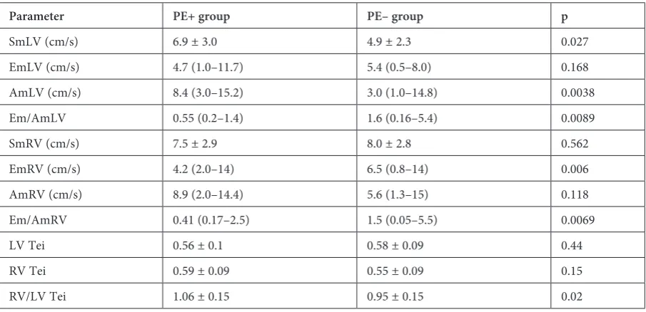
Table 2. Parameters assessed by tissue Doppler echocardiography in Pe+ group (n-20) and in Pe–group (n-19)
Parameter PE+ group PE– group p
SmLV (cm/s) 6.9 ± 3.0 4.9 ± 2.3 0.027
emLV (cm/s) 4.7 (1.0–11.7) 5.4 (0.5–8.0) 0.168
amLV (cm/s) 8.4 (3.0–15.2) 3.0 (1.0–14.8) 0.0038
em/amLV 0.55 (0.2–1.4) 1.6 (0.16–5.4) 0.0089
SmRV (cm/s) 7.5 ± 2.9 8.0 ± 2.8 0.562
emRV (cm/s) 4.2 (2.0–14) 6.5 (0.8–14) 0.006
amRV (cm/s) 8.9 (2.0–14.4) 5.6 (1.3–15) 0.118
em/amRV 0.41 (0.17–2.5) 1.5 (0.05–5.5) 0.0069
LV Tei 0.56 ± 0.1 0.58 ± 0.09 0.44
RV Tei 0.59 ± 0.09 0.55 ± 0.09 0.15
RV/LV Tei 1.06 ± 0.15 0.95 ± 0.15 0.02
for the parameter determining RV diastolic func-tion, i.e. em/amRV, was 0.84. The surface area under the ROc curve for the em/amRV param-eter was 0.753 with a 95% cI (0.589–0.876). The obtained value differentiated aPe patients from patients without aPe with a sensitivity of 80.0% and a specificity of 63.2%; PPV and NPV reached 72.7% and 76.5%, respectively.
Mitral annular lateral systolic velocities (SmLV) were higher in patients with confirmed aPe than in patients without aPe, whereas tricus-pid annular lateral systolic velocities did not differ for both groups.
Discussion
The assessment of diastolic function in pa-tients with cHf on sinus rhythm is based, among other factors, on the determination of both ear-ly and late diastolic mitral annular lateral veloci-ties and early and late diastolic tricuspid annular lateral velocities (emLV, amLV and emRV, am-RV), and early to late diastole ratio in both exam-ined annuli, i.e. em/amLV and em/amRV. Pa-tients with confirmed aPe differed from paPa-tients with excluded aPe in terms of the diastolic ve-locity profile both in the LV and RV. The ratio of early to late diastole in aPe patients was < 1 and amounted to 0.55 (0.2–1.4) in the lateral mitral annulus and 0.41 (0.17–2.5) in the lateral tricus-pid annulus as compared to patients with exclud-ed aPe, whose values were 1.6 (0.16–5.4) and 1.5 (0.05–5.5), respectively.
The abnormal diastolic velocity profile in aPe patients as compared to patients without aPe
most likely was due to different loading condi-tions in each ventricle. The reversal of the em/ /amLV ratio < 1 with regard to the mitral annulus was caused by a significant increase in the velocity during late diastole, that is amLV, in aPe patients as compared to patients without aPe, median 8.4 cm/s (3.0–15.2) cm/s and 3.0 cm/s (1.0–14.8) cm/s, respectively, p = 0.0038. However, as regards the lateral tricuspid annulus, the reversal of the em/amRV ratio < 1 was due to a decrease in the velocity during early diastole, that is emRV, in pa-tients with confirmed aPe as compared to papa-tients without aPe, median 4.2 cm/s (2.0–14.0) cm/s and 6.5 cm/s (0.8–14.0) cm/s, respectively, p = 0.0061.
from a pathophysiological point of view, an in-crease in the velocity during late diastole in the left part of the heart in patients with confirmed aPe, probably occurred analogously to an increased mi-tral annular systolic velocity (SmLV) in this group of patients as compared to patients without aPe (6.9 ± 3.0 cm/s vs. 4.9 ± 2.3 cm/s, p = 0.027), which confirms our previous observations [19]. Due to the presence of embolic material within the pul-monary arteries, a decrease in the preload on the left side of the heart occurs. This can be indicat-ed by a rindicat-educindicat-ed early diastolic mitral flow veloc-ity in patients with confirmed aPe as compared to patients with pulmonary hypertension (PH) and healthy subjects [15]. Due to a decreased preload, the left ventricle and the left atrium become rel-atively “empty”, and consequently their walls are more hyperkinetic. This can explain a significant increase in the late diastolic mitral annular later-al velocity, corresponding to the left atrilater-al con-traction, and a smaller LVDD in patients with confirmed aPe as compared to patients without aPe. additionally, an increase in these mitral an-nular velocities can be affected by the excitation of the adrenergic system as a result of stress caused by pulmonary embolism. Our results are only partly confirmed by the study of Hsiao et al. [16] that an-alyzed mitral diastolic velocities in aPe patients as compared to patients with PH. They observed only a tendency towards higher late diastolic mitral an-nular lateral velocities in aPe patients as compared with patients without aPe (9.9 ± 3.0 and 8.9 ± 3.2, p = 0.08), with similar early diastolic velocities.
as regards the right side of the heart, in both groups of patients RV overload occurs, as evidenced by the mean values of TRPG above 40 mm Hg, both in patients with confirmed aPe and in pa-tients with no evidence of aPe. In aPe papa-tients due to a decreased preload on the left side of the heart associated with the embolic material within the pulmonary arteries pressure and volume load increase on the right side. This is evidenced by the increased RV Tei index value in aPe patients as
compared to patients with PH and healthy indi-viduals, obtained in other studies [15, 17]. an in-creased RV Tei index value in aPe patients results from a prolonged isovolumetric contraction time (IVcT), and most of all, from a prolonged isovol-umetric diastolic time (IVDT) with a simultaneous shortening of ejection time (eT).
Due to the pressure overload, and mostly, vol-ume overload of the RV in aPe patients, the filling of the RV via the right atrium (Ra) and venae ca-vae is more difficult than normal. This can lead to a reduced early diastolic velocity (emRV), the so-called passive phase, as compared to patients with no evidence of embolic material in the pulmonary arteries. This can explain the mechanism of a sig-nificant decrease of emRV within the lateral tricus-pid annulus among our aPe patients as compared to the control group. Our results are supported by the study of Radman et al. [20], who also observed a reduced early diastolic tricuspid annular veloci-ty in aPe patients; however, in another study such a correlation was not observed [15].
The analysis of the diastolic function by tissue Doppler echocardiography in our research popu-lation is, however, more complex. We studied pa-tients with retained sinus rhythm, and LV diastolic and systolic dysfunctions. additionally, these pa-tients have an advanced stage of cHf and coexist-ing PH or confirmed aPe. Potential explanation for the mechanism of different diastolic velocities profiles in both patient groups is difficult. Due to potential interactions between the ventricles, an
increased LV end-diastolic pressure, characteristic for LV failure, negatively influences the RV systol-ic and diastolsystol-ic functions, consequently leading to pulmonary venous hypertension. a counter-rela-tionship also occurs: right heart dysfunction due to a sudden secondary RV overload, or due to isch-emia, leads to a decreased LV ejection.
Limitations of the Study
The major limitation of this study is the fact that it is a one-center study conducted with a small group of patients. another limitation is associat-ed with the presence of patients with both retainassociat-ed and disturbed LV systolic function, which might have affected the results obtained. another limi-tation of our study is the lack of analysis of mitral and tricuspid flow in a standard echocardiograph-ic examination.The authors concluded that Tissue Doppler echocardiography reveals changes in mitral and tricuspid annular velocities in cHf patients with confirmed aPe. These patients exhibit a reduced early diastolic tricuspid annular velocity and in-creased late diastolic mitral annular velocity. The high sensitivity of amLV and em/amLV param-eters, amounting to 95% in aPe diagnostics, is probably overestimated due to the small number of patients. consequently, this study requires fur-ther research to be conducted with a larger study group in order to confirm our preliminary results.
References
[1] Torbicki A, Perrier A, Konstantinides S, Agnelli G, Galie N, Pruszczyk P, Bengel F, Brady AJ, Ferreira D, Janssens U, Klepetko W, Mayer E, Remy-Jardin M, Bassand JP: eSc committee for Practice Guidelines (cPG). Guidelines on the diagnosis and management of acute pulmonary embolism. eur Heart J 2008, 29, 2276–2315.
[2] Kasper W, Gelbel A, Tiede N, Bassenge D, Kauder E, Konstantinides S, Meinertz T, Just H: Distinguishing between acute and subacute massive pulmonary embolism by conventional and Doppler echocardiography. br Heart J 1993, 70, 352–356.
[3] Kasper W, Meinertz T, Henkel B, Eissner D, Hahn K, Hofmann T, Zeiher A, Just H: echocardiographic findings in patients with proved pulmonary embolism. am Heart J 1986, 112, 1284–1290.
[4] Jardin F, Dubourg O, Gueret P, Delorme G, Bourdarias JP: Quantitative two-dimensional echocardiography in massive pulmonary embolism: emphasis on ventricular interdependence and leftward septal displacement. J am coll cardiol 1987, 10, 1201–1206.
[5] McConnell MV, Solomon SD, Rayan ME, Come PC, Goldhaber SZ, Lee RT: Regional right ventricular dysfunc-tion detected by echocardiography in acute pulmonary embolism. am J cardiol 1996, 78, 469–473.
[6] Miniati M, Monti S, Pratali L, Di Ricco G, Marini C, Formichi B, Prediletto R, Michelassi C, Di Lorenzo M, Tonelli L, Pistolesi M: Value of transthoracic echocardiography in the diagnosis of pulmonary embolism: Results of a prospective study in unselected patients. am J Med 2001, 110, 528–535.
[7] Nazeyrollas P, Metz D, Jolly D, Maillier B, Jennesseaux C, Maes D, Chabert JP, Chapoutot L, Elaerts J: Use of transthoracic Doppler echocardiography combined with clinical electrocardiographic data to predict acute pulmo-nary embolism. eur Heart J 1996, 17, 779–786.
[8] Perrier A, Tamm C, Unger PF, Lerch R, Sztajzel J: Diagnostic accuracy of Doppler echocardiography in unselect-ed patients with suspectunselect-ed pulmonary embolism. Int J cardiol 1998, 65, 101–109.
[9] Grifoni S, Olivotto I, Cecchini P, Pieralli F, Camaiti A, Santoro G, Pieri A, Toccafondi S, Magazzini S, Berni G, Agnelli G: Utility of an integrated clinical, echocardiographic, and venous ultrasonographic approach for triage of patients with suspected pulmonary embolism. am J cardiol 1998, 82, 1230–1235.
Burakowski J, Wawrzyńska L: Disturbed right ventricular ejection pattern as a new Doppler echocardiographic sign of acute pulmonary embolism. am J cardiol 2002, 90, 507–511.
[11] Torbicki A, Kurzyna M, Ciurzyński M, Pruszczyk P, Pacho R, Kuch-Wocial A, Szulc M: Proximal pulmonary embolism modifies right ventricular ejection pattern. eur Respir J 1999, 13, 616–621.
[12] Gromadzinski L, Ciurzynski M, Januszko-Giergielewicz B, Targoński R, Cygański P, Pruszczyk P: Diagnostic value of mitral and tricuspid annular excursion in the diagnostics of acute pulmonary embolism patients with chronic heart failure. Int J cardiol 2011, 149, 118–119.
[13] Pruszczyk P, Torbicki A, Pacho R, Chlebus M, Kuch-Wocial A, Pruszynski B, Gurba H: Noninvasive diagnosis of suspected severe pulmonary embolism: transesophageal echocardiography vs spiral cT. chest 1997, 112, 722–728.
[14] Kjaergaard J, Krogdgaard BK, Lund JO, Hassager C: Quantitative Measures of right ventricular dysfunction by echocardiography in the diagnosis of acute nonmassive pulmonary embolism. J am Soc echocardiography 2006, 19, 1264–1271.
[15] Hsiao SH, Chang SM, Lee ChY, Yang SH, Lin SK, Chiou KR: Usefulness of tissue Doppler parameters for iden-tifying pulmonary embolism in patients with signs of pulmonary hypertension. am J cardiol 2006, 98, 685–690.
[16] Hsiao SH, Lee CY, Chang SM, Yang SH, Lin SK, Huang WC: Pulmonary embolism and right heart function: Insights from myocardial Doppler tissue imaging. J am Soc echocardiography 2006, 19, 822–828.
[17] Hsiao SH, Yang SH, Wang WC, Lee CY, Lin SK, Liu CP: Usefulness of regional myocardial performance index to diagnose pulmonary embolism in patients with echocardiographic signs of pulmonary hypertension. am J cardiol 2006, 98, 1652–1655.
[18] Lang RM, Bierig M, Devereux RB, Flachskampf FA, Foster E, Pellikka PA, Picard MH, Roman MJ, Seward J, Shanewise JS, Solomon SD, Spencer KT, Sutton MS, Stewart WJ: chamber Quantification Writing Group; american Society of echocardiography’s Guidelines and Standards committee; european association of echocardiography. Recommendations of chamber quantification: a report from the american Society of echocardiography’s Guidelines and Standards committee and the chamber Quantification Writing Group, devel-oped in conjunction with the european association of echocardiography, a branch of the european Society of cardiology. J am Soc echocardiogr 2005, 18, 1440–1463.
[19] Gromadziński L, Targoński R: The role of tissue colour Doppler imaging in diagnosis of segmental pulmonary embolism in congestive heart failure patients. Kardiol Pol 2007, 65, 1433–1439.
[20] Rydman R, Larsen F, Caidahl K, Alam M: Right ventricular function in patients with pulmonary embolism: early and late findings using Doppler tissue imaging. J am Soc echocardiogr 2010, 23, 531–537.
Address for correspondence:
Leszek Gromadziński
Department of Internal Medicine, Gastroenterology and Hepatology University clinical Hospital, University of Warmia and Mazury Warszawska 30
10-082 Olsztyn Poland
e-mail: [email protected] Tel: +48 89 52 453 89
conflict of interest: None declared Received: 16.01.2013