This is an open access journal, and articles are distributed under the terms of the Creative Commons Attribution-Non Commercial-ShareAlike 4.0 License, which allows others to remix, tweak, and build upon the work non-commercially, as long as appropriate credit is given and the new creations are licensed under the identical terms.
© 2018 Journal of Advanced Pharmacy Education & Research | Published by SPER Publication
85
Expression of Her2 and Topo II Alpha in breast cancer
Diab M.
1*, Shaker O.
2, Nassar Y.
2, Abd El-Aziz G.
1, El-Marzoki M.
31Medical Biochemistry, Faculty of Medicine, Beni_Suef University, Beni_Suef, Egypt, 2Medical Biochemistry, Faculty of Medicine, Cairo University, 3General surgery, Faculty of medicine, Cairo University, Egypt.
Correspondence: Marwa Diab, Medical Biochemistry, Faculty of Medicine, Beni_Suef University, Beni_Suef, Egypt. E mail:[email protected]
ABSTRACT
Background: When some oncogenes as human epidermal growth factor receptor and Topoisomerase IIA get overexpressed, they have been indicated to cause the development and growth of special aggressive kinds of breast cancer (BC). In recent years, these proteins have become an important biomarkers. Objective: This examination aimed at identifying the expression of Her2 and TopoIIA genes in both metastatic and non-metastatic groups of cancer breast patients. Subject and Methods: The current study was carried out on 230 participants who were divided into 3groups: Group I (control) which included 50 healthy subjects; Group II which included 40 patients of metastatic breast cancer cases; and Group III which contained 140 patients with non-metastatic breast. A detailed history taking and thorough physical and clinical examination, and 5 ml of blood were collected from each subject, and biochemical tests were done. Total RNA was extracted from whole blood using RNA extraction kit, and then reverse transcription was done followed by real time PCR. Results: The results of the current examination indicated that there was a remarkable correlation between control and non-metastatic cancer groups considering Her2 and TopoIIA expression (p<0.001*) & (p<0.001*) respectively, and between control and metastatic groups considering Her2 and TopoIIA expression (p<0.001*) & (p<0.001*), and also between each other. Conclusion: Her2 and TopoIIA gene expressions can be considered as the most significant diagnostic markers to diagnose non-metastatic, and predict the metastatic breast cancer.
Keywords:Breast cancer, Her2, TopoIIA, gene expression.
Introduction
Breast cancer is the most prevalent cancer among females, and is second only to lung cancer as a cause of cancer death among women in the developed world. Breast cancer incidence rates have increased gradually since the 1940s in many industrialized countries. Western Europe, the United States, and Canada have had the highest incidence of breast cancer, with the lowest rates found in Asia [1]. In Egypt, breast cancer has been reported as
the most prevalent cancer among women in 2008. It has also been the pioneer at leading to cancer-related death including 29.1% of their total mortality. There has been a poor ratio of incidence to mortality (1.9:1). These estimates have been certified in several regional Egyptian cancer registries as well as in hospital-based frequencies [2]. There are several clinically
recognized types of breast cancer, with ductal carcinoma the most common, followed by lobular carcinoma, inflammatory breast cancer, medullary carcinoma of the breast, and the remaining less prevalent types [3].
Early diagnosis and treatment, including breast self-examination, clinical breast self-examination, and mammography can reduce the death caused by BC. In developing countries, the lack of early detection programs has increased the rate of mortality caused by BC [4]. One of the members of the
epidermal growth factor receptor (EGFR/ERBB) family is Her2. By amplification or overexpression of this oncogene, it has been shown that special aggressive kinds of breast cancer was developed or progressed. Recently, protein has been regarded as an important biomarker and target of therapy for almost 30% of breast cancer patients [5]. The Her2 gene is
located at chromosome 17q21 and thegene is amplified in 20-30% of breast carcinomas. The gene encoding for topoisomerase IIa (TopoIIA) at 17q21-q22 is located close to the Her2 locus. The enzyme TopoIIA is a significant element constituting DNA replication because of being able to make a double-stranded break in the DNA helix, and it also causes DNA coils to unwind before sealing the strandstogether again for progressive replication [6].
DNA topoisomerases are enzymes that adjust the over winding or under winding of DNA, and are required for the survival of
Access this article online
Website: www.japer.in E-ISSN: 2249-3379
How to cite this article: Diab M., Shaker O., Nassar Y., Abd El-Aziz G.,
El-Marzoki M.Expression of Her2 and TOPO II Alpha in Breast Cancer. J Adv
Pharm Edu Res 2018;8(2):85-91.
86 Journal of Advanced Pharmacy Education & Research | Apr-Jun 2018 | Vol 8 | Issue 2
all organisms. They are of high importance in adjusting cellular processes like replication, transcription, and chromosomal segregation by changing DNA topology [7]. DNA topoisomerase
IIA (TopoIIA) is a well-known anticancer target. Agents that target TopoIIA are among the most effective anticancer drugs presently accessible to treat the human cancers. TopoIIA gene is located close to the Her2 oncogene at the chromosome 17. TopoIIA is amplified in almost 90% of Her2 amplified initial breast tumors [8]. Hence, this study aimed at detecting the
expression of Her2 and TopoIIA genes in both metastatic and non-metastatic groups of breast cancer patients compared with healthy subjects.
Subjects and Methods
One hundred eighty (180) Egyptian women aged between (20-65) were included in the current study. They were recruited from National Cancer Institute in the period from January 2011 to December 2013. Patients were known as breast carcinoma according to the history taking, clinical examination, which was confirmed by mammography and surgical biopsies, and fifty (50) healthy women with no family history of breast cancer, who were recruited during routine checkups, were considered as controls. A written informed consent from patients was taken in order to participate in the study in accordance with the ethical guidelines of the Declaration of Helsinki.
The studied subjects were divided into three groups: Group I: (n =50) healthy females as a control ; Group II (n =40) patients with metastatic breast cancer cases (30 invasive ductal and 10 invasive lobular carcinoma, all 40 patients with bone metastasis, 36 with axillary and 4 supraclavicular lymphnode involvement, and according to grading system, 38 cases were grade 2, and 2 were grade 3); and Group III: (n =140), patients with non-metastatic breast cancer (classified into 12 invasive lobular and 128 invasive ductal carcinoma; and 88 cases with T2 , and 52 cases with T3, and 108 cases were grade 2 and 32 cases were grade 3 according to TNM grading system into).
Analytic procedure:
Venous blood samples were collected from the patients and controls in sterile EDTA and centrifuge tubes. Part in the centrifuge tube was incubated at 37°c for 15 minutes, then centrifuged at 3,000 rpm for 10 minutes at room temperature, the serum was separated and used for biochemical tests, and the rest of blood was taken for RNA extraction and detection of gene expression of Her2 and TopoIIA.
Quantitation of Her-2 and topoisomerase II
α
by real time PCR
1.
Total RNA extraction:Total RNA was extracted from the whole blood using RNA extraction kit provided by Qiagene extraction kit. The RNA
purity and concentration were quantified by NanoDrop ND-1000 (Nanodrop, USA).
2.
Reverse transcription:Reverse transcription for the extracted RNA was done using
SuperScript™ II reverse transcriptase (Invitrogen Life
Technologies Inc., Carlsbad, CA). The reaction was carried out for 60 min at 42°C, and the reaction mixture was subsequently inactivated for 15 min at 70°C. The cDNA was stored at -70°C
till used for quantitative PCR.
3.
Primers probe kits:Primers and TaqMan probes for Her-2 and the GAPDH control reference gene were designed and synthesized according to Taqman Gene Expression Assay (assays Hs00170433_m1 and 4326317E, respectively) (Applied Biosystems, Foster City, CA, USA). The sequence of the primers of topoisomerase II A was
Forward primer.
TTGAAGACGCTTCGTTATGGG-3', Reverse primer 5'-CCATCACAACTGGCCCTCTC-3' and Probe 5'-ACAGATCAGGACCAAGATGGTTCCCACAT-3'
4.
PCR for Her-2 mRNA andtopoisomerase II α:
Polymerase chain reaction (PCR) was conducted in the final volume of 20µL, in which there was 1.25ul primer probes mixture, 1.25 ul GAPDH, 5ul cDNA and 2.5 ul H2O. The reaction was performed using real time PCR for 45 cycles of:
95˚C for 15 sec and 60◦C for 30 sec. By calculating the ratio between the mean value of the target gene and the mean value of the reference gene (GAPDH) in each sample, the relative quantification (RQ) was measured. Considering the cycle threshold (Ct) value, the relative amount of PCR product was assessed for each primer set. The RQ was assessed by 2-
ΔΔCT. HER-2 and topoisomerase II α relative expressions level
were compared with the healthy controls.
Statistical analysis:
Journal of Advanced Pharmacy Education & Research | Apr-Jun 2018 | Vol 8 | Issue 2 87
Results
Totally, two hundred thirty (230) Egyptian women aged between (20-65 years) were included in the study, showing that there was a significant difference between metastatic cases in breast cancer considering grade, type and lymphnode involvement (p<0.001*, p < 0.05 and p < 0.05); respectively. Also, a significant relationship between non metastatic cases in breast cancer regarding type (p<0.05) and estrogen/progesterone receptors (p<0.05) was observed, while there was no significant relationship considering the grade, and family history (Table 1).
Table 1: Grade, type, family history, ER/PR and Lymphnode in both metastatic and non-metastatic breast
cancer groups.
Parameters Metastasis
Non-metastasis
Count % P value Count % P value
Grade 2 38 95%
<0.001*
108 78.4% 0.215
3 2 5% 32 21.6%
Type Invasive duct 30 75%
<0.05*
128 91.8% <0.05*
invasive lobular 10 25% 12 8.2%
Family history
No - -
- 68 45.5%
0.572
Yes - - 72 54.5%
ER/PR Negative - - - 118 84.1%
<0.05*
Positive - - 22 15.9%
Lymphnode Axillary 36 90%
<0.05*
- - -
Supraclavicular 4 10% - -
* indicates a statistical significant difference.
Regarding the grade, type and lymphnode involvement (in p<0.001*, p < 0.05 and p < 0.05; respectively) (Table 2) shows the significance in metastatic cases in breast cancer.
Table 2: Her2 and TopoIIA expression in non-metastatic, metastatic breast cancer and control groups.
Parameters Control group Non-metastatic group Metastasis group P value
Her-2 Mean 0.70 8.45 26.10 <0.001*
Standard Deviation 0.29 11.97 20.92
TopoIIA Mean 0.64 5.80 12.26 <0.001*
Standard Deviation 0.28 6.58 9.26
* indicates a statistical significant difference
Considering ER/PR (estrogen/progesterone receptors) expression and menstrual history, there was a statistically significant difference (median =12.8, minimum =0.8, maximum =25.3), (median =0.8, minimum =0.1, maximum =22.7; respectively), (p value =0.002 & 0.004; respectively), and TopoIIA expression (Table 3). Also, it was indicated that, there was a statistically significant difference between Her2 expression regarding ER/PR (estrogen/progesterone receptors) (median =18.6, minimum =1.05, maximum = 63.4) and (p< 0.0001*) and menstrual history (median =14.3, median = 0.7, maximum =75.5) and (p=0.007*) (Table 3).
Table 3: TopoIIA and Her2 expression in relation to ER/PR and menstrual history in all breast cancer groups.
Parameters
TopoIIA
P value
M
edian
Her2
M
edian
M
inimum
M
aximum
M
inimum
M
aximum P value
ER/PR -ve 2.3 0.30 25.70
0.002*
3.4 0.50 51.45
<0.0001*
+v 12.8 0.80 25.30 18.6 1.05 63.40
Menstrual history
post 6.4 0.50 42.20
0.004* 14.3 0.70 75.50
0.007*
pre 0.8 0.10 22.70 0.85 0.10 66.70
* indicates a statistical significant difference
As shown in (Table 4), there was a non-significant difference between Her2 expression considering the grade, lymph nodes, type, and family history. While, there was a significant relationship in TopoIIA regarding the family history, but there was not a significant relationship considering the grade, LN and type in metastatic group of breast.
Table 4: Her2 and TopoIIA expression in relation to grade, lymph nodes, type and family history in metastatic breast
group.
Parameters
Her-2 TopoIIA
M
edian
M
inimum
M
aximum P value Median Minimum Maximum P value
Grade 2 7.77 0.56 75.50 0.767 4.30 0.40 42.20 0.628
3 4.15 0.50 60.40 3.10 0.30 22.70
Lymph nodes
Axillary 18.40 6.30 75.50 0.801
12.60 2.40 42.20 0.208 supraclavicu
lar 38.50 10.30 66.70 5.15 4.80 5.50
Type Invasive
duct 5.77 0.50 75.50
0.492
4.00 0.30 42.20 0.780 invasive
lobular 9.83 0.80 18.60 2.80 0.40 14.20
Family history
No 4.98 0.56 63.40
0.157 4.30 0.40 25.30 0.042*
Yes 3.40 0.50 51.45 2.20 0.30 20.70
* indicates a statistical significant difference
Clinical size for (p <0.04*), indicated a significant correlation in TopoIIA (Fig. 1), while there was no significant correlation regarding Her2 and clinical size in metastatic group (Table 5).
Table 5: Correlation between Her2 and topoisomerase IIA regarding clinical size in metastatic cancer breast group.
Parameters Clinical size
N P value r
Her2 20 0.99 0.379
TopoIIA 20 <0.04* 0.463
88 Journal of Advanced Pharmacy Education & Research | Apr-Jun 2018 | Vol 8 | Issue 2
Figure 1: A significant correlation between TopoIIA considering clinical size in metastatic group.
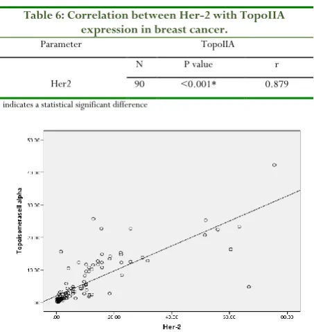
Also, as (Table 6) and (Fig. 2) represent, there was a statistically significant difference between TopoIIA expression regarding Her-2 expression (p <0.001), and also there was a statistically significant difference between Her-2 expression considering TopoIIA expression (p <0.001).
Table 6: Correlation between Her-2 with TopoIIA expression in breast cancer.
Parameter TopoIIA
Her2
N P value r
90 <0.001* 0.879
* indicates a statistical significant difference
Figure 2: A significant correlation between Her2 and TopoIIA expression in breast cancer patients.
Figure 3: ROC curve of her2 & TopoIIA in metastatic breast cancer.
Table 7: Her2 & TopoIIA expression in metastatic breast cancer.
Test Result Variable(s)
Area under
curve P value
95% Confidence Interval
Her-2 0.865 <0.001* 0.790-0.940
TopoIIA 0.782 <0.001* 0.685-0.879
The best cutoff value of her-2 = 6.05 was observed with the sensitivity of 100% and specificity of 62.3 %; and the best cutoff value of topoisomerase IIA = 3.2 was obtained with the sensitivity of 90% and the specificity of 55.1% (Table 8).
Table 8: The sensitivity and Specificity Her2 & TopoIIA
Parameters Sensitivity Specificity
Her2 100% 62.30%
TopoIIA 90% 55.1%
Discussion
Her2 is a member of the epidermal growth factor receptor (EGFR/ERBB) family. It has been revealed that this oncogene if amplified or overexpressed, can be a very significant factor in the development and progression of special dangerous kinds of breast cancer. In recent years, the protein has become an important biomarker and target of therapy for approximately 30% of breast cancer patients [5]. DNA topoisomerase IIA
(TopoIIA) is a famous anticancer target. Agents targeting TopoIIA can be classified as the most influential anticancer drugs which are presently used for the treatment of human cancers [8]. The enzyme TopoIIA is a remarkable element
constituting DNA replication which can make a double-stranded break in the DNA helix, leading to the unwinding of DNA coils before the strands get sealed together again in the progressive replication [6].
The present study showed a significant difference in non-metastatic cases in breast cancer regarding the type (p<0.05) and estrogen/progesterone receptors (p<0.05), meanwhile, there was no significant difference regarding the grade and family history, also,there was a significant difference between metastatic cases in breast cancer considering the grade (p<0.001), type (p<0.05) and lymph nodes (p<0.05). This agreed with Putti et al. (2005), who stated that, as estrogens play such a significant role in mammary gland and tumor development, breast tumors are frequently characterized as ERα positive or negative [9]. This has been explained by
Gupta and Kuperwasser (2006), who stated that, normal growth and development of mammary epithelial tissues were under the regulation of estrogens [10]. Estrogens are steroid
Journal of Advanced Pharmacy Education & Research | Apr-Jun 2018 | Vol 8 | Issue 2 89 receptor isoforms such that PR-B activates transcription and
PR-A is transcriptionally inactive and acts as a transdominant repressor of PR-B [11]. Further, PR-A is involved in repression
of ER, androgen receptor (AR), and PR-B dependent gene expression.
This disagreed with Foulkes and Narod (1995), who stated that, about 5 to 10% of breast cancers in the general population have a hereditary basis [12]. In affected families, however, risk is
particularly high if a first-degree relative has premenopausal bilateral breast cancer, or two first-degree relatives have any form of breast cancer. The major known breast cancer genetic mutations, BRCA1 and BRCA2, are involved in DNA repair and transcriptional regulation. In this study, there was no significant correlation between Her2 and TopoIIA and clinicopathological parameters (grade, LN, TNM grading system), however; there was a significant correlation between TopoIIA and tumor size (p<0.04). These results coincided with Todorovi et al. (2009), who detected that neither Her2 nor TopoIIA amplification has shown a correlation with the available clinicopathological parameters such as age, menopausal status, steroid receptor status (estrogen and progesteron receptors), tumor size, nodal status, distant metastases, histologic type and stage at the time of diagnosis [13]. These
results also agreed with Lindemann et al. (2007), who stated that there was no correlation between Her2 overexpression of tumor tissue and other clinicopathological findings [14]. While
Zhu et al. (2008), has reported that, the high TopoIIA expression was associated with poor histological grade, and high proliferative activity [15]. Several studies analyzed these
correlations with different subsets and achieved the same results.
The results of this study disagreed with Depowski et al. (2000), who reported that increased Topo IIA expression was correlated with clinicopathological features such as (decreased patient survival (p 5 .001), advanced tumor stage (p= 0 .034), lymph node metastasis (p= 0 .018), Tumor stage (p < .0001), node-positive status (p < .0001) and tumor grade (p 0 .025)
[16]. Also, these results disagreed with Gianni et al. (2011), who
stated that the prognosis of patients with TN tumors remained poor, as no new therapeutic agents targeting TN tumors were available, and also patients with luminal tumors exhibited a relatively good prognosis [17]. No metastasis, other than brain
metastasis, was observed in patients with the Her2 disease subtype. This has been indicated by Park et al. (2006), who stated that Her2 amplification is an early incidence in human breast tumorigenesis. Her2 amplification has been observed in almost half of the in situ ductal carcinomas without any trace of invasive diseases. Her2 status remains the same during progression to invasive disease, nodal metastasis and distant metastasis. The findings of this examination showed that there was a significant correlation between Her2 and TopoIIA regarding HR status in (ER/PR) (< 0.0001* and 0.002*); respectively.
This finding agreed with Zaczek et al. (2012), who found a strong association between TopoIIA copy number change and
HR and Her2status [18]. Tokiniwa et al. (2012) evaluated
ER-positive, Her2-negative breast cancer or Topo IIA expression
[19]. They found that 46% cases were positive for Topo IIA
overexpression. Rody et al. (2009) reported that 48% of cases were positive in ER-positive subset [20]. In contrast, Depowski
et al. (2000), reported that there was no correlation between topo IIA expression and tumor size, tumor grade, ER status, PR status, or disease recurrence [16].
In the current examination, a remarkable examination was observed between the control and both non metastatic and metastatic groups considering Her2 and TopoIIA expression (p<0.001*). Regarding TopoIIA, the findings of this study agreed with Depowski et al. (2000), who stated that increased topo II expression was related to an aggressive form of breast cancer indicating Her-2 expression and anticipating disease-related mortality, lymph node metastasis, and advanced tumor stage [16]. And also, this agreed with Woessner et al. (1991),
who said that TopoIIA was essential for cell growth which has been typically expressed at high levels in rapidly growing cancer cells, whereas TopoIIβ has been expressed in quiescent cells in virtually all tissues throughout the whole cell cycle, and has been dispensable for cell survival [21]. This was explained by the
fact that TopoIIA is a ubiquitous ribozyme that changes the instantaneous cleavage of double-stranded DNA, and the chromosomal topological structure, facilitating subsequent double-strand break (DSB) religation.
Considering Her2, the findings of this study agreed with Owens et al. (2004), who stated that Human epidermal growth factor receptor 2 (Her2) is a gene amplified or overexpressed in 20%–
25% of all the breast cancers, and Her2 positivity is associated with an aggressive disease course,a poor prognosis, and relative sensitivity to anthracyclines [22]. Lindemann et al. (2007), also
performed Immunohistochemistry for Her2, and revealed overexpression in 20 cases (51.3%), whereas 19 cases were scored negative [14]. No Her2 expression was observed in
normal ducts. Immunofluorescent staining for Her2 showed positive immunofluorescence intensities in both normal and tumor tissues.
This was explained by Holbro et al. (2003), who stated that increased Her2 homodimers disrupted cell polarity [23].
Increased dimers drove proliferative, survival, invasive and metabolic functions. Increased Her2 expression resulted in an increase in the rare DHer2 isoform withmore potent signaling characteristics. And, several transcription factors were induced in Her2-overexpressing cells resulting in a plethora of gene expression changes.
The present examination also showed that there was a remarkable correlation between expression of both Her2 and TopoIIA with each other in the all the groups of breast cancer cells (P<0.001٭). This finding agreed with Depowski et al. (2000), who reported that there was an increased TopoIIA expression correlated with Her2 expression (p < 0.0001) [16].
This finding also agreed with Rody et al. (2009), Zaczek et al. (2012) and Sparano et al. (2012) [18, 20, 24]. The most important
90 Journal of Advanced Pharmacy Education & Research | Apr-Jun 2018 | Vol 8 | Issue 2
between TopoIIA copy number change and HR and Her2 status.
Also, the obtained results agreed with Hicks and Tubbs (2005), who reported that in Her2-positive breast cancer, the amplification of TopoIIA varied from 25% to 42% [25]. While
Jacot et al. (2013), assessed that, 29.1% of the Her2-amplified cases were co-amplified with TopoIIA [26]. The proportion of
TopoIIA positive tumors in this study was lower than what obtained in other studies. However, the frequency of Her2 amplification was comparable with others, and this weighed against methodological problems. TopoIIA aberrations were strongly associated with HR and Her2 status, in contrast to Jarvinen et al. (2000) and Karen et al. (2004), who had shown that the amplifications of HER2 and TopoIIA were independent events [27, 28].
The results of this study were coincided with Jacot et al. (2013), who stated that TopoIIA is one of the genes close to HER2 and its protein product, topoisomerase II A, is the molecular target of anthracycline treatment [26]. TopoIIA
amplification status has been thought to be linked to the response to the treatment. However, the data were conflicting and, as yet, unresolved, and also agreed with Karen et al. (2004), who examined the status of the TopoIIA gene as a predictive marker for the treatment with anthracyclines, and for the close relationship to HER2, both topographically on the chromosome 17, and pathologically with frequent co-expression [28].
This study showed a significant value of Her2 and TopoIIA for the anticipation of metastatic group of breast cancer (p<0.001*& p<0.001*; respectively) with 95% confident interval of (0.790-0.940&0.685-0.879; respectively), with the best cutoff value of Her2 = 6.05 with the sensitivity of 100% and the specificity of 62.3 %. The best cutoff value of TopoIIA = 3.2 with the sensitivity of 90%, and the specificity of 55.1%. Also, the significant values of Her2 and TopoIIA were the predictors of non-metastatic group of breast cancer (p<0.001*& p<0.001*; respectively) with 95% confident interval (0.926-0.992&0.894-0.979; respectively), with the best cutoff value of Her2 = 1.225 with the sensitivity of 88.8%, and the specificity of 100 %. The best cutoff value of TopoIIA was 1.15 with the sensitivity of 84% and the specificity of 100%.
This agreed with Depowski et al. (2000) who stated that the correlation between TopoIIA expression and other known prognostic parameters has been inconsistent [16]. Indeed,
TopoIIA was confirmed as a strict proliferation marker in breast cancer, mainly for its role and high expression of proliferating cells.
Also, the results of this study agreed with Coon et al. (2002), who identified the correlation between Her2 amplifications and TopoIIA genecopy number changes inthe preclinical and early clinical studies [6]. It has been speculatedthat TOPIIA is in fact
the predictive marker for chemotherapy with anthracyclines. Karen et al. (2004), showed that the status of the TopoIIA gene has been examined as a predictive marker for the treatment
with anthracyclines and for the close relationship with Her2, both topographically on the chromosome 17, and pathologically with frequent co expression [28].
In contrast, Jacot et al. (2013) stated that as a prognostic marker, TopoIIA is probably of limited values [26]. TopoIIA
aberrations have been strongly related to HR and Her2 status, and the significance of these markers in prognostication has still been unchallenged.
This was explained by the fact that over expression of the Her2 protein, either through gene amplification or through transcriptional deregulation has been seen in approximately 25–
30% of breast and ovarian cancers, conferring worse biological behavior. Using new methodologies has resolved the primarily existing conflicting reports on the predictive relevance of Her2, and the existing abundant data now proves this primary significant genetic-biologic result. There can be up to 25–50 copies of the Her2 gene in breast cancers, and up to 40- to 100-fold increase in Her2 protein expression leading to up to 2 million receptors located at the tumor-cell surface (Lohrisch and Piccart, 2001) [29].
Conclusion
Her2 and TopoIIA genes are over expressed in both metastatic and non-metastatic breast cancer groups, and also have significant relationship regarding the expression of each other, so Her2 and TopoIIA genes expression can be defined as remarkable markers for detecting non-metastatic breast cancer cases, and a good marker for predicting the metastatic breast cancer cases.
List of abbreviations:
BC: Breast cancerHer2: Human epithelial growth factor receptor TopoII A: Topoisomerase 2 alpha
EGRF: Epidermal growth factor receptor PCR: Polymerase chain reaction ER: Estrogen receptor
HR: Hormonal receptor PR: Progesterone receptors AR: Androgen receptor
ERE: Estrogen releasing elements BRCA1: Breast cancer gene one DSB: Double strand break
DHer2: Dimer human epidermal growth factor receptor RQ: Relative quantitation
ELISA: Enzyme- Linked-ImmunosorbentAssay. EDETA: Ethelene diamine tetra acetic acid.
Acknowledgments:
Journal of Advanced Pharmacy Education & Research | Apr-Jun 2018 | Vol 8 | Issue 2 91 General surgery at Qasr El Aini hospital for contributing to the
data collection and health examinations.
References:
1. Parkin, D., Bray, F. and Devesa, S. 2001. Cancer burden in the year 2000. The global picture. Eur. J. Cancer., 37: S4-S66.
2. The National Cancer Registry Program of Egypt (NCRPE) 2012. Reports and Statistics. Journal of the Egyptian National Cancer Institute, 27: 1-5.
3. Turashvili, G., Bouchal, J., Baumforth, K., Wei, W., Dziechciarkova, M., Ehrmann, J., Klein, J., Fridman, E., Skarda, J., Srovnal, J., Hajduch, M., Murray, P., and Kolar, Z. 2007. Novel markers for differentiation of lobular and ductal invasive breast carcinomas by laser microdissection and microarray analysis. BioMed Central (BMC) Cancer, 7: 55.
4. Sadler, G.R., Dhanjal, S.K., Shah, N.B., Ko, C.M. and Anghel, M. 2001. Asian India women: Knowledge, attitudes and behaviours toward breast cancer early detection. Public Health Nurs., 18: 357-363.
5. Mitri, Z., Constantine, T. and O'Regan, R. 2012. "The Her2 Receptor in Breast Cancer: Pathophysiology, Clinical Use, and New Advances in Therapy". Chemother. Res. Pract. Article ID 743193, 7 pages. doi:10.1155/2012/743193.
6. Coon, J.S., Marcus, E. and Gupta-Burt, S. 2002. Amplification and overexpression of topoisomerase IIalpha predict response to anthracycline-based therapy in locally advanced breast cancer. Clin. Cancer Res., 8: 1061-1067. 7. Schoeffler, A.J. and Berger, J.M. 2008. DNA
topoisomerases: Harnessing and constraining energy to govern chromosome topology. Q Rev. Biophys., 41: 41-101.
8. Azarova, A.M., Lyu, Y.L., Lin, C.P., Tsai, Y.C., Lau, J.Y., Wang, J.C. and Liu, L.F. 2007. Roles of DNA topoisomerase II isozymes in chemotherapy and secondary malignancies. Proc. Natl. Acad. Sci. USA., 104: 11014-11019.
9. Putti, T. C., El-Rehim, D. M., Rakha, E. A., Paish, C. E., Lee, A. H., Pinder, S. E. and Ellis, I. O. 2005. Estrogen receptor-negative breast carcinomas: A review of morphology and immunophenotypical analysis. Mol. Pathol., 18: 26-35.
10. Gupta, P. B., and Kuperwasser, C. 2006. Contributions of estrogen to ER-negative breast tumor growth. J. Steroid Biochem. Mol. Biol., 102: 71-78.
11. Zhou, H., Luo, M. P., Schönthal, A. H., Pike, M. C., Stallcup, M. R., Blumenthal, M., Zheng, W. and Dubeau, L. 2002. Effect of reproductive hormones on ovarian epithelial tumors: I. Effect on cell cycle activity. Cancer Biol. Ther., 1: 300-306.
12. Foulkes, W., and Narod, S. 1995. Hereditary breast and ovarian cancer: Epidemiology, genetics, screening and predictive testing. Clin. Invest. Med., 18: 473-483. 13. Todorovi, C., Rakovi, Z. and Nikoli-Vukosavljevi. 2009.
Metastatic breast cancer survival according to HER2 and Topo2a gene status. Disease Markers, 26: 171–180. 14. Lindemann, L., Resau., Na-hrig., Leeser., Annecke.,
Welk, Scha., Lengyel and Harbeck, N. 2007. Histopathology, 51: 54-62.
15. Zhu, L., Li, Y.F., Chen, W.G., He, J.R., Peng, C.H. and Zhu, Z.G. 2008. HER2 and topoisomerase IIa: possible predictors of response to neoadjuvant chemotherapy for breast cancer patients. Chin. Med. J., 121: 1965–68. 16. Depowski, S.L., Rosenthal, T.M., Brien, S., Stylis, R.L.,
Johnson and J.S. Ross 2000. Topoisomerase 2 alpha expression in breast cancer: correaltion with clinical variables, Mod Pathol., 13: 542-547.
17. Gianni, L., Dafni, U. and Gelber, R.D. (2011): Treatment with trastuzumab for 1 year after adjuvant chemotherapy in patients with Her2.positive early breast cancer: a 4year follow up of a randomised controlled trial. Lancet Oncol 12: 236-244.
18. Zaczek, A, Markiewicz A, Supernat A. 2012. Prognostic value of TOP2A gene amplification and chromosome 17 polysomy in early breast cancer. Pathol. Oncol. Res., 18: 885–94.
19. Tokiniwa, Horiguchi., Takata., Kikuchi., Rokutanda., Katsunori, T., Tetsunari, O. and Izumi, Takeyoshi 2012. Breast Cancer, 19: 309-314.
20. Rody, A, Karn T, Ruckhaberle E, 2009. Gene expression of topoisomerase II alpha (TOP2A) by microarray analysis is highly prognostic in estrogen receptor (ER) positive breast cancer. Breast Cancer Res Treat., 113: 457-66.
21. Woessner, R.D., Mattern, M.R., Mirabelli, C.K., Johnson, R.K. and Drake, F.H. 1991. Proliferation- and cell cycle-dependent differences in expression of the 170 kilodalton and 180 kilodalton forms of topoisomerase II in NIH-3T3 cells. Cell Growth Differ., 2: 209-214.
22. Owens, M.A., Horten, B.C. and Da Silva, M.M. 2004. Her2 amplification ratios by fluorescence in situ hybridization and correlation with immunohistochemistry in a cohort of 6556 breast cancer tissues. Clinical Breast Cancer, 5: 63-69. doi: 10.3816.
23. Holbro, T., Beerli, R.R., Maurer, F., Koziczak, M., Barbas, C.F. and Hynes, N.E. 2003. The ErbB2/ErbB3 heterodimer functions as an oncogenic unit: ErbB2 requires ErbB3 to drive breast tumor cell proliferation. Proc. Natl. Acad. Sci. USA, 100: 8933-8938.
24. Sparano, J.A., Goldstein, L.J., Davidson, N.E. 2012. TOP2A RNA expression and recurrence in estrogen receptor-positive breast cancer. Breast Cancer Res. Treat., 134: 751–7.
25. Hicks, D.G. and Tubbs, R.R. 2005. Assessment of the Her2 status in breast cancer by fluorescence in situ hybridization: a technical review with interpretive guidelines. Hum Pathol., 36: 250-261.
26. Jacot, W., Fiche, M. and Zaman, K. 2013. The Her2 amplicon in breast cancer: Topoisomerase IIA and beyond. Biochim. Biophys. Acta., 1836: 146–57.
27. Jarvinen, T.A., Tanner, M and Rantanen, V. 2000. Amplification and deletion of topoisomerase IIalpha associate with ErbB-2 amplification and affect sensitivity to topoisomerase II inhibitor doxorubicin in breast cancer. Am. J. Pathol., 156: 839-847.
28. Karen, E., Olsen., Helle- Knudsen., Birgitte, B., Rasmussen, Eva- Balslev., Ann-Knoop., Bent- Ejlertsen, Kirsten, V. and Jens- Overgaard. 2004. for the Danish Breast Cancer Co-operative Group Acta Oncologica, 43(1): 35-42.