Original Article
Evaluation of Anatomic Variations in Maxillary Sinus with the Aid of Cone Beam
Computed Tomography (CBCT) in a Population in South of Iran
Shoaleh Shahidi 1, Barbad Zamiri 2, Shahla Momeni Danaei 3, Setareh Salehi 4, Shahram Hamedani 5
1
Biomaterials Research Center, Dept. of Oral and Maxillofacial Radiology, School of Dentistry, Shiraz University of Medical Sciences, Shiraz, Iran.
2
Dept. of Oral and Maxillofacial Surgery, School of Dentistry, Shiraz University of Medical Sciences, Shiraz, Iran.
3 Orthodontics Research Center, Dept. of Orthodontics, School of Dentistry, Shiraz University of Medical Sciences, Shiraz, Iran. 4
Undergraduate Student, Student Research Committee, School of Dentistry, Shiraz University of Medical Sciences, Shiraz, Iran.
5
Dental Research Development Center, School of Dentistry, Shiraz University of Medical Sciences, Private Practice, Shiraz, Iran.
KEY WORDS
Cone-beam Computed
Tomography;
Normal Variations;
Maxillary Sinus;
Antrum;
Pneumatization
Received October 2014;
Received in revised form June 2015 ; Accepted July 2015;
ABSTRACT
Statement of the Problem: Anatomic variations of the maxillary sinus can be detect-ed in cone-beam computdetect-ed tomography (CBCT) and may assist to locate the posterior
superior alveolar artery (PSAA) and define the maxillary sinus morphology more
accu-rately for a more strict surgical treatment plan.
Purpose: The study aimed to determine normal variations of the maxillary sinus with the aid of CBCT in a sample population in south of Iran.
Materials and Method: This cross-sectional prevalence study was based on evalua-tion of 198 projecevalua-tion data of CBCT scans of some Iranian patients aged 18-45,
re-ferred to a private oral and maxillofacial radiology center in Shiraz from 2011 to 2013.
CBCT scans were taken and analyzed with NewTom VGi device and software. The
anatomic variations which were evaluated in the axial images included the presence of
alveolar pneumatization, anterior pneumatization, exostosis, and hypoplasia.
Moreo-ver, the location and height of sinus septa and the location of PSAA were assessed.
SPSS software (version 17.0) was used to analyze the data.
Results: In a total of 396 examined sinuses, maxillary sinus alveolar pneumatization was the most common anatomic variation detected. Anterior pneumatization was
de-tected in 96 sinuses (24.2%). Antral septa were found in 180 sinuses (45.4%) and were
mostly located in the anterior region. Meanwhile, PSAA was mostly detected
intra-osseous in 242 sinuses (65.7%).
Conclusion: Anatomic variations of the maxillary sinus were common findings in CBCT of the maxilla. Preoperative imaging with CBCT seems to be very helpful for
assessing the location of PSAA and the maxillary sinus morphology; Its data might be
used to adjust the surgical treatment plan to yield more successful treatments.
Corresponding Author: Hamedani Sh., School of Dentistry, Ghasrodasht Ave, Shiraz-Iran. P.O Box: 71345-1836 Email: [email protected] Tel and Fax: +98-713-6280458
Cite this article as: Shahidi Sh., Zamiri B., Momeni Danaei Sh., Salehi S., Hamedani Sh. Evaluation of Anatomic Variations in Maxillary Sinus with the Aid of Cone Beam Computed
Tomography (CBCT) in a Population in South of Iran. J Dent Shiraz Univ Med Sci., 2016 March; 17(1): 7-15.
Introduction
The maxillary sinus in adults is composed of a
pyramid-shaped cavity in the facial skull with its base at the
lat-eral nasal wall and its apex extending up to the
zygo-matic process of the maxilla. [1] It can exhibit anatomic
variations such as pneumatization, hypoplasia, antral
septa, exostosis, and variations in location of the
arter-ies. [2] All the surgical interventions in the posterior
maxillary region require detailed knowledgeofthe
max-illary sinus anatomy and possible anatomical variations.
[3]
development of the maxillary sinus, which can occur
during embryological development or later in life due to
trauma, iatrogenic, or structural causes. [4] The narrow
infundibular passage associated with the absence of a
natural ostium would result in mucosal thickening of the
hypoplastic sinus. [5] Furthermore, MSH causes the
proximal extension of the lateral nasal wall and
subse-quently makes the surgical procedures difficult. [2, 6]
Maxillary sinus septa are barriers of cortical bone.
The shape is described as an inverted gothic arch arising
from the inferior or lateral walls of the sinus that divide
the maxillary sinus floor into multiple compartments,
known as recesses. [1, 3] These septa were first
ana-lyzed by Arthur S. Underwood, an anatomist who
re-ported their prevalence and characteristics and these
septa were afterwards, referred to as Underwood’s
sep-ta. [1]
In a systematic review published by Pommer et
al., [7] electronic and hand searching of English
litera-ture were employed to identify the studies published
from 1995 to 2011.They reported that the observed
sep-ta were at least 2-4 mm in height, and 7.5 mm on
aver-age. They were present in 28.4% of 8923 investigated
sinuses (95% confidence interval: 24.3–32.5%). Septa
were located in premolar, molar, and retromolar regions
in 24.4%, 54.6% and 21.0% of cases, respectively. Their
orientation was transverse in 87.6%, sagittal in 11.1%,
and horizontal in 1.3% of the studied cases. Complete
septa (dividing the sinus into two separate cavities) were
found only in 0.3% of samples. Other rare conditions
included multiple septa in one sinus (4.2%) and bilateral
septa (17.2%). Moreover, the diagnosis of septa by
us-ing panoramic radiographs yielded incorrect results in
29% of cases. [7]
Septum removal before sinus augmentation is a
preferred procedure, as with the septum in place, there is
a high possibility of membrane perforation that results
in maxillary sinusitis. [3] Dental panoramic
radiog-raphy, computed tomography (CT), and cone beam
computed tomography (CBCT) have all been used to
identify the maxillary sinus septa. [8-15] CBCT is a
technique that has been proposed for maxillofacial
im-aging duringthe lastdecade and wasfirst reported by
Mozzo et al. [1, 16]
The posterior superior alveolar artery (PSAA) and
infraorbital artery (IOA) are the branches of maxillary
artery that supply the lateral sinus wall and the
overly-ing membrane. The blood supply of the maxillary sinus
and Schneiderian membrane comes from the maxillary
artery. [17] The presence of this artery was first
men-tioned by Strong in 1934. [18]
The branches of maxillary artery should be taken
into consideration because of the potential risk of
bleed-ing durbleed-ing the procedures such as open sinus lift
sur-gery, horizontal osteotomy of the maxilla, Le Fort I
fracture treatment, and Caldwell-Luc surgeries. [17-20]
In a study done by Rahpeyma et al., thirty five
CBCT scans from 35 dentate patients were selected in
coronal sections of the second premolar (P2), first molar
(M1), and second molar (M2). The presence of alveolar
antral artery in each situation was determined and the
bone thickness in the region of alveolar antral artery was
measured perpendicular to the lateral wall of the
maxil-la. The alveolar antral artery was present in 67.1% of
CBCTs. [19]
Many imaging techniques such as panoramic,
wa-ters, Caldwell, CT, MRI, and CBCT can be used to
study the maxillary sinuses region. For a long period,
skull projections including Waters, Caldwell and lateral
sinus were used for evaluation of the paranasal sinuses.
Waters view is useful for gross evaluation of the
maxil-lary sinus especially for localized mucosal thickening
along the sinus floor, generalized thickening of the
mu-cosal lining around the entire wall of the sinus, and
near-complete or complete radiopacification of the
si-nus. Plain films are no longer considered to be a part of
the primary imaging modalities. At best, they give only
an overview of the anatomy and underlying pathoses, as
they are limited to display three-dimensional (3D)
struc-tures in a two-dimensional (2D) plane. CT and MR
im-aging have the advantage of being able to show fine
anatomic details in serial topographic sections, and thus
excluding the gross volume averaging which is a
char-acteristic feature in plain films. In fact, in most cases,
when a plain-film study shows the probable presence of
the disease, a CT or MR imaging is consequently
ob-tained. [21-22]
CBCT uses a cone- or pyramidal-shaped beam to
acquire multiple projections in only one rotation. On the
other hand, multislice computed tomography (MSCT)
employs fan-shaped beams rotating around the patient
CBCT may be recommended as a low-cost
dose-sparing technique compared with standard medical
computed tomography scans (MDCT), though CBCT
has slightly more radiation exposure than routine
pano-ramic radiography for dentomaxillofacial imaging. [1,
16, 25-31]
The effective dose from a standard dental protocol
scan with MDCT is 1.5 to 12.3 times greater than
com-parable medium–field of view dental CBCT scans
ac-cording to International Commission on Radiological
Protection (ICRP 2007). [16] Moreover,
beam-hardening artifacts due to dental materials (like
amal-gam and crowns) and implants are weaker at CBCT
than at MSCT. [32]
To minimize the risk of postoperative
complica-tions of maxillary sinus floor lift and other surgeries in
this region, it is crucial to be familiar with different
ana-tomic and pathologic findings in sinus. [1, 8-13, 29, 33]
As the maxillary sinuses are significant anatomic
struc-tures in dental practice that their exact and definitive
radiological assessment is necessary, and considering
CBCT as an important diagnostic image modality in
dentistry, the recognition of anatomic variations of the
maxillary sinuses in CBCT is noteworthy. [1-2]
Several studies have been performed on the
preva-lence of anatomic variations in different populations;
however, our information is insufficient regarding the
Iranian population. Therefore, the aim of the present
study was to determine the maxillary sinus normal
vari-ations with the aid of CBCT in a sample of population
resident in south of Iran.
Materials and Method
This cross-sectional prevalence study was based on
evaluation of CBCT scans of some Iranian patients aged
18-45, who referred to a private oral and maxillofacial
radiology center in Shiraz, from 2011 to 2013. To this
end, 198 CBCT images of originally Iranian patients
were selected from the archive of adults who needed
those images for other justified reasons.
All CBCT images of the adult patients which
showed maxillary sinuses were included in the study
sample. The CBCT images of patients with systemic
problems and evidence of previous trauma or
manipula-tion of the maxillary sinuses, as well as those images
with any sign of pathologic changes in maxillary sinuses
were excluded from the study.
CBCT scans were taken with NewTom VGi
de-vice (covering the maxillary region, focal spot=0.3 or
0.15mm, scanning time=90s). They were analyzed by
the related NewTom software on a multiplanar
recon-struction window in which the axial, coronal, and
sagit-tal planes could be visualized in 0.3 mm intervals.
To standardize the reading and interpreting of the
CBCT images, two researchers were trained and
cali-brated by using 10% of the samples in a one-week pilot
study before the data collection began.
The anatomic variations evaluated in the axial
im-ages were alveolar pneumatization, anterior
pneumatiza-tion, location and height of sinus septa, exostosis,
hypo-plasia, and location of the PSAA.
The septa height more than 2 mm (the important
factor in sinus floor elevation) was registered and
loca-tion of the septa was divided into 3 groups of anterior,
middle, and posterior. The distances from the artery to
the medial sinus wall were determined and the locations
of the artery were categorized as intra-osseous (A),
be-low the membrane (B), and on the outer cortex of the
lateral sinus wall(C). In the presence of two alveolar
antral arteries in a coronal section, the larger one was
considered. The presence of septa was evaluated in the
coronal and sagittal images.
The SPSS software (Ver. 17.0) was used to
ana-lyze the data. The descriptive analysis was presented as
frequency, mean±SD, 95% confidence intervals (CI),
and the range.
Results
In a total of 198 CBCT images, 396 sinuses were
evalu-ated in which 130 cases belonged to females (65.7%)
and 68 to males (34.3%).
Maxillary sinus alveolar pneumatization
(maxil-lary sinus extension into alveolar process) was the most
common anatomic variation detected, observed in 228
sinuses (57.5%). The pneumatization sites were
multi-ple in 90 (65.2%) and single in 48 cases (34.8%). (Table
1)
The anterior pneumatization was detected in 96
sinuses (24.2%), 40 single (58.8%) and 28 multiple
(41.2%). Scalloped margin between teeth roots was
observed in 100 sinuses (25.2%).
Table 1: Frequency of normal variations
Hypoplasia Exostosis Scalloped Margin Septa Alveolar
Pneumatization
Anterior Pneumatization
All sinuses 26 (6.5%) 13 (3.2%) 100 (52.2%) 180 (45.4%) 228 (57.5%) 96 (24.2%)
Patients
Unilateral 14 (70%) 3 (37.5) 44 (61.1%) 70 (56%) 48 (34.8%) 40 (58.8%)
Bilateral 6 (30%) 5 (62.5) 28 (38.9%) 55 (44%) 90 (65.2%) 28 (41.2%)
Total 20 (100%) 8 (100%) 72(100%) 125 (100%) 138 (100%) 68 (100%)
26 sinuses (6.5 %) that included 14 unilateral (70%) and
6 bilateral (30%) cases. (Table 1)
Exostosis was identified in 13 sinuses (3.28%).
Antral septa were found in 180 sinuses (45.4%);
bilat-eral in 55 (44%) and unilatbilat-eral in 70 cases (56%).
(Ta-ble 1)
Sinus septum was in anterior region in 106
(58.9%), middle in 38 (21.1%), and posterior in 36
(20%) of sinuses containing septa. (Figure 1) Sixty four
(35.5%) of the septa were also detected in coronal slices
and 112 (62.2%) were viewed in sagittal sections, as
well. Ninety eight (54.4%) of all septa divided the sinus
into 2 cells and 8 (4.4%) into 3 separate cells.
Figure 1: Locations of septa
The minimum and maximum height of the right
sinus septum was measured to be respectively 2.1 and
23 with the mean±SD=8.17±3.6. These numbers for the
left sinus septum were 3 and 25.6, respectively, with the
mean±SD=8.28±4.29. (Table 2)
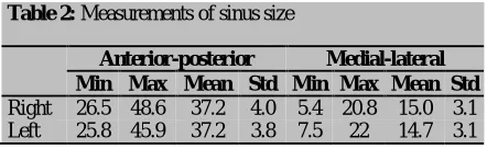
Table 2: Measurements of sinus size
Anterior-posterior Medial-lateral Min Max Mean Std Min Max Mean Std
Right 26.5 48.6 37.2 4.0 5.4 20.8 15.0 3.1
Left 25.8 45.9 37.2 3.8 7.5 22 14.7 3.1
PSAA was absent in 28 sinuses (7%). Figure 2
shows the percentage of different locations of the artery
in those images in which artery was detected. The artery
was located on the outer cortex of the sinus wall in 50
cases (13.5%). Moreover, the artery was intra-osseous
in 242 sinuses (65.7%), and below the membrane in 76
sinuses (20.6%). The minimum and maximum distance
from the artery to the medial wall on the right sinus was
10.60 and 37.50, respectively (mean±SD=24.8657±
4.94112), and 13.20 and 36.60 on the left side (mean±
SD=24.8214±4.71998).
Figure 2: Location of posterior superior alveolar artery (PSAA)
Discussion
Preoperative imaging is very important and clinically
relevant for the detection of maxillary sinus variations
and pathologic problems. In 3D imaging, the treatment
plan can be modified and the outcome of pre-prosthetic
surgery in posterior maxilla can become more
predicta-ble. [29]
The alveolar antral artery is an important
anatomi-cal structure within the lateral maxillary sinus wall. The
presence of this artery was first mentioned in an article
by Strong published in 1934. [18]
According to Ilgüy et al. [17] and Khajehahmadi
et al., [34] an important point especially in the
Cald-well-Luc and open sinus lift surgery is that the maxillary
sinus wall has a considerable vascular anastomosis.
Damage to the vessels of the bone can cause bleeding,
may obscure the physician’s line of sight, and may lead
to perforation of the Schneiderian membrane, all of
which prolong the operation and assessment of the sinus
membrane reflection. [17, 34]
In the present study, the presence and location of
the PSAA was observable through CBCT scans. The
artery was observed in 93% of the sinuses and was
mostly intraosseous (65.7%). The success rate for
iden-tifying the artery was higher than that reported by Ilgüy
al. (52.9%), [36] Mardinger et al. (55%), [37]and Kim
et al. (52%). [8] This may be related to the method and
resolution of the advanced images that were used to
detect and describe the artery.
CBCT provides accurate and reliable linear
meas-urements for reconstruction and imaging of dental and
maxillofacial structures. In the study by Ilgüy et al. in
2013, the distance of the artery from the medial sinus
wall was 13.92±2.84 mm; [17] while in our study, this
number was 24.8657±4.94mm on the right sinus and
24.8214±4.71mm on the left sinus. These differences
may be explained by the anatomic variation in the
posi-tions of arteries and the populaposi-tions that were examined.
According to Naitoh et al., antral septa was
de-fined as a pointed bone structure and maxillary sinus
exostosis as a rounded bone structure, both of which
originated from any maxillary sinus wall. [12] Van den
Bergh et al. emphasized that antral septa, detected in
almost half of the CBCT exams, might increase the risk
of sinus membrane perforation during the maxillary
sinus floor elevation surgery. [39] Abrahams et al. and
Aimetti et al. reported that the accidental perforation of
this membrane could lead to development of acute or
chronic sinusitis, and subsequent bone graft resorption.
[40-41] Furthermore, antral septa should be considered
in lifting the bone plate and sinus membrane during
surgery. [42]
With normal sinus anatomy, preparation and
hori-zontal rotation of a trap door in the maxillary sinus wall
is a common procedure and is possible when the
Schneiderian membrane is sufficiently lifted. The most
frequent complication in this procedure would be the
tearing of the sinus membrane, which is in turn
correlat-ed to the presence of septa in the maxillary sinus. [39,
42-43]
The presence of maxillary sinus septa can be
de-tected in panoramic radiographs. However, CT and
CBCT are definitely the preferred imaging techniques
for the assessment of this anatomic variation. Krennmair
et al. found that panoramic radiograph can lead to false
diagnosis regarding the positive or negative
identifica-tion of septa in 21.3% of cases. They stated that CT
scanning was the preferred imaging method for
detect-ing the presence (or absence) of sinus septa since it
al-lows the high-resolution imaging of delicate bony
struc-tures. [10]
According to Pommer et al., [7] diagnosis of sinus
septa by using 2D panoramic radiographs compared
with 3D computed tomography produced incorrect
re-sults in 29% of cases. They claimed that panoramic
radiographs may not image those sinus septa with
sagit-tal orientation and might, thus, lead to false assumption
of narrow internal sinus anatomy and subsequent
non-augmentation of the medial portion of the sinus cavity.
The pre-operative radiographic imaging of sinuses
should be made concerning the surgical complications
and following modifications that can be possibly made
to avoid these consequences. [7] In case of sinus floor
augmentation; it ranges from modification in the
surgi-cal access strategy (or window design) to change in
implant positions or even complete avoidance of bone
graft surgery. Considering the high prevalence and
sig-nificant morphologic variability in sinus septa in the
above-mentioned investigation, 3D radiological imaging
prior to sinus floor augmentation surgery may help to
reduce complications in the presence of maxillary sinus
septa. [7]
The range of septa prevalence was found to be
24-33% in a review article (four studies) published by
Katranji et al. [44] and 13-35% in a review of 11 studies
performed by Maestre-Ferrín et al. [45] which included
investigations that used panoramic radiographs. The
prevalence of sinus septa was found to be 16.1% in
Güncü et al.’s study, [35] 16% in Krennmair et al.’s
study, [10-11] and 26.5% in Kim et al.’s study when CT
was used to assess the sinuses. [8]
In a study performed in Iran with spiral CT-scan,
the prevalence of at least one septum was 29.5%. [3]
The results of the present study revealed that sinus septa
were observed in 45.4% of the 396 sinuses. The results
of the CT evaluation of the maxillary sinus septa in the
reported articles are not consistent with those of the
present study. On the other hand, much higher
percent-ages have been reported with CBCT, and they are close
to the results obtained by the current study.
The prevalence of sinus septa was reported to be
55.2% by Ilgüy et al., [17] 58% as found by Orhan et
al., [1] and 47% as reported by Neugebauer et al. [46]
In another study, Lana et al. stated that the prevalence
of antral septa was 44.4%. [2] These differences could
be attributed to the different imaging modalities
Complete septa (dividing the sinus into two
sepa-rate cavities) were found only in 0.3% as reported in the
systemic review done by Pommer et al., [7] and in 25.2
% (n=100) of the sinuses in our study.
The analysis of the position of septa showed that
sinus septum was in anterior region in 106 (58.9%),
middle in 38 (21.1%), and posterior in 36 (20%) of
si-nuses containing septa.
In the study carried out by Faramarzie et al. in
Iran, most of the septa (53.84%) were reported to be in
the middle region. [3] In some studies, a greater number
of incidence was found in the middle regions; [1, 10-11,
47] while, several other studies detected them in the
anterior [9, 13] or posterior regions. [1, 9, 13] Selcuk et
al. found that the distribution of septa in the anterior
region was higher than in the posterior region (20.3%
and 2.5%, respectively). [48] Hadchiti et al. reported no
statistically significant difference in the antero-posterior
location; that is, 55 septa were posteriorly located in the
molar region (28.65%), 75 were near the first and
se-cond premolar (middle area) (39.06%), and 67 septa
were detected in the anterior area (32.29%). [49]
Ac-cording to Faramarzie et al.’s study, the sequence of
tooth extraction can also affect the formation of antral
septa in different regions of the sinus. [3]
In our measurements, the mean height of septa
was 8.22mm; while, previous studies reported different
heights for the septa ranging from 5.6 to 20.6 mm. [1, 9,
11-13, 46-47]
Alveolar pneumatization was reported in
approx-imately 50% of the population in the study by Schuh et
al., [50] 100% of the patients in Lana et al.’s research,
[2] and was present in 228 sinuses (57.5%) in our study.
Gosau et al. stated that atrophy of the maxilla caused by
edentulism was characterized by vertical and horizontal
bone loss. [51] The maxillary sinus pneumatization,
particularly the alveolar extension, can intensify the
problem of reminiscent bone caused by atrophy of the
maxilla, leaving only few millimeters of bone for
im-plant insertion. [2, 52]
The frequency of maxillary sinus hypoplasia was
reported to be 4% in Shiki et al.’s study, [53] 4.8% in
Lana et al.’s research, [2] and 6.5% in the current
inves-tigation. Shiki et al., [53] found the antral exostosis in
3% of the population, Lana et al. [2] reported it to be
2.6%, and it was 3.2% in our study. These differences
may be due to different sample sizes, the resolution of
CBCT units which were used, as well as the anatomic
variations in different populations.
Fernandes reported that the size of maxillary sinus
differed among various ethnics in different populations.
[54] They experienced that 48.6% of European
maxil-lary sinuses had larger maxilmaxil-lary sinus volumes than
Zulu sinuses. Moreover, Butaric et al. reported that the
Peruvian samples had lower antral volume than the
Australian samples. [55] Another study reported that the
mean maxillary sinus volume in girls was larger than
that in boys aged 4-9 in a Japanese population. [56]
Therefore, the current study seems to be justified
con-cerning these differences observed in different ethnics.
Investigating the prevalence of these important
anatom-ical features in Iranian population, especially in
differ-ent parts of the country would be helpful for young
sur-geons in this population.
Conclusion
The anatomic variations of maxillary sinus are common
findings in CBCT of the maxilla. Since some of these
conditions can modify the surgery planning to more
specialized procedures, they are crucial to be recognized
in dental practice. Inevitably, preoperative imaging with
CBCT is helpful for assessing the location of the PSAA,
maxillary sinus morphology, and normal variations
which may be used to adjust the surgical treatment plan
to yield more successful treatments.
Conflict of Interest
The authors of this manuscript certify that they have no
conflict of interest regarding this research.
References
[1] Orhan K, Kusakci Seker B, Aksoy S, Bayindir H,
Ber-beroğlu A, Seker E. Cone beam CT evaluation of
maxil-lary sinus septa prevalence, height, location and
morphol-ogy in children and an adult population. Med Princ Pract.
2013; 22: 47-53.
[2] Lana JP, Carneiro PM, Machado Vde C, de Souza PE,
Manzi FR, Horta MC. Anatomic variations and lesions of
the maxillary sinus detected in cone beam computed
to-mography for dental implants. Clin Oral Implants Res.
2012; 23: 1398-1403.
rzie M. Prevalence, height, and location of antral septa in
Iranian patients undergoing maxillary sinus lift. J Perio
Imp Dent. 2009; 1: 43-47.
[4] Stammberger H. Endoscopic endonasal surgery--concepts
in treatment of recurring rhinosinusitis. Part I. Anatomic
and pathophysiologic considerations. Otolaryngol Head
Neck Surg. 1986; 94: 143-147.
[5] Weed DT, Cole RR. Maxillary sinus hypoplasia and
ver-tical dystopia of the orbit. Laryngoscope. 1994; 104(6 Pt
1): 758-762.
[6] Kapoor PK, Kumar BN, Watson SD. Maxillary sinus
hypoplasia. J Laryngol Otol. 2002; 116: 135-137.
[7] Pommer B, Ulm C, Lorenzoni M, Palmer R, Watzek G,
Zechner W. Prevalence, location and morphology of
maxillary sinus septa: systematic review and
meta-analysis. J Clin Periodontol. 2012; 39: 769-773.
[8] Kim MJ, Jung UW, Kim CS, Kim KD, Choi SH, Kim
CK, et al. Maxillary sinus septa: prevalence, height,
loca-tion, and morphology. A reformatted computed
tomogra-phy scan analysis. J Periodontol. 2006; 77: 903-908.
[9] Koymen R, Gocmen-Mas N, Karacayli U, Ortakoglu K,
Ozen T, Yazici AC. Anatomic evaluation of maxillary
si-nus septa: surgery and radiology. Clin Anat. 2009; 22:
563-570.
[10]Krennmair G, Ulm C, Lugmayr H. Maxillary sinus septa:
incidence, morphology and clinical implications. J
Crani-omaxillofac Surg. 1997; 25: 261-265.
[11]Krennmair G, Ulm CW, Lugmayr H, Solar P. The
inci-dence, location, and height of maxillary sinus septa in the
edentulous and dentate maxilla. J Oral Maxillofac Surg.
1999; 57: 667-671.
[12]Naitoh M, Suenaga Y, Kondo S, Gotoh K, Ariji E.
As-sessment of maxillary sinus septa using cone-beam
com-puted tomography: etiological consideration. Clin Implant
Dent Relat Res. 2009; 11 Suppl 1: e52-e58.
[13]Velásquez-Plata D, Hovey LR, Peach CC, Alder ME.
Maxillary sinus septa: a 3-dimensional computerized
tomographic scan analysis. Int J Oral Maxillofac
Im-plants. 2002; 17: 854-860.
[14]Kasabah S, Slezák R, Simůnek A, Krug J, Lecaro MC.
Evaluation of the accuracy of panoramic radiograph in
the definition of maxillary sinus septa. Acta Medica
(Hradec Kralove). 2002; 45: 173-175.
[15]Almog DM, Romano PR. CT-based dental imaging for
implant planning and surgical guidance. N Y State Dent J.
2007; 73: 51-53.
[16]Mozzo P, Procacci C, Tacconi A, Martini PT, Andreis IA.
A new volumetric CT machine for dental imaging based
on the cone-beam technique: preliminary results. Eur
Ra-diol. 1998; 8: 1558-1564.
[17]Ilgüy D, Ilgüy M, Dolekoglu S, Fisekcioglu E. Evaluation
of the posterior superior alveolar artery and the maxillary
sinus with CBCT. Braz Oral Res. 2013; 27: 431-437.
[18]Strong C. The Innervation and Vascular Supply of the
Antrum: (Section of Laryngology). Proc R Soc Med.
1934; 27: 745-751.
[19]Rahpeyma A, Khajehahmadi S, Amini P. Alveolar Antral
Artery: Does its Diameter Correlate with Maxillary lateral
wall Thickness in Dentate Patients? Iran J
Otorhinolaryn-gol. 2014; 26: 163-167.
[20]Ella B, Sédarat C, Noble Rda C, Normand E, Lauverjat
Y, Siberchicot F, et al. Vascular connections of the lateral
wall of the sinus: surgical effect in sinus augmentation.
Int J Oral Maxillofac Implants. 2008; 23: 1047-1052.
[21]Fatterpekar GM, Delman BN, Som PM. Imaging the
paranasal sinuses: where we are and where we are going.
Anat Rec (Hoboken). 2008; 291: 1564-1572.
[22]White S, Pharoah M. Oral radiology: Principles and
in-terpretation. 7th ed. St. Louis: Mosby; 2009. p. 472-492.
[23]Boeddinghaus R, Whyte A. Current concepts in
maxillo-facial imaging. Eur J Radiol. 2008; 66: 396-418.
[24]Koong B. Cone beam imaging: is this the ultimate
imag-ing modality? Clin Oral Implants Res. 2010; 21:
1201-1208.
[25]Liang X, Jacobs R, Hassan B, Li L, Pauwels R, Corpas L,
et al. A comparative evaluation of Cone Beam Computed
Tomography (CBCT) and Multi-Slice CT (MSCT) Part I.
On subjective image quality. Eur J Radiol. 2010; 75:
265-269.
[26]Loubele M, Bogaerts R, Van Dijck E, Pauwels R,
Vanheusden S, Suetens P, et al. Comparison between
ef-fective radiation dose of CBCT and MSCT scanners for
dentomaxillofacial applications. Eur J Radiol. 2009; 71:
461-468.
[27]Ludlow JB, Davies-Ludlow LE, Brooks SL, Howerton
WB. Dosimetry of 3 CBCT devices for oral and
maxillo-facial radiology: CB Mercuray, NewTom 3G and i-CAT.
Dentomaxillofac Radiol. 2006; 35: 219-226.
[28]Ludlow JB, Ivanovic M. Comparative dosimetry of dental
CBCT devices and 64-slice CT for oral and maxillofacial
radiology. Oral Surg Oral Med Oral Pathol Oral Radiol
[29]Dobele I, Kise L, Apse P, Kragis G, Bigestans A. Radiog-
raphic assessment of findings in the maxillary sinus using
cone-beam computed tomography. Stomatologija. 2013;
15: 119-122.
[30]Okano T, Harata Y, Sugihara Y, Sakaino R, Tsuchida R,
Iwai K, et al. Absorbed and effective doses from cone
beam volumetric imaging for implant planning.
Den-tomaxillofac Radiol. 2009; 38: 79-85.
[31]Xu J, Reh DD, Carey JP, Mahesh M, Siewerdsen JH.
Technical assessment of a cone-beam CT scanner for
oto-laryngology imaging: image quality, dose, and technique
protocols. Med Phys. 2012; 39: 4932-4942.
[32]Carrafiello G, Dizonno M, Colli V, Strocchi S, Pozzi
Taubert S, Leonardi A, et al. Comparative study of jaws
with multislice computed tomography and cone-beam
computed tomography. Radiol Med. 2010; 115: 600-611.
[33]González-Santana H, Peñarrocha-Diago M,
Guarinos-Carbó J, Sorní-Bröker M. A study of the septa in the
max-illary sinuses and the subantral alveolar processes in 30
patients. J Oral Implantol. 2007; 33: 340-343.
[34]Khajehahmadi S, Rahpeyma A, Hoseini Zarch SH.
Asso-ciation between the lateral wall thickness of the maxillary
sinus and the dental status: cone beam computed
tomog-raphy evaluation. Iran J Radiol. 2014; 11: e6675.
[35]Güncü GN, Yildirim YD, Wang HL, Tözüm TF.
Loca-tion of posterior superior alveolar artery and evaluaLoca-tion of
maxillary sinus anatomy with computerized tomography:
a clinical study. Clin Oral Implants Res. 2011; 22:
1164-1167.
[36]Elian N, Wallace S, Cho SC, Jalbout ZN, Froum S.
Dis-tribution of the maxillary artery as it relates to sinus floor
augmentation. Int J Oral Maxillofac Implants. 2005; 20:
784-787.
[37]Mardinger O, Abba M, Hirshberg A, Schwartz-Arad D.
Prevalence, diameter and course of the maxillary
in-traosseous vascular canal with relation to sinus
augmenta-tion procedure: a radiographic study. Int J Oral
Maxillo-fac Surg. 2007; 36: 735-738.
[38]Betts NJ, Miloro M. Modification of the sinus lift
proce-dure for septa in the maxillary antrum. J Oral Maxillofac
Surg. 1994; 52: 332-333.
[39]van den Bergh JP, ten Bruggenkate CM, Disch FJ,
Tuinzing DB. Anatomical aspects of sinus floor
eleva-tions. Clin Oral Implants Res. 2000; 11: 256-265.
[40]Abrahams JJ, Hayt MW, Rock R. Sinus lift procedure of
the maxilla in patients with inadequate bone for dental i-
mplants: radiographic appearance. AJR Am J Roentgenol.
2000; 174: 1289-1292.
[41]Aimetti M, Romagnoli R, Ricci G, Massei G. Maxillary
sinus elevation: the effect of macrolacerations and
micro-lacerations of the sinus membrane as determined by
en-doscopy. Int J Periodontics Restorative Dent. 2001; 21:
581-589.
[42]ten Bruggenkate CM, van den Bergh JP. Maxillary sinus
floor elevation: a valuable pre-prosthetic procedure.
Peri-odontol 2000. 1998; 17: 176-182.
[43]Hatano N, Shimizu Y, Ooya K. A clinical long-term
radi-ographic evaluation of graft height changes after
maxil-lary sinus floor augmentation with a 2:1 autogenous bone/
xenograft mixture and simultaneous placement of dental
implants. Clin Oral Implants Res. 2004; 15: 339-345.
[44]Katranji A, Fotek P, Wang HL. Sinus augmentation
com-plications: etiology and treatment. Implant Dent. 2008;
17: 339-349.
[45]Maestre-Ferrín L, Carrillo-García C, Galán-Gil S, Peñ
ar-rocha-Diago M, Peñarrocha-Diago M. Prevalence,
loca-tion, and size of maxillary sinus septa: panoramic
radio-graph versus computed tomoradio-graphy scan. J Oral
Maxillo-fac Surg. 2011; 69: 507-511.
[46]Neugebauer J, Ritter L, Mischkowski RA, Dreiseidler T,
Scherer P, Ketterle M, et al. Evaluation of maxillary sinus
anatomy by cone-beam CT prior to sinus floor elevation.
Int J Oral Maxillofac Implants. 2010; 25: 258-265.
[47]Ulm CW, Solar P, Krennmair G, Matejka M, Watzek G.
Incidence and suggested surgical management of septa in
sinus-lift procedures. Int J Oral Maxillofac Implants.
1995; 10: 462-465.
[48]Selcuk A, Ozcan KM, Akdogan O, Bilal N, Dere H.
Var-iations of maxillary sinus and accompanying anatomical
and pathological structures. J Craniofac Surg. 2008; 19:
159-164.
[49]Hadchiti W, Nasseh I, Hayek E, Mora F, Bouchard P.
Prevalence, location and orientation of maxillary sinus
septa. Annal Oral Maxillofac Surg 2014; 2: 9.
[50]Schuh E, Schmiedl R, Vogel G. Anatomic limits of
en-dosseous dental implantation. Z Stomatol. 1984; 81:
81-90.
[51]Gosau M, Rink D, Driemel O, Draenert FG. Maxillary
sinus anatomy: a cadaveric study with clinical
implica-tions. Anat Rec (Hoboken). 2009; 292: 352-354.
[52]Blake FA, Blessmann M, Pohlenz P, Heiland M. A new
oor augmentation. Int J Oral Maxillofac Surg. 2008; 37:
183-185.
[53]Shiki K, Tanaka T, Kito S, Wakasugi-Sato N, Matsumoto
Takeda S, Oda M, et al. The significance of cone beam
computed tomography for the visualization of anatomical
variations and lesions in the maxillary sinus for patients
hoping to have dental implant-supported maxillary
resto-rations in a private dental office in Japan. Head Face
Med. 2014; 10: 20.
[54]Fernandes CL. Volumetric analysis of maxillary sinuses
of Zulu and European crania by helical, multislice
com-puted tomography. J Laryngol Otol. 2004; 118: 877-881.
[55]Butaric LN, McCarthy RC, Broadfield DC. A preliminary
3D computed tomography study of the human maxillary
sinus and nasal cavity. Am J Phys Anthropol. 2010; 143:
426-436.
[56]Ikeda A, Ikeda M, Komatsuzaki A. A CT study of the
course of growth of the maxillary sinus: normal subjects
and subjects with chronic sinusitis. ORL J