4°C: The influence of sodium octanoate
Vahid Baghdadi PhD1, Fatemeh Yari PhD1,*, Negar Rezaei MD PhD 1
, Mohammad Hessam Rafiee
PhD1
1- Blood Transfusion Research Center, High Institute for Research and Education in Transfusion Medicine, Tehran, Iran *Corresponding author: Dr Fatemeh Yari,, Associate professor, Blood Transfusion Research Center, High Institute for Research and Education in Transfusion Medicine, IBTO bldg, Hemmat. Exp.Way. Next to the Milad Tower, Tehran, Iran, P.O. Box: 14665-1157. E-mail: [email protected].
Received: 28 January 2019 Accepted: 01 March 2019
Abstract
Background: Storage of platelet concentrates (PCs) at room temperature (20-24°C) limits its storage time to 5 days due to the destructive effects of platelet storage lesion (PSL) and bacterial contamination. Although prolonged storage of platelets (PLTs) at 4°C reduces the likelihood of bacterial contamination and PSL levels, it is accompanied by an increase in the clearance rate and changes in the surface markers of PLTs. The goal of this study was to evaluate the effects of sodium octanoate (SO) as a stabilizer on PLTs during storage at 4°C.
Materials and Methods: In this experimental study, PCs were divided into three portions and stored for 5 days at 3 different conditions, including 20-24°C, 4°C temperature, and 4°C in presence of SO. PLTs enumeration was performed using an automated hematology analyzer. To measure the metabolic activity and survival rate of PLTs, the water-soluble tetrazolium salt (WST-1) assay was performed. The activity of lactate dehydrogenase enzyme (LDH) was measured by a biochemical analyzer. Additionally, the levels of PLT glycoprotein Ibα (GPIbα) and CD62P (P-selectin) were measured on PLTs by flow cytometry technique.
Results: PLTs count was higher in SO-treated (4°C) PLTs than two other studied samples. Additionally, the viability was higher in the SO-treated PLTs than that in other groups. LDH amount was lower in the SO-treated PLTs than that in other groups (P>0.05). GPIbα expression was significantly higher in SO-treated PLTs than that other groups (P<0.05). On the other hand, the expression of CD62P was lower at 4°C in PLTs in the presence of SO (P>0.05).
Conclusions: SO could modulate the effects of cold temperatures on PLTs. Furthermore, we found that the survival of platelets was better maintained in the presence of SO at 4°C.
Keywords: CD62P, GPIbalpha, LDH, Platelet, Sodium Octanoate
Introduction
Today, platelets (PLTs) are stored at 20-24°C for 5 to 7 days because of the likelihood of bacterial contamination (1-3). Platelet storage lesion (PSL) is another deleterious effect of 20-24°C temperature storage on PLTs, which is associated with reduced recovery, survival and, the hemostatic activity of PLTs (4). During the storage at 20-24°C, alteration in the shape and size of PLTs is occurred. In this condition, the form of PLTs alters from discoid to sphericity thus leading to reduction of the mean platelet volume (MPV) (5-6). During the storage of PLTs at 20-24° C, due to the release of proteins from the alpha granules, the expression of
CD62P increases on the PLTs surface (7, 8). In addition, the expression of the surface markers such as GPIIb, GPIIIa, CD41, and CD61 increases during storage at 20-24°C. Studies have shown that PLTs storage at 20-24°C leads to the clustering of α-subunits of GPIb. Moreover, the processes of cell apoptosis are activated during storage at 20-24°C (9-10). Agitation of platelet concentrate (PC) during storage can lead to the release of lactate dehydrogenase enzyme (LDH) from the cytosol that is associated with PLT lysis (11). The new approaches for the better quality of PLTs are included cold storage of PLTs, usage of PLT additive solutions, and improvement in the
106 Iran J Ped Hematol Oncol. 2019, Vol 9. No 2, 105-116 processing and preparation of PLTs (12).
Cold storage could increase the shelf life of PLTs by reducing the risk of bacterial contamination, diminishing the PLT metabolism, and preserving their functionality potential (13). Despite the benefits of 4ºC storage of PLTs, transfusion of cold-stored PLT was stopped in 1970. This was due to the rapid removal of cold-stored PLTs after transfusion (14). Based on the recent studies, hepatic macrophages selectively detect the clustered β-N-acetylglucosamine (β-GlcNAc) glycan residues on PLTs when stored for short time at cold. In this condition, the rapid clearance of PLTs is occurred from the circulation (15, 16). Interestingly, after long-time storage at cold, PLTs are removed by a different mechanism. Removing is performed by hepatocytes via their Ashwell-Morell receptors. It was shown that cold storage of PC induces significant alterations in the expression level and localization of both surface and intracellular proteins (17-19). Several methods have been suggested to overcome the problems related to PLTs stored at cold. Capping of β-GlcNAc residues by enzymatic galactosylation improves the circulation of PLTs kept for a short time in the cold (20). Another suggested method is to use the cryoprotective reagents. Studies have shown that trehalose can inhibit the phagocytosis of PLTs stored at cold by a human monocytic cell line (THP1 cells). Trehalose can also inhibit the apoptosis of PLTs stored at cold temperature (21). N-acetylcysteine can also preserve the refrigerated PLTs by inhibiting the PLTs activation, preserving sialidase activity, and decreasing their metabolic activity (22). On the other hand, octanoic acid is a saturated medium-chain fatty acid with an eight-carbon backbone (C8H15O2-). Octanoic acid exists in the human serum at the amount of 0.2 μM. It was shown that sodium octanoate (SO) had not negative effects on embryonic and fetal growth at the concentration of 400 µM or lower (23).
Besides, it was known that the medium-chain fatty acids and medium medium-chain triglycerides had no toxicological effects on animals and humans (24). SO was previously used for the production of infusible PLT membranes as a heat stabilizer (25). In this study, we intended to investigate the effects of SO as a stabilizer on the metabolic activity, viability, and the expression levels of markers enrolled on the PLTs function during storage at cold temperature (4º C).
Materials and Methods
Preparation of platelet concentrates (PCs)
This experimental study was approved by the Ethics College's Bioethics Committee (IR. TMI.REC.1396.004). Twelve PC bags (Macopharma, France) were prepared from Iranian Blood Transfusion Organization (IBTO).
Prior to the addition of SO to the bags, all the parameters of the study including MPV, PDW, PLT count, CD62P, GPIbα (CD42b), LDH, and WST-1) were evaluated. Then, PC bags were divided into three equal portions (A, B, C) using a digital balance (Sartorius, Germany) and a Terumo Sterile Connecting Device (TSCD - II, Terumo Tubing welder, Japan). SO was added to one of the bags. The SO containing bag and the second control bag were transferred to 4°C refrigerator, and the third bag was stored at 20°-24°C in a shaker- incubator.
Determination of the effective
concentration of SO
Different concentrations of SO (100, 200, 400, 800 µM) were examined. The selection of the concentrations of SO was based on the previous studies (23, 25). The PLT bags were placed at 4°C for 5 days in the refrigerator without agitation. The parameters of PLT count, MPV, PDW, and PS were examined on the storage days of 1, 3 and 5.
Iran J Ped Hematol Oncol. 2019, Vol 9. No 2, 105-116 107 PLT enumeration and MPV and PDW
measurement
After diluting the PCs in phosphate buffered saline (PBS), the PLT count, PLT volume (MPV), and PLT distribution (PDW) measurements were done using an automated hematology analyzer (Sysmex XT- 2000i, Kobe, Japan). This hematology analyser uses unique fluorescence flow cytometry technology.
Evaluation of the PLT metabolic activity using WST-1 assay
To measure the mitochondrial activity of PLTs, we used the WST-1 cell proliferation assay kit (WST-1, Cayman, USA). In this method, the tetrazolium salt is altered to formazan in viable cells by cellular mitochondrial dehydrogenases. For this purpose, PLTs were diluted with PBS and 10×10⁶ PLTs (100 µl) were added into each well. Then, 10 µl of WST-1 mixture was added to each well and the plate was incubated for 4 hours at 37°C in a CO₂ incubator. The absorbance of the wells was measured using a microplate reader at the wavelength of 450 nm.
Lactate dehydrogenase (LDH)
measurement
The activity of LDH enzyme was measured at 340 nm using the pyruvate-Lactate method using LDH kit (Pars Azmoon, Iran) by a biochemical analyzer (Hitachi 911, Japan) and the amount of this parameter was evaluated by a standard curve.
Evaluation of GPIbα (CD42b) expression CD42b expression level was determined by flow cytometry technique. To the tube containing 1.5 × 10⁶ PLTs, 5 μl of PE-conjugated anti-CD42b antibody) Biolegend, US (was added. The tube was placed at 4°C for 45 minutes. After 3 times washing, the cells were analyzed by flow cytometry technique using the CyFlow® Space (Partec, Germany).
Evaluation of CD62P expression
CD62P was considered as a PLT activation marker. The levels of CD62P were determined using 3×10⁵ PLTs, which stained with FITC-conjugated anti-CD62P (BioLegend, US (for 45 minutes at 4°C. We used an IgG1 isotype control. The samples were then analyzed rapidly using flow cytometry technique.
Statistical analysis
SPSS (version 22) was used to analyze and process the data. For the evaluation of the effects of each treatment at different time points, we used two-way repeated measure ANOVA with two within-subject factors (3 paired group×3 times).
Results
Determination of the effective
concentration of SO
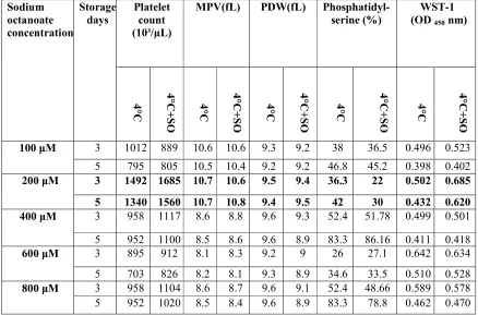
The optimum concentration of SO was determined. In this concentration (200 µM), PS exposure was lower on the third and fifth days of storage in comparison to other concentrations. Additionally, based on WST-1 assay, better viability of PLTs was observed at this concentration of SO (Table I).
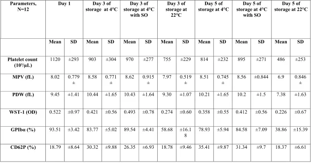
PLT enumeration, MPV, and PDW The number of PLTs was decreased during storage in all the groups of study. A decreasing trend for platelet count was seen as follows: SO (4°C) PLTs > 4°C PLTs > 22°C PLTs during storage (Table II, Figure 1). The differences between the groups were not statistically significant. MPV and PDW levels were increased in the both groups of 4°C and SO (4°C) in comparison to the group of 22°C PLTs, but the differences were not statistically significant. At the temperature of 22°C, the amount of MPV was decreased on day 5 compared to day 1 and the difference was statistically significant (P< 0.05).
108 Iran J Ped Hematol Oncol. 2019, Vol 9. No 2, 105-116 PLTs survival rates using WST-1 assay
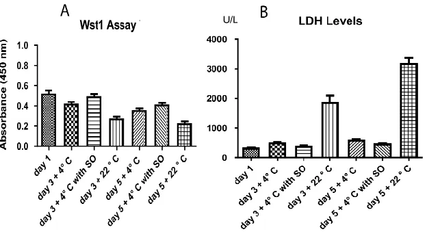
The metabolic activity and the survival rate of PLTs were decreased during storage and the highest reduction was related to 22°C-kept PLTs. The differences with other groups were statistically significant (P <0.05). The metabolic activity of PLTs was well-maintained at SO (4°C) in comparison to 4°C (Fig.2A), but the differences were not statistically significant.
The activity of LDH
The LDH amount of PLTs was significantly increased during storage and it was higher at 22°C compared to 4°C (p< 0.05). It is of note that the amount of LDH was lower in PLTs in the presence of SO (4°C) (Fig.2B), but the difference with 4°C group was not statistically significant.
The level of GPIbα expression
GPIbα expression on PLTs was higher in the group of SO (4°C) during storage time
in comparison to other groups. GPIbα on PLTs had the lowest expression during storage at 22°C in comparison to cold storage condition (Table II, Figure 3). The difference in the GPIbα expression between the SO group and 4°C group was significant (P= 0.046). Besides, this difference between the SO group and 22°C group was also significant (P= 0.001).
The levels of CD62P expression
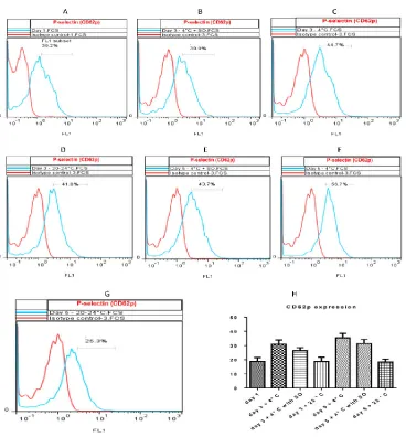
The level of CD62P expression was evaluated on PCs during storage. The expression level of CD62P on PLTs was lower in the presence of SO (4°C) in comparison to 4°C during storage, but the differences were not statistically significant. Additionally, the expression of CD62P on PLTs was lower at 22°C than 4°C during storage and the differences were statistically significant between the groups (P< 0.05) ( Table II, Figure 4).
Figure1. Effects of temperature and sodium octanoate on the PLT enumeration, MPV and PDW. A) PLTs count was decreased during storage in all the groups of study. But, the decline was higher in PLTs stored at room temperature in comparison with the PLTs stored at 4°C. The lowest decrease of PLTs count was observed at 4°C+SO. B) MPV amount was increased in both groups of 4°C and SO (4°C) and decreased at room temperature during storage. C) PDW amount was increased in both groups of 4°C and SO (4°C) and decreased at room temperature during storage.
Iran J Ped Hematol Oncol. 2019, Vol 9. No 2, 105-116 109 Figure2. Effects of temperature and SO on the survival rate (WST-1) and LDH amount of platelets in different days of storage. A) The viability was reduced in all the groups of study during storage time, specifically in PLTs stored at room temperature. The highest viability was observed in SO- treated PLTs. The differences weren’t statistically significant. B) The level of LDH enzyme was lower at 4ºC than that of room temperature. SO could reduce the amount of enzyme compared with 4ºC.
Figure 3. Flow cytometry plot for GPIbα staining. Effects of temperature and SO on the expression level of GPIbα on PLTs. The decreased expression of GPIbα was observed in all the groups of study during storage. The expression of GPIbα on SO-treated PLTs (4°C) was higher than other groups during storage and there was the lowest expression of GPIbα on PLTs stored at room temperature. A) The expression level on day 1. B) The expression level at 4°C on day 3. C) The expression level at 4°C+SO on day 3. D) The expression level at 4°C on day 5. E) The expression level at 20-24°C on day 3. F) The expression level at 4°C+SO on day 5. G) The expression level at 20-24°C on day 5. H) A diagram for showing the expression level of GPIbα on PLTs at different days of storage in all the groups of study .The difference in the GPIbα expression between the SO group and 4°C group was significant (P= 0.046). Besides, this difference between the SO group and 22°C group was also significant (P= 0.001).
110 Iran J Ped Hematol Oncol. 2019, Vol 9. No 2, 105-116 Figure 4. Flow cytometry plot for CD62P staining. Effects of temperature and SO on the expression level of CD62P during storage. There was an increase in the expression level of CD62P in PLTs of PCs stored at 4°C. The expression of CD62P was lower in the SO-treated PLTs during storage at 4°C compared to 4°C alone. A) The expression of CD62P on day 1. B) The expression level at 4°C on the third day. C) The expression level at 4°C+SO on the third day. D) The expression level at 20-24°C on the third day. E) The expression level at 4°C+SO on the fifth day. F) The expression level at 4°C on the fifth day. G) The expression level at 20-24°C on day 5. H) A diagram for showing the expression level of CD62P levels on PLTs at different days of storage in all the groups of study.
Iran J Ped Hematol Oncol. 2019, Vol 9. No 2, 105-116 111 Table I: Determination of more suitable concentration of sodium octanoate for platelets during storage at 4ºC
Sodium octanoate concentration
Storage days
Platelet count (10³/µL)
MPV(fL) PDW(fL) Phosphatidyl- serine (%)
WST-1 (OD 450 nm)
4
°C
4°C+
SO
4
°C
4°C+
SO
4
°C
4°C+
SO
4
°C
4°C
+SO 4°C
4°C+
SO
100 μM 3 1012 889 10.6 10.6 9.3 9.2 38 36.5 0.496 0.523
5 795 805 10.5 10.4 9.2 9.2 46.8 45.2 0.398 0.402
200 μM 3 1492 1685 10.7 10.6 9.5 9.4 36.3 22 0.502 0.685
5 1340 1560 10.7 10.8 9.4 9.5 42 30 0.432 0.620 400 μM 3 958 1117 8.6 8.8 9.6 9.3 52.4 51.78 0.499 0.501
5 952 1100 8.5 8.6 9.6 8.9 83.3 86.16 0.411 0.418
600 μM 3 895 912 8.1 8.3 9.2 9 26 27.1 0.642 0.634
5 703 826 8.2 8.1 9.3 8.9 34.6 33.5 0.510 0.528
800 μM 3 958 1104 8.6 8.7 9.6 9.1 52.4 48.66 0.589 0.578
5 952 1020 8.5 8.4 9.6 8.9 83.3 78.8 0.462 0.470
112 Iran J Ped Hematol Oncol. 2019, Vol 9. No 2, 105-116
Table II: The mean ± standard deviation for different parameters of platelets during the storage days at different conditions
Day 5 of storage at 22°C Day 5 of
storage at 4°C with SO Day 5 of
storage at 4°C Day 3 of
storage at 22°C Day 3 of
storage at 4°C with SO Day 3 of
storage at 4°C Day 1 Parameters, N=12 SD Mean SD Mean SD Mean SD Mean SD Mean SD Mean SD Mean ±253 486 ±271 895 ±232 814 ±229 755 ±277 970 ±304 903 ±293 1120 Platelet count (10³/µL) 0.846 ± 6.9 0.844 ± 8.56 0.745 ± 8.51 0.519 ± 7.97 0.915 ± 8.62 0.771 ± 8.58 0.779 ± 8.02 MPV (fL) ±1.63 7.38 ±1.5 10.2 ±1.65 10.21 ±1.07 9.30 ±1.64 10.43 ±1.65 10.44 ±1.41 9.45 PDW (fL) 0.67 ± 0.226 0.56 ± 0.412 0.55 ± 0.358 0.60 ± 0.274 0.78 ± 0.493 0.56 ± 0.421 0.97 ± 0.522 WST-1 (OD) ±15.39 38.86 ±7.09 84.58 ±5.94 78.93 ±16.1 8 58.68 ±4.41 89.54 ±5.02 83.77 ±3.42 93.51 GPIbα (%) ±6.61 18.37 ±9.7 31.34 ±9.87 35.41 ±9.46 18.78 ±6.93 26.35 ±9.88 30.32 ±8.64 18.79 CD62P (%)
Iran J Ped Hematol Oncol. 2019, Vol 9. No 2, 105-116 113
Discussion
In this study, the potential of SO was evaluated on the viability and the expression level of PLTs’ surface markers during storage at 4°C. For this purpose, PCs were kept for 5 days under the following conditions: 20-22°C, 4°C, and 4°C + SO. It was observed that PLT count decreased during storage in all the groups of study. However, the highest reduction was observed in PLTs of PCs at room temperature in comparison with the PLTs stored at 4°C. The differences were statistically significant. Our study showed that SO could keep the PLT count at 4°C and it also led to better survival rate of PLTs. The results of this study were correlated with Wood et al.,'s study (2016), indicating PLTs count was reduced in both groups of PLTs stored at room and cold temperature during storage. However, despite the higher count of PLTs at cold temperature, the difference was not statistically significant (19). In another study by Handigund et al.,'s on PLTs of PCs at cold temperature, similar results were observed (22).
Further, higher MPV and PDW were observed in PLTs at cold (4°C and 4°C + SO) than 22°C which could be due to PLT deformation from discoid to sphericity. At room temperature, the MPV and PDW of PLTs were lower during storage compared to the day one; this may also be due to the formation of microvesicles from PLTs similar to the results reported by Seghatchian et al.,'s (12). Similar to this study, Johnson and Becker showed significant changes in the morphology and increased PLT volume when PCs were kept at cold (26).
WST-1 test was done to unravel the mitochondrial metabolic activity and the survival rate of PLTs during storage. The survival of PLTs was decreased during storage and one of the reasons for this reduction was the production of lactate and reducing the pH (27, 28). In this study, the viability was reduced in all the groups during storage time and the highest
reduction in viability was observed in PLTs stored at room temperature. It is of note that the lowest reduction in viability was observed in PLTs treated with SO. The results of this study indicated that SO could be effective in the survival rate of PLTs.
According to the results of this study, the level of LDH enzyme was decreased at 4ºC than the room temperature. Moreover, SO could reduce the level of the enzyme in comparison to other groups. The reduction was not statistically significant. Based on the results, the highest level of enzyme activity was observed in PLTs stored at room temperature on the third and fifth days of storage. According to the previous studies, part of the activity of LDH is due to the release of the enzyme during PC agitation (15). One of the important factors in maintaining the quality and survival of PLTs of PC is the low level of LDH released in the plasma during storage (17-18). With evaluating the effects of LDH, SO-treated PLTs have less damage in their membrane than other groups during storage, which could indicate the positive effect of SO on the maintenance and survival rate of PLTs during storage of PC. Like this study, previous reports stated that reducing the GPIbα expression occurred during the storage of PLTs regardless of the storage temperature (29). The prolonged storage of PLTs at cold temperature leads to increased exposure of galactose residues. Subsequently, PLTs are removed by liver hepatocytes using their asialoglycoprotein receptors (30). According to the previous studies, storage of PLTs at cold leads to the reduction of surface glycoproteins (GPIb, GPIX, GPIIb, and GPIV) on PLTs (19). In parallel, the results of this study showed decreased expression of GPIbα on PLTs during storage in all the groups of study. Additionally, GPIbα expression was higher on SO-treated PLTs at 4°C than other groups during storage. This can be due to the protective effect of SO. Studies indicated that the loss of GPIbα could be
114 Iran J Ped Hematol Oncol. 2019, Vol 9. No 2, 105-116 (31-32). The lowest expression of GPIbα
was observed in PLTs stored at room temperature. This reduction can affect the function and survival of PLTs after transfusion.
It has been suggested that CD62P acts as a mediator in the clearance of PLTs in the circulation (33-35). However, some studies did not agree and stated that despite the binding of PLTs to leukocytes via CD62P, this molecule did not have a role in the clearance of PLTs (36). It was previously reported that the expression of CD62P on PLTs increased in low-temperature conditions (19،29). In this study, similarly, an increase in the expression of CD62P on PLTs was observed for 4°C-stored PLTs. Moreover, the expression of CD62P was lower in the SO-treated PLTs during storage at 4°C compared with 4°C alone. In this study, the lowest expression of CD62P on PCs was observed at room temperature. Based on the previous studies, the level of CD62P expression on the surface of the PLTs was increased during storage at room temperature and this could imply the continuous release of CD62P from the surface of PLTs subsequent to their activation (10). Altogether, it could be concluded that SO had the potential to enhance PLTs survival and modulate the effects of cold temperatures on PLTs. Futher studies are required to unravel the other effects of SO on PLTs kept at 4ºC.
Conclusion
According to results of this study, SO could modulate the effects of cold temperatures on PLTs. Furthermore, we found that the survival of platelets was better maintained in the presence of SO at 4°C.
Acknowledgments
This study was the result of a PhD thesis which was financially supported by Blood Transfusion Research Center, High Institute for Research and Education in
Transfusion Organization, Tehran.
Conflict of interest
None of the authors have any conflicts of interest to declare.
References
1.Acker JP, Marks DC, Sheffield WP. Quality assessment of established and emerging blood components for transfusion. J blood transfus 2016; 2016:4860284.
2.Reddoch-Cardenas KM, Bynum JA, Meledeo MA, Nair PM, Wu X, Darlington DN, et al. Cold-stored platelets: A product with function optimized for hemorrhage control. Transfus Apher Sci 2018; 1473-0502(18):30482-8.
3.Wood B, Johnson L, Hyland RA, Marks DC. Maximising platelet availability by delaying cold storage. Vox Sang 2018; 113(5):403-11.
4.Ezuki S, Kanno T, Ohto H, Herschel L, Ito T, Kawabata K, et al. Survival and recovery of apheresis PLTs stored in a polyolefin container with high oxygen permeability. Vox Sang 2008; 94:292-8. 5.Thon JN, Schubert P, Devine DV. PLT storage lesion: a new understanding from a proteomic perspective. Transfus med rev 2008; 22:268-79.
6.Manasa K, Vani R. Influence of oxidative stress on stored platelets. Adv hematol 2016; 2016:4091461.
7.Hornsey VS, McColl K, Drummond O, McMillan L, Morrison A, Morrison L, et al. Extended storage of platelets in SSP+ platelet additive solution. Vox Sang 2006; 91(1):41-6.
8.Sperling S, Vinholt PJ, Sprogøe U, Yazer MH, Frederiksen H, Nielsen C. The effects of storage on platelet function in different blood products. Hematology 2019; 24(1):89-96.
9.Wandall HH, Hoffmeister KM, Sørensen AL, Rumjantseva V, Clausen H, Hartwig JH, Slichter SJ. Galactosylation does not prevent the rapid clearance of long-term,
Iran J Ped Hematol Oncol. 2019, Vol 9. No 2, 105-116 115 4° C-stored platelets. Blood 2008;
111(6):3249-56.
10.Izadpanahi HA, Yari F, Khorramizadeh MR, Maghsudlu M. Evaluation of biochemical parameters of platelet concentrates stored in plasma or in a platelet additive solution (Composol). Iran J Ped Hematol Oncol 2011; 1(3):83-9. 11.Ng MS, Tung JP, Fraser JF. Platelet storage lesions: What more do we know now? Transfus med rev 2018; 7963(17):30189
12.Getz TM, Montgomery RK, Bynum JA, Aden JK, Pidcoke HF, Cap AP. Storage of platelets at 4 C in platelet additive solutions prevents aggregate formation and preserves platelet functional responses. Transfusion 2016; 56(6):1320-8.
13.Klein HG, Spahn DR, Carson JL. Red blood cell transfusion in clinical practice. Lancet 2007; 370(9585):415-26.
14.Vostal JG, Gelderman MP, Skripchenko A, Xu F, Li Y, Ryan J, et al. Temperature cycling during platelet cold storage improves in vivo recovery and survival in healthy volunteers. Transfusion 2018; 58(1):25-33.
15.Hoffmeister KM, Felbinger TW, Falet H, Denis CV, Bergmeier W, Mayadas TN, et al. The clearance mechanism of chilled blood PLTs. Cell 2003; 112(1):87-97. 16.Li R, Hoffmeister KM, Falet H. Glycans and the platelet life cycle. Platelets. 2016; 27(6):505-11.
17.Quach ME, Chen W, Li R. Mechanisms of platelet clearance and translation to improve platelet storage. Blood 2018; 131(14):1512-21.
18.Rumjantseva V, Grewal PK, Wandall HH, Josefsson EC, Sørensen AL, Larson G, et al. Dual roles for hepatic lectin receptors in the clearance of chilled PLTs. Nat med 2009; 15(11):1273-80.
19.Wood B, Padula MP, Marks DC, Johnson L. Refrigerated storage of PLTs initiate’s changes in PLT surface marker expression and localization of intracellular proteins. Transfusion 2016; 56(10):2548-59.
20.Hoffmeister KM, Josefsson EC, Isaac NA, Clausen H, Hartwig JH, Stossel TP. Glycosylation restores survival of chilled
blood PLTs. Science
2003;301(5639):1531-4.
21.Liu Q, Xu L, Jiao SX, Wang TX, Song Y, Wen ZK. Trehalose inhibited the phagocytosis of refrigerated PLTs in vitro via preventing apoptosis. Transfusion 2009; 49(10):2158-66.
22.Handigund M, Bae TW, Lee J, Cho YG. Evaluation of in vitro storage characteristics of cold stored PLT concentrates with N acetylcysteine (NAC). Transfus Apher Sci 2016; 54(1):127-38. 23.Fredrickson J, Krisher R, Morbeck DE. The impact of the protein stabilizer octanoic acid on embryonic development and fetal growth in a murine model. J assist reprod genet 2015; 32(10):1517-24. 24.Lemarie F, Beauchamp E, Legrand P, Rioux V. Revisiting the metabolism and physiological functions of caprylic acid (C8: 0) with special focus on ghrelin octanoylation. Biochimie 2016; 120:40-8. 25.Ahmadzadeh N, Yari F, Amirizadeh N, Khorramizadeh MR. Production and characterization of liquid‐stored and lyophilized reconstituted human infusible PLT membranes. Int J Lab Hematol 2011; 33(6):586-92.
26.Johnson L, Tan S, Wood B, Davis A, Marks DC. Refrigeration and cryopreservation of PLTs differentially affect PLT metabolism and function: a comparison with conventional PLT storage conditions. Transfusion 2016; 56(7):1807-18.
27.Deyhim MR, Mesbah-Namin SA, Yari F, Taghikhani M, Amirizadeh N. L-carnitine effectively improves the metabolism and quality of PLT concentrates during storage. Ann Hematol 2015; 94(4):671-80.
28.Maekawa Y, Yagi K, Nonomura A, Kuraoku R, Nishiura E, Uchibori E, et al. A tetrazolium-based colorimetric assay for metabolic activity of stored blood PLTs. Thrombosis research 2003; 109(5-6):307-14.
116 Iran J Ped Hematol Oncol. 2019, Vol 9. No 2, 105-116 W, Wagner DD, Kaufman RM, Silberstein
LE, et al. In vitro function and phagocytosis of galactosylated PLT concentrates after long‐term refrigeration. Transfusion 2007; 47(3):442-51.
30.Rumjantseva V, Hoffmeister KM. Novel and unexpected clearance mechanisms for cold PLTs. Transfus Apher Sci 2010; 42(1):63-70.
31.Li R, Josephson C, Chen W, Liang X, Syed AK, Jessup P, et al. Inhibiting GPIb Shedding Preserves Post-Transfusion Recovery and Hemostatic Function of PLTs After Prolonged Storage. Arterioscler Thromb Vasc Biol 2017; 37(12): 2271–2279.
32.Canault M, Duerschmied D, Brill A, Stefanini L, Schatzberg D, et al. p38 mitogen-activated protein kinase activation during PLT storage: consequences for PLT recovery and hemostatic function in vivo. Blood 2010; 115(9):1835-42.
33.Chen Y, Davis-Gorman G, Watson RR, McDonagh PF. Platelet CD62p expression and microparticle formation in murine acquired immune deficiency syndrome and chronic ethanol consumption. Alcohol 2003; 38(1):25-30.
34.Ramström S, Södergren AL, Tynngård N, Lindahl TL. Platelet function determined by flow cytometry: new perspectives? Semin Thromb Hemost 2016; 42(3):268-81.
35.Vandendries ER, Furie BC, Furie B. Role of P-selectin and PSGL-1 in coagulation and thrombosis. Thromb Haemost 2004; 92(3):459-66.
36.Faraday N, Rosenfeld B. In vitro hypothermia enhances PLT GPIIb-IIIa activation and P-selectin expression. Anesthesiology: Anesthesiology 1998; 88(6):1579-8