Original Research Article
EPIDEMIOLOGICAL PROFILE AND HISTOPATHOLOGICAL ASPECTS OF WILMS TUMOR (NEPHROBLASTOMA) IN CAMEROON
Jean Paul Ndamba Engbang *1,2, Zacharie Sando 3,4, Roxane Odyssée Ze Afane 1, Godefroy Simo5, André Moune 6, , Bruno Djimeli Djougmo 2, Roger Gilbert Ateba 7,8,
Amadou Fewou 4,9
1
Faculty of Medicine and Pharmaceutical Sciences, The University of Douala, Douala, Cameroon 2
Laquintinie Hospital of Douala, Douala, Cameroon 3
Yaoundé General Hospital, Yaoundé, Cameroon. 4
Faculty of Medicine and Biomedical Sciences, The University of Yaoundé I, Yaoundé, Cameroon 5
Bio-Medical and Cancer Center of Bafoussam, Bafoussam, Cameroon 6
Anapathos laboratory, Douala, Cameroon 7
Douala Gyneco-Pediatric Hospital, Douala, Cameroon 8
Anapathos Laboratory, Douala, Cameroon 9
Douala General Hospital, Douala, Cameroon Authors’ contributions This work was carried out in collaboration between all authors. Author JPNE gave the concept, designed the study and wrote the manuscript. Authors ROZA,did the data collection and compilation. Authors GS, AM, BDD and RGA managed the compilation and literature search. Authors ZS and AF did the final approval of manuscript. All authors read and approved the final manuscript
ABSTRACT
Objective: The main objective was to determine the epidemiologic profile and the histologic aspects of nephroblastoma in Cameroon.
Materials And Methods: This retrospective study was conducted over a period of 6 months. It included records of patients from 5 different hospitals in Cameroon that were approved by anatomical pathology laboratory unit. The age, gender, region of origin and histopathologic finding of the tumor were recorded from the laboratory registers for each patient. Data was assembled and stored using Excel 2013 software and was analyzed by 6.0 version of EPI info software.
Results: From this study, kidney cancer is the 2nd cause of urogenital cancers (with 9% of cases) after prostate cancer in Cameroon. Nephroblastoma was the commonest cause of kidney cancer in our country with 80 cases over a period of 14years (2004-2017). The mean pediatric age was 4,00±2,73 years while the mean adult was 30,25±19,91 years. Males were more affected with 60% predominance and most of them were origins from the west region of the country. The majority of tumors (53.22%) had a favorable histology.
Conclusion: In summary, nephroblastoma has a non-negligible impact in the population of Cameroon and has diverse histopathologic aspects
KEYWORDS : Kidney; nephroblastoma; Wilms Tumor; Cameroon
1. INTRODUCTION
Nephroblastoma or Wilms tumor (WT) is a malignant kidney tumor developed at the expense of kidney embryonic tissue [1]. Nephroblastoma is the fourth most common malignant disease in children after leukemia, lymphoma and brain tumors [2]. It mainly affects children from 1 to 5 years old with a peak between 2 - 3 years and may be associated with several types of congenital malformations in 7 to 10% of cases [1, 3]. The incidence of WT varies among racial and ethnic groups. Blacks have a high incidence, Caucasians seem to have an intermediate incidence and Asians have a low incidence [4]. In the United States the age-standardized rate of nephroblastomas is 8.5 per million Caucasian children per year (with a slight female predominance), the figure for black American children is 10.9 [5]. In the UK, the incidence (age-standardized) is 0.8 per 100,000 population. In Poland approximately 70 new cases of nephroblastoma are recorded each year with rare extra-renal localization in adults and children [6, 7]. The WT in France has an incidence of 1/10000 births [8]. In 2006 according to Ekenze et al one had an incidence between 2 and 5 years with a ratio men / women of 1.1 [9]. WT overall has a very favorable prognosis, the survival rate is 90% over 5 years. [10] Clinically the main sign of call that alert the surrounding remains the appearance of an abdominal mass in 80% of cases, we also noted other manifestations such as abdominal pain 37%, fever23 %, hematuria 21% and arterial hypertension [4, 11, 12]. The paraclinical diagnosis is currently facilitated by the advent of ultrasound, computed tomography (CT) and magnetic resonance imaging (MRI) which allow the detection of the tumor despite the inevitable place of histology insofar as it provides the diagnosis of certainty [7]. The management of nephroblastoma is done according to the recommendations of the International Society of Pediatric Oncologists (SIOP) which recommends preoperative chemotherapy, followed by surgical treatment and postoperative chemotherapy or according to the national study group. The Wilms tumor in North America,
which recommends primary nephrectomy followed by chemotherapy, radiotherapy is reserved for patients with stage 3 disease [6]. In Cameroon, although few studies have been done on this subject, the work done by Engbang et al on urogenital cancers in the Cameroon littoral region shows a predominance of 54.84% of nephroblastoma over a period of 10 years, as long as the study conducted by Angwafo III et al on primary renal masses in children in Yaounde shows a predominance of 72.4% over a 15-year period [13, 14]. From this analysis it appears that the studies have not been devoted specifically to nephroblastoma, hence the interest aroused by this theme towards this pathology, particularly in the control of epidemiological and histopathological aspects at the national level.
2. MATERIAL AND METHODS
The study is a descriptive and retrospective hospital based type, concerning patients from urology or oncology services in different health centers in five regions of Cameroon, diagnosed of nephroblastomas between January 2004 and December 2015. The study protocol was approved by Institutional Ethics Committee for Research on Human Health (No 1346 CEI-Udo/04/2018/T). The samples examined were mainly composed of biopsies and surgical specimens fixed in 10% formalin and processed according to the usual techniques of paraffin embedding, microtome cutting and staining with hematoxylin-eosin. Only patients for whom the diagnosis was confirmed by the histology were included in the study. Files with incomplete data were excluded. The parameters studied were frequency, age, sex and histopathological type of the tumor. Data entry was done using computer based statistical Package for Social Sciences (SPSS) version 20. The descriptive statistic elements were used to calculate the frequencies and proportions
3. RESULTS
3.1. Frequency of urogenital cancers in Cameroon
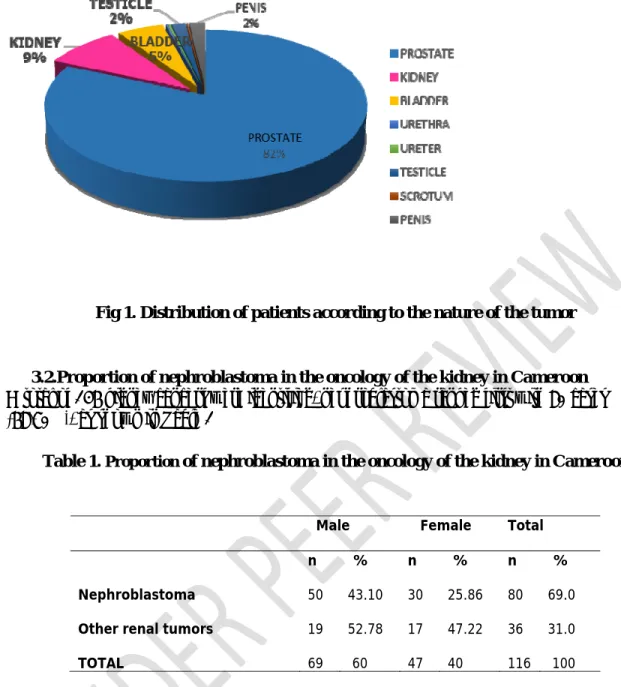
During our study period, 1665 urogenital cancers were recorded. Kidney cancer was in the second position of urogenital tumors at 9%, after prostate cancer - 82% (Fig. 1)
Fig 1. Distribution of patients according to the nature of the tumor
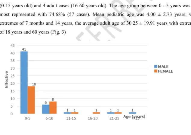
3.2.Proportion of nephroblastoma in the oncology of the kidney in Cameroon A total of 146 kidney cancers were identified, nephroblastoma ranked first with 80 cases (68.70%), as shown in Table 1
Table 1. Proportion of nephroblastoma in the oncology of the kidney in Cameroon
Male Female Total
n % n % n %
Nephroblastoma
50 43.10 30 25.86 80 69.0
Other renal tumors 19 52.78 17 47.22 36 31.0
TOTAL 69 60 47 40 116 100
3.3.Distribution of patients by sex
In a total of 80 cases, 79 had sex identification. Of the 79 cases, 75 were pediatric and 4 were adult. 50 cases (63%) were male and 29 cases (37%) female, with a sex ratio H / F 1.7: 1 In the pediatric proportion, out of the 75 cases, 48 were male (64%) and 27 (36%) female; with a sex ratio H / F of 1.8: 1 (Fig. 2)
Fig 2. Distribution of patients by sex
3.4.Age distribution
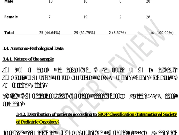
Of the 80 cases identified, 79 were with age precision. Of the 79 cases, 75 were pediatric cases (0-15 years old) and 4 adult cases (16-60 years old). The age group between 0 - 5 years was the most represented with 74.68% (57 cases). Mean pediatric age was 4.00 ± 2.73 years; with extremes of 7 months and 14 years, the average adult age of 30.25 ± 19.91 years with extremes of 18 years and 60 years (Fig. 3)
Fig 3. Distribution of patients by age
3.3. Patient distribution by tumor localization
In 51.79% of cases (29 cases) the tumor involved the right kidney and in 44.64% of cases the left kidney The bilateral form was present in 2 patients or 3.57% as shown below in Table 2.
According to this table, In regards to the mono-lateral localization, the tumor sits more on the left in the male sex and on the right in female, the bilateral form is essentially feminine.
Table 2. Distribution of patients by tumor localization
Left kidney Right kidney Bilateral Total
Male 18 10 0 28 Female 7 19 2 28 Total 25 (44.64%) 29 (51.79%) 2 (3.57%) 56 100.00%) 3.4. Anatomo-Pathological Data
3.4.1. Nature of the sample
The type of sample was documented in 59 reports of the 80 collectors. The majority of the pieces were from nephrectomy in 83.05% of cases (49 cases), and biopsy in 17% of cases (10 cases).
Similarly in the pediatric population nephrectomy accounted for 78% (46 cases), 15.25% biopsy or (9 cases).
3.4.2. Distribution of patients according to SIOP classification (International Society of Pediatric Oncology)
In our study stage II and III were the most represented with respectively 44.78% (30 cases) and 31.58% (18 cases). Stage I had 6 cases (10.53%), Stage IV was less frequent, represented 1 case (1.75%). Stage V (bilateral) was observed in only 3.5% (2 cases) of patients (Table 3)
Table 3. Distribution of patients according to SIOP classification (International Society of Pediatric Oncology)
STAGE I STAGE II STAGE III STAGE IV STAGE V TOTAL
n % n % n % n % n % n % MALE 3 5.26 20 35.09 12 21.05 1 1.75 1 1.75 37 64.92 FEMALE 3 5.26 10 17.54 6 10.53 - - 1 1.75 20 35.08 TOTAL 6 10.52 30 52.63 18 31.58 1 1.75 2 3.5 57 100.00
The majority of patients were in stage II and the most represented age group was between 0-5 years (30cas) or 52.63%. In the adult population, there were 2 cases (3.51%) in stage III and 1 case (1.75%) in stage IV (table 4)
Table 4: Relationship between classification and age group 0 -5 n % 6 -10 n % 11 - 15 n % >16 n % Total n % STAGE I 3 5.26 - 3 5.26 - 6 10.52 STAGE II 18 31.58 12 21.05 - - 30 52.63 STAGE III 11 19.30 5 8.77 - 2 3.51 18 31.58 STAGE IV - - - 1 1.75 1 1.75 STAGE V 2 3.51 - - - 2 3.51 TOTAL 34 59.65 17 29.82 3 5.26 3 5.26 57 100.00
3.4.4. Distribution of patients by seat of histolopathological type
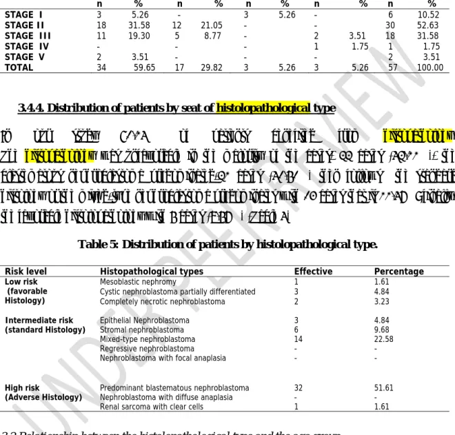
In our study 71.25% of patients benefited from histopathology. The histopathology was unfavorable in the majority of the cases, 33 cases (53.22%), the blastematous nephroblastoma predominated-32 cases (51.61%) then follows the standard histology or the mixed-type nephroblastoma predominates with 14 cases that is 22.58%; finally, the favorable histolopathology with 6 cases (9.68%) (Table 5)
Table 5: Distribution of patients by histolopathological type.
Risk level Histopathological types Effective Percentage
Low risk (favorable Histology)
Mesoblastic nephromy 1 1.61
Cystic nephroblastoma partially differentiated 3 4.84
Completely necrotic nephroblastoma 2 3.23
Intermediate risk
(standard Histology) Epithelial Nephroblastoma Stromal nephroblastoma 3 6 4.84 9.68
Mixed-type nephroblastoma 14 22.58
Regressive nephroblastoma - -
Nephroblastoma with focal anaplasia - -
High risk
(Adverse Histology) Predominant blastematous nephroblastoma Nephroblastoma with diffuse anaplasia 32 - 51.61 -
Renal sarcoma with clear cells 1 1.61
3.2 Relationship between the histolopathological type and the age group
As schown in Table 6, the majority of patients with adverse histology were male (17 cases)
Table 6: Relationship Between Histopathological Type and Age Group
0-5 6-10 11-15 ≥16 TOTAL
M F T % M F T % M F T % M F T %
Favorable 4 1 5 8.0 1 - 1 1.6 - - - - - - 9.67
Standard 11 5 16 25.8 3 4 7 11.3 - - - - - - 37.11
4. DISCUSSION
In our study, kidney cancer was the second most common urogenital cancer with a prevalence of 9% (80 cases). Our result is quite different from that of Sow et al of Cameroon in 1994 from his findings urogenital cancers was in the third position after prostate and bladder with a prevalence of 1.74% (75 cases) over 13 years, while Engbang et al in the same country in 2016 found a prevalence of 8.55% (110 cases) over a 12-year period [14, 15]. This shows that the number of cases of nephroblastoma in the country is increasing yearly; which could be explained by the evolution of diagnostic infrastructural conditions, equipment and personnel to reach a larger layer of the population.
From our view, WT is the most common kidney cancer in Cameroon 68.70% with 80 cases of nephroblastoma over a period of 14 years this result is similar to that found by Sow et al that nephroblastoma was most common in kidney cancer with 75 cases in 13 years [14]. These data are similar to those of Atteby et al in Côte d'Ivoire, 56 cases over 10 years and Ekenze et al in Nigeria, 35 cases over 10 years [16, 17]. This difference in size could be explained on the one hand by the fact that our study is multicentric and on the other hand our study duration which was relatively long.
Some syndromes are associated with WT. As with many cancer genes, the first clue to the location of a Wilms tumor gene came from the cytogenetic analysis of DNA from patients with Wilms tumor in whom there were commonly associated, genetically determined anomalies. Because of the association of seemingly unrelated phenotypic abnormalities, a large chromosomal disruption would be predicted. This strategy revealed a constitutional deletion of band 13 of the short arm of chromosome 11 in patients with the rare congenital syndrome consisting of Wilms tumor, aniridia, genitourinary abnormalities or gonadoblastoma, and mental retardation (WAGR syndrome). Loss of genetic material from this region was associated with tumorigenesis, suggesting that the critical loss was of a tumor-suppressor gene [18]. The Denys-Drash syndrome is also associated with constitutional abnormalities of the WT1 gene. In this syndrome, patients have severe genitourinary abnormalities (eg, male pseudohermaphroditism) and renal failure secondary to progressive, diffuse glomerular nephropathy. Fifty percent to 90% of these patients will develop Wilms tumor [19]. The alteration of the WT1 gene in patients with this syndrome seems to be a missense mutation within the DNA-binding region of this transcription factor. The Beckwith-Wiedemann syndrome (BWS) is another syndrome of which Wilms tumor is a common component. It is characterised by pre- and postnatal overgrowth, macroglossia, anterior abdominal wall defects, ear lobe creases and posterior helical pits, neonatal hypoglycaemia and hemihypertrophy (growth asymmetry). Non-malignant renal tract abnormalities also occur, including nephromegaly, renal cysts, medullary sponge kidney, medullary dysplasia and hydronephrosis.The overall risk of childhood cancer associated with Beckwith-Wiedemann syndrome has been estimated to be 4–21 % [20].
The average pediatric age found in our study was 4 ± 2.73 years; with the extremes of 7 months and 14 years. These results are similar to those of Abuidris et al in Sudan who found a mean age of 4.1 with extreme 2 months and 13 years [21]. D C Stefan et al observed an average age of 39 (range 3 to 167) months [21]. The young age of patients has been found by many authors around the world and ranged from 2.8 to 5.1 years [16, 22, 23, 24, 25]. These results show that nephroblastoma is a tumor of infancy.
The age group between 0 - 5 years was the most represented with 74.68% (57 cases). Shehu et al in Nigeria also found the same age group as the most affected [26]. Memon et al in Pakistan and Li et al in China found that the 0-4 age group was the most represented [24, 27].
In most cases of Wilms tumors involving one kidney and nearly all cases involving both kidneys, the tumors are thought to arise from immature kidney tissue that never developed properly. These immature tissues are known as nephrogenic rests. Nephrogenic rests (NR) are precursor lesions found within WT-bearing and WT predisposition syndrome-associated kidneys, which are composed of remnants of embryonal cells [28]. NRs are categorized into two major types: intralobar nephrogenic rests (ILNRs) and perilobar nephrogenic rests (PLNRs) [28]. ILNRs occur at deeper sites within the renal lobe and ILNR-derived tumours normally show mesenchymal (rhabdomyogenic) differentiation. PLNRs are located at the periphery of the renal lobe and PLNR-derived tumours typically have blastemal- or epithelial-predominant histology lacking myogenic differentiation. The localization of NRs and histologic elements of their associated tumours suggests the origins of precursor cells [29].
Our sample consisted of 80 cases of nephroblastoma with 29 cases (37%) for the female population and a male predominance with 50 cases (63%) that is a sex-ratio H / F of 1.17: 1.These data is closer to that of Stefan et al in South Africa who found a male predominance with 75 men and 71 women with a sex ratio of 1.06: 1 and those of Ekenze et al with 22 boys and 20 girls with a male ratio to women of 1,1: 1 [22, 23]. Crump an al shown that high fetal growth was associated with increased risk of Wilms tumor with onset before age 5 years among girls born. These findings appear consistent with previous evidence for the role of perilobar nephrogenic rests and early-life growth factor pathways, which may be more common among girls than boys. Further investigation of these mechanisms is warranted and ultimately may reveal better targets for prevention or treatment of specific subtypes of Wilms tumor [30].
Olshan suggested that high birth weight could be associated with changes in expression in the IGF2 (insulin-like growth factor 2 ) gene or other genes that regulate growth on the short end of chromosome 11 where the Wilms tumor 1 gene is also located[31]. Breslow et al. defined two “ideal” types of Wilms tumor, one characterized among other features by the presence of ILNR (intralobar nephrogenic rests) and with WT1 mutation as the critical molecular event and the other characterized by PLNR with molecular events involving dysregulated expression of genes in the region of IGF2 [32]. They suggested that PLNR might be the result of excessive or prolonged exposure to IGF2 of nephrogenic blastema during the period of nephron formation. The right kidney was the most affected with 51.79% (29 cases) followed by the left kidney with 44.64% (25 cases). These results are similar to those of Sow et al who found that the right kidney with 39 cases against 36 cases for the left kidney which was the most affected by Ekenze et al [14, 17] in Nigeria, with 21 cases against 14 cases for the right kidney. There are differences from one series to the next with respect to the side most often attained. The right kidney is as frequently affected as the left kidney [10].
Wilms tumour (WT) is an embryonal tumour whose histology can consist of three major components (epithelial, blastemal and stromal) [33; 34] which recapitulates the developmental phases of nephrogenesis [35], but in a disorganised manner. Some WTs additionally show myogenic and other mesodermal differentiation. In the WT that contain epithelial elements, there are at least two different types of epithelial structures based on the morphology and WT1 immunohistochemistry [36]. One of these is a not fully differentiated epithelial structure, which is positive for WT1 immunostaining, and which might be equivalent to early glomerular epithelia of the developing kidney [37]. The other has the appearance of a fully formed epithelial
structure, which is negative for WT1 immunostaining, suggesting that this type of epithelial structure is equivalent to the UB and its derivative, the collecting duct [37].
In our study, stages II and III were the most represented with respectively 44.78% (30 cases) and 31.58% (18 cases), stage I had 6 cases or 10.53%, stage IV was very less represented with 1 case (1.75%); stage V (bilateral) was observed in only 3.5% (2 cases) of patients. A study conducted by Ladjadj et al in Algeria showed that Stage II (39%) was predominant [38]. Stefan et al in South Africa found that the most common stages were stage I (50%) and stage IV (23%), while stages II and III had a similar incidence of around 10% and stage V (bilateral) was observed in only 4% of patients [17]. A study conducted by Kanyamuhunga et al in Rwanda showed approximately half 14/25 of the patients (56%) were in advanced stages, 7 children (28%) had stage IV, 7 stage III children, 6 patients ( 24%) with stage II, while the other five (20%) had stage I with a high-risk tumor [39]. In our study 71.25% of patients had histology. Of the 57 cases that received histology 33 cases or 53.22% are of high risk group, 14 cases or 22.58% are of intermediate risk group and 6 cases or 9.68% are low risk.
One of the major contributions of some studies was a report that separated Wilms tumors into distinct histopathologic categories based on prognosis [40]. Since that study, two distinct histopathologic types of Wilms tumor have become recognized—favorable and unfavorable. The unfavorable group comprises Wilms tumors with anaplasia (extreme nuclear and cytologic atypia). Anaplasia is present in about 5% of Wilms tumors and is more common in older children, reaching a peak at approximately 5 years of age [41]. In our study, the histopathology was unfavorable in the majority of the cases either 33 cases (53,22%) the blastematous nephroblastoma predominated-32 cases (51,61%) then comes the standard histology or the mixed-type nephroblastoma predominates with 14 cases that is 22.58%; finally, the favorable histolopathology with 6 cases (9.68%)
5. CONCLUSION
At the end of this study, it appears that renal cancer is the second most common urogenital cancer with 9%. Nephroblastoma is the first kidney cancer in our country with 80 cases observed over 14 years. Mean pediatric age was 4.00 ± 2.73 years; while the average adult age of 30.25 ± 19.91 years. There is a predominance of masculine at 64%. Histology was unfavorable in most of our sample (53.22%).
CONSENT It is not applicable. ETHICAL APPROVAL
Institutional Ethics Committee for Research on Human Health (No 1346 CEI-Udo/04/2018/T) REFERENCES
1. Gilles V. Néphroblastome ou tumeur de Wilms. IGR.2003; 2: 1-12.
2. Bergeron C. Cancer de l’enfant. Institut mère enfant, annexe pédiatrique, Hôpital sud 3. Renn.2000: 1- 10.
3. Govender D, Beckwith J, Wilms M, Beckwith M, Webber JB, Parham B et al. The pathology of nephroblastoma .current diagnostic pathology. 2000 ; 6 : 45- 54.
4. Stiller CA, Parkin DM. International variations in the incidence of childhood renal tumours. Br J Cancer 1990; 62: 1026–1030.
6. Apoznański W, Sawicz-Birkowska K, Palczewski M, Szydełko T. Extrarenal nephroblastoma. Cent European J Urol. 2015;68(2):153-6.
7. Perlman E, Boccon-Gibod L. [Kidney tumors in childhood]. Ann Pathol. 2004; 24(6):516-35.
8. Ekenze SO, Agugua-Obianyo NEN, Odetunde OA. The challenge of nephroblastoma in a developing country. Annals of Oncology.2006 ; 17: 1598–1600
9. Poole JE. Wilms’ tumour (nephroblastoma).CME.2010; 28 : 324-326
10.Aron BS. Wilms’ tumor: A clinical study of eighty-one patients. Cancer. 1974; 33: 637– 646.
11.Mohr RR, Murphy GP. Wilms’ tumor. NY State J Med. 1974; 74: 660–665.
12.Engbang NJP, Sala B, Moby H, Fonkwa C, Essomba B, Essam Sime JD, Ateba G, Fewou A. Cancers urogénitaux dans la région du littoral-Cameroun: Epidémiologie et histopathologie. Revue de Médecine et de de pharmacie. 2014;4(2):440-446
13.Angwafo III FF, Long DD. Primary renal masse in children in Cameroon: a plea for pre-treatment histology. African Journal of Urology. 2001 ; 7(2): 51-56
14.Sow M, Mbakop A, Obama M-T, Tedjoua E, Abondo A. Les tumeurs du rein en milieu africain. Incidence et aspects anatomo-cliniques.A propos de 123 cas observés à l'Hôpital Central et au C.H.U. de Yaoundé (Cameroun). Progrès en Urologie.1994 ; 4 : 214-218 15.Engbang JPN, Sala B, Fonkwa C, Ligan Y, Djimeli BD, Simo G et al.
Histo-Epidemiology of Kidney Cancer in Cameroon: About 110 Cases. Journal of Cancer and Tumor International. 2016 ; 5(1) : 1- 10
16.Atteby Y, Couitchéré L, Atimere Y, Ouattara J, Armah S, Oulai S. Le néphroblastome à Abidjan : aspects épidémiologiques, cliniques et évolutifs. rev int sc méd -rism-2016;18,1:47-50.
17.Ekenze SO, Ekwunife H, Eze BI, Ikefuna A, Amah CC, Emodi IJ. The Burden of Pediatric Malignant Solid Tumors in a Developing Country. J Trop Pediatr. 2010 Apr;56(2):111-4
18.Davidoff AM. Wilms tumor. Adv Pediatr. 2012;59(1):247-67
19.Alomari AI, Tham JC. Denys-Drash syndrome (DDS) Pediatr Nephrol. 2006;21:1237– 40.
20.Pritchard-Jones K, Dome JS (eds.). Renal Tumors of Childhood: Biology and Therapy, Pediatric Oncology. 2014. Springer-Verlag Berlin Heidelberg
21.Abuidris DO, Elimam ME, Nugud FM, Elgaili EM, Ahmed ME, RS Arora. Wilms Tumour in Sudan. Pediatr Blood Cancer 2008;50:1135–1137
22.Stefan DC, Stones DK, van Zyl A, Uys R. The cost of nephroblastoma treatment in South Africa: A very cost-effective investment with guidelines for the rest of Africa.SAJCH.2014 ; 8(4) : 2-6
23.Ekenze SO, Agugua-Obianyo NEN, Odetunde OA. The challenge of nephroblastoma in a developing country. European Society for Medical Oncology.2006 ; 17 : 1598 – 1600 24.Memon F, Rathi SL, Memon MH. Pattern of solid paediatric malignant neoplasm at
25.Odunvbun ME, Akenzua GA. Assessment of the Pattern of Childhood Malignant Diseases seen at the University of Benin Teaching Hospital (2004-2008), Benin City, Nigeria. Journal of community health & primary health care.2015 ; 27(2) :67- 72.
26.Shehu UA, Adegoke SA, Abdulsalam U, Ibrahim M, Oyelami OA, Adeodu OO. Pattern of childhood malignant tumours in two tertiary teaching hospitals in Nigeria: comparative study. Niger J Paed 2013; 40 (2): 175 – 178.
27.Li CK, Mang OWK, Foo W. Epidemiology of paediatric cancer in Hong Kong, 1982 to 1991. Hong Kong Cancer Registry. HKMJ.1999;5:128-34
28.Beckwith JB. Precursor lesions of Wilms tumor: clinical and biological implications. Med Pediatr Oncol. 1993; 21(3):158±68.
29.Fukuzawa R, Anaka MR, Morison IM, Reeve AE. The developmental programme for genesis of the entire kidney is recapitulated in Wilms tumour. PLoS ONE; 2017, 12(10): e0186333.
30.Crump C, Sundquist J, Sieh W, Winkleby MA, Sundquist K. Perinatal risk factors for Wilms tumor in a Swedish national cohort. Eur J Epidemiol. 2014;29(3):191-7.
31.Olshan AF. Wilms' tumor, overgrowth, and fetal growth factors: a hypothesis. Cancer Genet Cytogenet 1986; 21: 303–7.
32.Breslow NE, Beckwith JB, Perlman EJ, Reeve AE. Age distributions, birth weights, nephrogenic rests, and heterogeneity in the pathogenesis of Wilms tumor. Pediatr Blood Cancer 2006; 47: 260–7.
33.Beckwith JB, Kiviat NB, Bonadio JF. Nephrogenic rests, nephroblastomatosis, and the pathogenesis of Wilms' tumor. Pediatr Pathol. 1990; 10(1±2):1±36.
34.Schüz J, Schmidt LS, Kogner P, Lähteenmäki PM, Pal N, Stokland T, Schmiegelow K. Birth characteristics and Wilms tumors in children in the Nordic countries: a register-based case-control study. Int J Cancer. 2011 May 1;128(9):2166-73. doi: 10.1002/ijc.25541.
35.Mierau GW, Beckwith JB, Weeks DA. Ultrastructure and histogenesis of the renal tumors of childhood: an overview. Ultrastruct Pathol. 1987; 11(2±3):313±33
36.Fukuzawa R, Heathcott RW, Sano M, Morison IM, Yun K, Reeve AE. Myogenesis in Wilms' tumors is associated with mutations of the WT1 gene and activation of Bcl-2 and the Wnt signaling pathway. Pediatr Dev Pathol. 2004; 7(2):125±37.
37.Fukuzawa R, Heathcott RW, Sano M, Morison IM, Yun K, Reeve AE. Myogenesis in Wilms' tumors is associated with mutations of the WT1 gene and activation of Bcl-2 and the Wnt signaling pathway. Pediatr Dev Pathol. 2004; 7(2):125±37.
38.Ladjadj Y, Ahmed MS. Aspects épidemiologiques des néphroblasomes.Thèse de médecine; Alger ; 2005 ; 8 :15-22
39.Kanyamuhunga A, Tuyisenge L, Stefan DC. Treating childhood cancer in Rwanda: the nephroblastoma example.Pan African Medical journal. 2015 ; 8688 : 1- 6.
40.Beckwith JB, Palmer NF. Histopathology and prognosis of Wilms tumors: results from the First National Wilms’ Tumor Study. Cancer. 1978;41:1937–48.
41.Green DM, Beckwith JB, Breslow NE, et al. Treatment of children with stages II to IV anaplastic Wilms’ tumor: a report from the National Wilms’ Tumor Study Group. J Clin Oncol. 1994;12:2126–31