POUR L'OBTENTION DU GRADE DE DOCTEUR ÈS SCIENCES
acceptée sur proposition du jury: Prof. J.-Ph. Thiran, président du jury
Prof. M. Unser, directeur de thèse Prof. C. Oscar Sánchez Sorzano, rapporteur
Prof. F. Peyrin, rapporteuse Prof. D. Van De Ville, rapporteur
High Performance Reconstruction Framework for Straight
Ray Tomography: from Micro to Nano Resolution Imaging
THÈSE N
O6621 (2015)
ÉCOLE POLYTECHNIQUE FÉDÉRALE DE LAUSANNE
PRÉSENTÉE LE 11 MAI 2015À LA FACULTÉ DES SCIENCES ET TECHNIQUES DE L'INGÉNIEUR LABORATOIRE D'IMAGERIE BIOMÉDICALE
PROGRAMME DOCTORAL EN GÉNIE ÉLECTRIQUE
Suisse 2015 PAR
Th`ese pr´esent´ee `a la facult´e des sciences et techniques de l’ing´enieur pour l’obtention du grade de docteur `es sciences
et accept´ee sur proposition du jury
Prof. Jean-Philippe Thiran, pr´esident
Prof. Michael Unser, directeur de th`ese
Prof. Francoise Peyrin, rapporteur
Prof. Carlos Oscar S´anchez Sorzano, rapporteur
Prof. Dimitri Van De Ville, rapporteur
´
Ecole polytechnique f´ed´erale de Lausanne—2015
Cover design by Annette Unser Printing and binding by Repro-EPFL Typeset with LATEX
Copyright © 2015 by Masih Nilchian Available athttp://bigwww.epfl.ch/
Abstract
We develop a high-performance scheme to reconstruct straight-ray tomographic scans. We preserve the quality of the state-of-the-art schemes typically found in traditional computed tomography but reduce the computational cost substantially. Our approach is based on 1) a rigorous discretization of the forward model using a generalized sampling scheme; 2) a variational formulation of the reconstruction problem; and 3) iterative reconstruction algorithms that use the alternating-direction method of multipliers. To improve the quality of the reconstruction, we take advantage of total-variation regularization and its higher-order variants. In addition, the prior information on the support and the positivity of the refractive index are both considered, which yields significant improvements.
The two challenging applications to which we apply the methods of our framework are grating-based x-ray imaging (GI) and single-particle analysis (SPA). In the context of micro-resolution GI, three comple-mentary characteristics are measured: the conventional absorption contrast, the differential phase contrast, and the small-angle scattering contrast. While these three measurements provide powerful insights on bi-ological samples, up to now they were calling for a large-dose deposition which potentially was harming the specimens (e.g., in small-rodent scanners). As it turns out, we are able to preserve the image quality of filtered back-projection-type methods despite the fewer acquisition angles and the lower signal-to-noise ratio implied by a reduction in the total dose ofin-vivograting interferometry. To achieve this, we first apply our reconstruction framework to differential phase-contrast imaging (DPCI). We then add Jacobian-type regular-ization to simultaneously reconstruct phase and absorption. The experimental results confirm the power of our method. This is a crucial step toward the deployment of DPCI in medicine and biology. Our algorithms have been implemented in the TOMCAT laboratory of the Paul Scherrer Institute.
In the context of near-atomic-resolution SPA, we need to cope with hundreds or thousands of noisy projections of macromolecules onto different micrographs. Moreover, each projection has an unknown ori-entation and is blurred by some space-dependent point-spread function of the microscope. Consequently, the determination of the structure of a macromolecule involves not only a reconstruction task, but also the deconvolution of each projection image. We formulate this problem as a constrained regularized reconstruc-tion. We are able to directly include the contrast transfer function in the system matrix without any extra computational cost. The experimental results suggest that our approach brings a significant improvement in the quality of the reconstruction. Our framework also provides an important step toward the application of SPA for thede novo generation of macromolecular models. The corresponding algorithms have been implemented in Xmipp.
Keywords:Discretization, variational formulation, iterative reconstruction, alternating-direction method of multipliers, grating-based x-ray imaging, single-particle analysis, phase-contrast imaging.
R´esum´e
Dans ce travail, nous d´eveloppons des m´ethodes de reconstruction de haute-performances pour la tomogra-phie sans diffraction. Nous obtenons des r´esultats comparables `a l'´etat-de-l'art en tomogratomogra-phie par ordina-teur en termes de qualit´e, tout en r´eduisant significativement le coˆut de calcul. Notre approche comporte trois aspects : 1) une discr´etisation rigoureuse du mod`ele d'analyse en suivant un sch´ema d'´echantillonnage g´en´eralis´e, 2) une formulation variationnelle du probl`eme de reconstruction, et 3) des algorithmes de recons-truction it´eratifs fond´es sur la m´ethode intitul´eealternating direction method of multipliers. Afin d'augmenter la qualit´e de la reconstruction, nous utilisons les techniques de r´egularisation bas´ees sur la variation totale ou des variantes `a des ordres sup´erieurs. De plus, la prise en compte des informationsa priorisur le support et la positivit´e de l'indice de r´efraction de l'objet d'´etude conduit `a des am´eliorations significatives.
Les m´ethodes que nous avons d´evelopp´ees sont mises en pratique sur deux applications importantes : l'imagerie par rayon X pour l'interf´erom´etrie `a r´eseau (en anglais GI pourGrating interferometry) et l'analyse de particule unique (en anglais SPA poursingle particle analysis). Dans le contexte de la micro-r´esolution GI, trois caract´eristiques compl´ementaires sont mesur´ees : le contraste d'absorption, le contraste de phase et le contraste de diffusion. Bien que ces trois grandeurs apportent des informations tr`es utiles pour l'´etude biologiques in vivo, jusqu`a maintenant leur obtention n´ecessitait une longue exposition qui pouvait ˆetre toxique pour le sp´ecimen ´etudi´e (par exemple lors des scanners r´ealis´es sur de petits rongeurs). Il apparaˆıt cependant que nous sommes capables de reproducer la qualit´e de l'image reconstruite par des m´ethodes de r´etroprojection filtr´ee, malgr´e le faible nombre de projections durant l'acquisition, ce qui implique une r´eduction de la dose totale de radiation. Pour atteindre ce but, nous appliquons d'abord notre m´ethode de reconstruction `a l’imagerie par contrast de phase differential. Nous effectuons ensuite une r´egularisation bas´ee sur le Jacobien afin de reconstruire simultan´ement la phase et l'absorption. Les r´esultats exp´erimentaux confirment les performances de notre m´ethode. Il s'agit d'une ´etape cruciale pour l'utilisation de la l’imagerie par contraste de phase differentiel en m´edecine et en biologie. Nos algorithmes ont ´et´e impl´ement´es et sont utilis´es au laboratoire TOMCAT de l'Institut Paul Scherrer.
Pour la SPA, r´ealis´ee `a une r´esolution quasi atomique, nous devons alors composer avec des centaines ou des milliers de projections de macromol´ecules, qui sont corrompues par du bruit. Chaque projection a une orientation inconnue et elles sont flout´ees par une fonction d'´etalement du point (en anglais PSF pourpoint spread function) qui peut ˆetre diff´erente pour chaque orientation. Ainsi, la d´etermination de la structure de la macromolecule nımplique pas seulement une tˆache de reconstruction, mais aussi la deconvolution de chaque image projet´ee, ce que nous formulons comme un probl`eme de reconstruction r´egularis´e sous contrainte. Nous sommes capables d'inclure directement la function du transfer de contraste dans le syst`eme matriciel sans coˆut de calcul suppl´ementaire. Les r´esultats exp´erimentaux confirment que notre approche augmente significativement la qualit´e de la reconstruction. Le cadre propos´e est de plus une ´etape importante pour l'application de la SPA pour l'interpr´etation de mod`eles de macromol´ecules. Les algorithmes correspondants ont ´et´e impl´ement´es sur Xmipp.
Mots-cl´es :Discr´etisation, formulation variationnelle, des algorithmes de reconstruction it´eratifs, alter-nating direction method of multipliers, l'imagerie par rayon X pour l'interf´erom´etrie `a r´eseau, l'analyse de particule unique, l’imagerie par contraste de phase.
Acknowledgement
This thesis would not have been completed without the help and support of many people. I take this oppor-tunity to express my gratitude to all of them.
I would like to express my deepest gratitude to my thesis advisor, Prof. Michael Unser. This thesis would not have been possible without his guidance and support. Michael is a great advisor and a fantastic group leader. His doors were always open for discussions. Working with him was a great opportunity for me and allowed me to grow, not only scientifically but also on a personal level. I am really indebted to him for what he taught me. I am also deeply indebted to my master thesis supervisor, Prof. Mohammad-Reza Aref, for his well-known wisdom, support, kindness, and generosity of heart. His invaluable pieces of advice have been extremely useful throughout my life.
I would like to express my sincere to the president of the thesis jury, Prof. Jean-Philippe Thiran and my committee members, Prof. Francoise Peyrin, Prof. Carlos Oscar S´anchez Sorzano, Prof. Dimitri Van De Ville, for accepting to assess my thesis and for their helpful comments and suggestions.
I have been very fortunate to collaborate with Prof. Marco Stampanoni, Dr. Zhential Wang and Dr. Peter Modregger from TOMCAT beamline in Paul Scherrer institute. Marco, Zhentian and Peter have a great personality, sense of humor, and a mind full of innovative ideas. Working with them was another unique experience, they taught me many interesting things about the problem of grating-based X-ray imaging. I hope that our collaboration will continue and we will finally step over the radiation dose limitation for in vivoimaging. In addition, I have been very fortunate to collaborate with Prof. Carlos Oscar S´anchez Sorzano for introducing and patiently teaching me the subject of single particle analysis. He has a great personality, and a mind full of innovative ideas. He is a super active and hardworking person and one of the best collaborators with who I have had experienced. I hope that our collaboration will continue and we will finally step over the reconstruction resolution of 2.5 ˚A.
During my PhD research, I was very fortunate to have been in the biomedical imaging group (BIG). I would like to offer my gratitude to my friends and colleagues in BIG for all the enjoyable moments and memories. I thank the fellow present and past lab members of the Biomedical Imaging Group (BIG), Prof. Arash Amini, Anais Badoual, Dr. Jean-Charles Baritaux, Ayush Bhandari, Emrah Bostan, Dr. Nicolas Chenouard, Dr. Ning Chu, Dr. Ricard Delgado Gonzalo, Prof. Adrien Depeursinge, Julien Fageot, Dr. Denis Fortun, Dr. Matthieu Guerquin-Kern, Dr. Ulugbek Kamilov, Dr. Hagai Kirshner, Dr. Florian Luisier, Junhong Min, Pedram Pad, Zsuzsanna P¨usp¨oki, Dr. Sathish Ramani, Dr. Daniel Sage, Daniel Schmitter, Prof. Chandra Sekhar Seelamantula, Dr. Tom´aˇs ˇSkovr´anek, Dr. Martin Storath, Dr. Pouya D. Tafti, Raquel Terr´es Cristofani, Dr. Philippe Th´evenaz, Virginie Uhlmann, Dr. C´edric Vonesch, and Dr. John Paul Ward. And more particularly, I thank my present and past office mates Dr. Jean-Charles Baritaux, Ayush Bhandari, Julien Fageot, and Zsuzsanna P¨usp¨oki. I am also grateful to Manuelle Mary, and Nadia Macor for helping me out with various administrative matters inside and outside EPFL.
I am grateful to all my Iranian friends in Lausanne who made the life in Switzerland so amazing and vii
memorable. In particular, I am so thankful to my friends known among iranians as Tir-Federal crew: Mo-hammad Parhizkar, Reza Parhizkar, Hamed Hassani, Farid Movahedi Naini and Saeid Haghighatshoar. We shared an apartment for more than a year and we had so many memories together. All the time that we spent together and all the friendship and dedication that we had for one another is beyond imagination. I hope the best wishes for them all. I am sure I will miss forever these amazing friends (to be honest, they are more than friends, like brothers) and the unforgettable moments that I spent with them.
I would have not succeeded either in life or in my research without the unconditional love, support and patience of my family. My deepest gratitude goes to my father for being an amazing father, a true friend and a great support all throughout my life. He taught me how to follow the path of honor, how to devote myself to others and how to always live away from pride and ignorance. I can not stop crying. Today is the father day, and I missed him too much (divanevar dooset daram baba!). I am deeply thankful to my mother for her unconditional love and all the sacrifices that she made in her life for her children and specially for me. There are no words that can express my deepest gratitude to her. Undoubtedly, she is the strongest woman I know. She was my mother and my father for the last ten years (binahaiat mamnunetun hastam madaram va aasheghetunam!). I would like to be specially thankful to my grandmother for her continuous supports, even more when we missed my lovely father. I would like to deeply thank my brother and his lovely wife. Having such a nice older brother is an exceptional experience. He was my brother, my friend and in one word everything for me. All the time that we spent together and dedication that we had for one another is beyond imagination. I am specially thankful to my aunt’s family, Majid, Farzaneh, Tohid and Tolou. They were kindly hosting me during my study in Tehran. Without any doubt, they are the best aunt’s family that anyone can have. They are like my second father and mother, and my lovely brother and sister. I am really indebted to my aunt’s husband, Majid, for what he taught me. Specially, I would like to thank Tohid for sharing his room with me for more than four years. I am also thankful to all my aunts, uncles and cousins. My special gratitudes go to my father-in-law and mother-in-law who always treated me like a son and trusted me in crucial occasions of my married life. I also deeply thank my sister-in-law who has always been like true sister to me.
Special thanks and recognitions go to my beloved wife for her deep love and heart-filling affection. I truly thank her for sticking by my side and believing in me even in those moments that I did not believe in myself. There are no words that can truly express my gratitude and love for her.
Contents
Abstract i
R´esum´e iii
Acknowledgement vii
1 Introduction 1
1.1 X-ray Grating Interferometry: Potentiallyin vivoImaging Modality . . . 3
1.2 Single Particle Analysis: A Step Towardsde novoGeneration of Atomic Models . . . 4
1.3 Main Contributions . . . 4
1.4 Thesis Outline . . . 6
2 Mathematical Preliminaries 7 2.1 X-ray transform . . . 7
2.1.1 Problem geometry . . . 8
2.1.2 Definition and properties . . . 9
2.2 Differential variants of the x-ray transform . . . 11
2.3 Direct inversion formula . . . 11
3 Discretization Scheme 15 3.1 Discretization Using Shift-Invariant Functional Spaces . . . 16
3.1.1 Matrix formulation . . . 16
3.1.2 Fast implementation . . . 17
3.1.3 Desirable properties of the basis functions . . . 17
3.1.4 Revisiting optimality in the projection domain . . . 19
3.1.5 Incompatible properties . . . 20
3.2 Basis functions . . . 22
3.3 Box splines . . . 23
3.3.1 Basic geometric definition . . . 23
3.3.2 Elementary box spline constituents . . . 24
3.3.3 x-ray projection of box splines . . . 25
3.3.4 Explicit formulae in 2-D . . . 27
3.4 Optimized Kaiser-Bessel window function . . . 29
3.4.1 Generalized Kaiser-Bessel window functions . . . 29
3.4.2 Measure of optimality of a basis function . . . 30 ix
3.4.3 Optimal parameters for the Kaiser-Bessel window function . . . 31
3.5 Numerical evaluation . . . 32
3.5.1 Box splines and Kaiser-Bessel window functions . . . 32
3.5.2 Optimality of the proposed taper parameter for KBWFs . . . 39
3.5.3 B-splines vs Kaiser-Bessel . . . 41
3.6 Discussion and Conclusion . . . 44
4 Reconstruction Algorithms 45 4.1 Reconstruction as an optimization problem . . . 46
4.2 Reconstruction algorithm . . . 47
4.2.1 Alternating direction method of multipliers . . . 48
4.2.2 Generalization of the proposed reconstruction scheme . . . 51
4.3 Memory efficient and fast 3D reconstruction in parallel-beam tomography . . . 54
5 FFT-cost implementation of HTH 59 5.1 Notations . . . 59
5.2 Computation ofHTH . . . 59
5.2.1 Review . . . 59
5.2.2 Generalized sampling based implementation . . . 60
5.3 FFT-cost implementation ofHTHin parallel beam geometry . . . 61
5.3.1 Error of approximation . . . 62
5.4 General cases . . . 65
5.4.1 Differential variants of x-ray projection . . . 65
5.4.2 Weighted norm and speed of convergence . . . 66
5.4.3 Extension to higher dimension . . . 67
5.5 Experimental validation . . . 67
5.5.1 One-by-one comparison . . . 67
5.5.2 Performance evaluation of the kernel in different reconstruction frameworks . . . 70
6 X-ray Grating Interferometry: potentiallyin vivoimaging modality 71 6.1 Motivation . . . 71
6.2 Physical Model . . . 71
6.3 Differential phase-contrast imaging . . . 73
6.3.1 Mathematical Consideration . . . 74
6.3.2 Imaging requirements . . . 76
6.4 Reconstruction framework using Alternating direction method of multipliers with precondi-tioned conjugate gradient . . . 78
6.4.1 Discretization of the Forward model . . . 78
6.4.2 problem specific regularization . . . 79
6.4.3 Parameter selection . . . 79
6.4.4 Convergence and inexact minimization . . . 80
6.4.5 Stopping criteria . . . 80
6.4.6 Convergence speed in practical problems . . . 80
6.5 Experimental Validation . . . 81
6.5.1 Performance metrics . . . 81
7 Improved Reconstruction Scheme for X-ray Grating Interferometry 85
7.1 Constrained regularized weighted norm . . . 85
7.1.1 Problem specific regularization . . . 85
7.1.2 Parameter setting . . . 86
7.1.3 Experimental result . . . 87
7.2 Joint phase unwrapping and radiation dose reduction in DPCI . . . 88
7.3 Complex refractive index reconstruction . . . 95
7.3.1 Model Fitting . . . 95
7.3.2 Optimization . . . 96
7.3.3 Experimental result . . . 98
8 Grating-Based Radiography: Enhanced Contrast Radiographs in Mamography 99 8.1 Phase Retrieval in Differential Phase contrast Imaging . . . 99
8.1.1 Methods . . . 100
8.1.2 Experiments . . . 103
8.2 Joint absorption and phase retrieval in Grating-based X-ray radiography . . . 109
8.2.1 Joint absorption and phase retrieval . . . 111
8.3 Conclusion . . . 112
9 Single Particle Analysis: A Step Towards Interpretation of Atomic Models 115 9.1 Physical Model . . . 115
9.1.1 The Weak-Phase Object Approximation in cryo-EM . . . 116
9.2 General Overview . . . 117
9.2.1 Iterative Refinement . . . 118
9.2.2 Literature review . . . 118
9.2.3 Contrast Transfer Function Correction . . . 120
9.3 Reconstruction framework . . . 120 9.3.1 Discretization scheme . . . 121 9.3.2 Image Reconstruction . . . 121 9.4 Experimental result . . . 123 9.4.1 Implementation details . . . 123 9.4.2 Performance metric . . . 123 9.4.3 Simulation-based analysis . . . 124
9.4.4 Real data experiment . . . 124
9.4.5 Implementation remarks . . . 124
9.5 Conclusion . . . 125
10 Conclusion 129 10.1 Summary of Results . . . 129
10.1.1 Reconstruction Framework . . . 129
10.1.2 Grating-based X-ray Imaging . . . 130
10.1.3 Single Particle Analysis . . . 131
10.2 Outlook . . . 131
List of Figures
1.1 Wave propagation . . . 2
1.2 Three complementary information retrieved by GI . . . 3
2.1 Problem geometry in two dimension . . . 8
2.2 problem geometry in three dimension . . . 9
3.1 Fast implementation . . . 18
3.2 X-ray transform of box spline . . . 26
3.3 Multiscale relationship of box splines . . . 27
3.4 Zwart-Powell box spline . . . 29
3.5 Residual error for Kaiser-Bessel window funcions . . . 31
3.6 Speed comparison . . . 33
3.7 Performance comparison of box spline family . . . 34
3.8 Accuracy of the discretization scheme . . . 34
3.9 Performance evaluation of the discretization scheme with respect to the grid size . . . 35
3.10 Reconstruction comparison of the analytical phantom . . . 36
3.11 Reconstruction performance with respect to the grid size . . . 37
3.12 Visualization of the rein of interest of the reconstructed object . . . 38
3.13 Reconstructed region of interest of the coronal section of human lung . . . 40
3.14 Optical Kaiser-Bessel window function taper parameter . . . 41
3.15 Reconstruction performance with respect to Kaiser-Bessel window function taper parameter 42 3.16 Kaiser-Bessel window function and Cubic B-spline . . . 43
4.1 Line artifacts in slice by slice reconstruction . . . 55
5.1 How band-limitted Kaiser-Bessel window function and cubic B-spline are? . . . 64
5.2 Frequency response of the digital filter used in the FBP method for conventional CT . . . 66
5.3 Reference images . . . 68
5.4 Comparison of theHTHusing the kernel implementation and direct application . . . . 69
5.5 Cost function versus iteration number . . . 70
6.1 Three dimensional setup of grating interferometry based X-ray imaging . . . 72
6.2 The in front view of the GI setup . . . 74
6.3 Talbot carpet . . . 77
6.4 Speed of convergence of different iterative techniques . . . 82
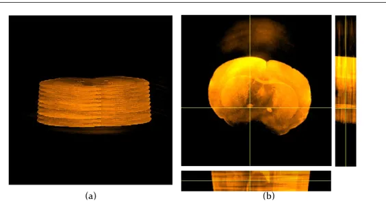
6.5 Comparison of the reconstruction results of rat brain . . . 83 xiii
6.6 The SNR and SSIM metrics for images reconstructed from a subset of projections . . . 84
7.1 Two reference samples . . . 87
7.2 Scaffold reconstruction with 250 projections . . . 89
7.3 Reconstruction performance comparison . . . 90
7.4 Itoh’s Phase Unwrapping . . . 91
7.5 Sinogram . . . 91
7.6 Phase wrapping in differential phase-contrast tomography . . . 92
7.7 Joint phase unwrapping and reconstruction . . . 93
7.8 Region of interest . . . 94
7.9 Complex refractive index reconstruction . . . 98
8.1 Quantitative comparison on phase tomographic data . . . 105
8.2 The phase retrieval results of the biopsy sample . . . 107
8.3 The phase retrieval results of the mastectomy breast sample . . . 108
8.4 The results of a selected region of interest from the whole breast phase retrieval . . . 110
8.5 Joint phase and absorption retrieval . . . 113
8.6 Joint phase and absorption interval (region of interest) . . . 114
9.1 Iterative refinement in single particle analysis . . . 118
9.2 Orientation and position estimation . . . 119
9.3 Ribosome 50S . . . 126
9.4 Performance comparison in single particle analysis . . . 127
List of Tables
3.1 Comparison of different discrete models of X-ray Transform . . . 33
3.2 Kaiser-Bessel window functions versus Cubic B-spline . . . 42
5.1 Optimal taper parameter of Kaiser-Bessel window functions with different supports . . . 63
5.2 Time ratio between the kernel implementation and direct application ofHTH . . . . 68
5.3 Signal-to-noise ratio between the kernel implementation and direct application ofHTHin conventional CT . . . 68
5.4 Signal-to-noise ratio between the kernel implementation and direct application ofHTHin the case of the first derivative of the x-ray transform . . . 69
6.1 List of notations . . . 75
7.1 Performance of different reconstruction techniques that have been applied on Phantom and Scaffold samples. . . 88
Chapter 1
Introduction
Computerized tomography (CT) revolutionized diagnostic medicine by enabling physicians to view the in-ternal structure of organs in 1970s. It aims at reconstructing the object using several images taken. The acquisition is performed by illuminating a specimen (or an organ) by an electromagnetic wave along differ-ent oridiffer-entations. In more detail, a monochromatic wave is represdiffer-ented by complex wave function
u(y,t) =a(y)exp(jφ(y))exp(j2π νt), (1.1)
wherea,φ, andν are the amplitude, phase, and frequency of the wave, respectively [1]. The parameter
y∈R2is a coordinate on a plane that is perpendicular to the direction of propagation of the wave, andt is the time parameter. The interaction of the wave with the specimen can be described by the complex refractive indexn(x) =1−δ(x) +jβ(x), wherex∈R3specifies a spatial coordinate. Then, the wave function over the exiting curve of the specimen in the context of diffraction-less electromagnetic plane wave (parallel beam with extremely small wavelength) is given by
uo(y,t) =u(y,t)exp j2π λ Z R n(sθ+PT θ⊥y)ds , (1.2)
whereλ is called the wavelength,θis the unit vector that specifies the direction of propagation of the wave,
andPT
θ⊥∈R
3×2is the transpose of the matrixP
θ⊥∈R2×3withPθ⊥xthe orthogonal projection ofxonto the plane perpendicular to θ. Equation (1.2) describes the phase shift and attenuation introduced by the specimen, as shown in Figure 1.1.
In conventional CT, the decay of the wave intensity (the attenuation) is inferred for several orienta-tions. This is linked to the X-ray transform of the attenuation coefficient µ(x) =4π β(x)/λ [2]. Since the mathematical model of CT is based on the x-ray transform, the specimen can be reconstructed using the analytical solution, particularly filtered back-projection-type algorithm (FBP). The drawback of FBP is the requirement of a large number of orientations with high signal-to-noise projection measurements for good-quality reconstruction which is equivalent to long exposure time. In order to reduce the radiation dose, several sophisticated algorithms have been developed for CT, including iterative coordinate descent (ICD) methods [3], block-based coordinate descent [4], ordered subset algorithms based on separable quadratic surrogates [5], preconditioned nonlinear conjugate-gradient methods [6], and alternating direction method of multipliers [7]. Precise modeling of the acquisition process in these methods offers a gain in quality with respect to FBP for the same data and degrades more gracefully than FBP when the data worsen; equiva-lently, it results in a notable radiation dose reduction. Moreover, specialized hardwares (CT vendors) have
illuminati on phase sh ift attenu ation x1 x2 x1 x2
y
illuminati on(a)
(b)
Figure 1.1: (a) The specimen introduces a phase shift and an attenuation. (b) Illustration of the intensity received by the detector.
also been manufactured to implement these techniques. GE Healthcare started with ASIR (adaptive statis-tical iterative reconstruction) in 2008; the Siemens company provided SAFIRE (sinogram affirmed iterative reconstruction) in 2010; the Toshiba company proposed AIDR (adaptive iterative dose reduction) in 2010.
The phase shift of the transmitted wave is given by the x-ray transform of the local phase shift per length,
φ(x) =2π δ(x)/λ. Note that, in practice, intensity is the only measurable quantity. Therefore, it is necessary to find a mechanism that transforms the phase into an intensity. This fact motivates the development of various phase-contrast imaging modalities (PCI) including analyzer based [8–10], interferometric [11–13], and free-space propagation methods [14–16]. These methods differ substantially in terms of the physical signal that is measured and their experimental setup. They often show higher contrast over the conventional imaging of biological samples and soft tissues [17–23]. The iterative reconstruction scheme in PCI has not been developed as much as the conventional CT.
In this thesis, we develop a unified and high-performance reconstruction scheme for straight-ray tomog-raphy. We achieve the same level of sophistication as the state-of-the-art iterative schemes in conventional CT and take profit of recent developments in the specialized area of straight-ray tomography, but at a much lower computational cost. After successive application of conventional CT for the visualization of the spec-imen with the resolution of lower than micro meter, several imaging modalities have been developed from micro to nano resolution. In these modalities, the state-of-the-art reconstruction until very recently has been using direct methods. We demonstrate the proposed framework in the context of grating-based x-ray imaging for micro resolution and single-particle analysis for near-atomic-resolution imaging.
Figure 1.2: [25] (a) Conventional x-ray image based on attenuation. (b) Differential phase-contrast image based on x-ray refraction. (c) Dark-field image based on x-ray scattering. All three images are intrinsically perfectly registered as they are extracted from the same data recorded with a grating interferometer. Examples of regions of enhanced contrast are marked with arrows, show-ing (b) the refraction of the trachea and (c) the scattershow-ing of the lungs. The white bars correspond to 1 cm.
1.1
X-ray Grating Interferometry: Potentially
in vivo
Imaging
Modal-ity
Phase-sensitive x-ray imaging using grating interferometry (GI) is a tomographic technique that was first proposed by David et al. [24] and Momose et al. [19]. A unique property of GI is to provide simultaneously three complementary information about the object of interest: 1) The absorption contrast, 2) the differential phase contrast, and 3) the small scattering angle which is called dark field or visibility-reduction contrast as demonstrated in Figure 1.2. Additional advantages are its compatibility with regular laboratory sources of x-rays and its high sensitivity to variations in the density of electrons, which offers further opportunities to probe the specimen.
The data provided by differential phase-contrast imaging (DPCI) corresponds to the first derivative of the x-ray transform of the real part of the refractive index of the sample. Thus, in practical applications, the common reconstruction scheme for DPCI is based on a variant of the filtered back-projection (FBP) algorithm. While FBP is a fast (non-iterative) method, it typically requires a large number of projections with high signal-to-noise ratio to achieve a good reconstruction quality [26]. This implies long exposure times which could damage the specimen. High doses of x-ray radiation can lead to an increased risk of developing cancer and may cause the genetic deffects [27–32]. In order to be able to use this technique for in vivoimaging, one requires reducing the radiation dose significantly.
Recently, several authors have proposed iterative techniques that exploit prior knowledge on the spec-imen to significantly reduce the number of required projections [33–36] at no cost in the quality. Their approaches are all based on a penalized maximum-likelihood formulation, with a standard`2-norm
iterative reconstruction algorithm for DPCI.
1.2
Single Particle Analysis: A Step Towards
de novo
Generation of
Atomic Models
The purpose of single-particle analysis is to combine images of similar particles, typically proteins or viruses, often acquired from transmission electron microscopy. The proper combination then provides high-resolution 3D reconstruction of the sample. The first use of this technique dates back to the reconstruction of human wart virus and bushy stunt virus in the 1970s [37, 38]. This technique has progressed significantly since then, currently reaching to 0.5 nanometer resolution [39]. This motivates researchers to improve its resolution even further to be able to use this technique forde novogeneration of an atomic model. The res-olution improvement can be achieved by developing precise, fast and robust particle determination scheme besides high-end microscopes and careful data acquisition.
Since the linear image-formation model of the cryo-electron microscopy (Cryo-EM) is based on the x-ray transform, one needs to reform an inversion equation for reconstructing the particle. Given the size of the data and the fact that the angular distribution of the views is uneven, this is already a non-trivial reconstruction problem. In single-particle analysis, the problem is further complicated owing to the lack of information about the orientations besides the structural variability in the particle. Moreover, the Cryo-EM images are extremely noisy because of the low electron exposure to limit the radiation damage. Then, the reconstruction problem suffers from overfitting, which means that the reconstruction express noise instead of the underlying particle details. In order to deal with this issue, It is convenient to introduce some prior information and formulate the problem as regularized inverse problem. In the literature, the smoothness concept is widely used as prior information in SPA [40–42]. This is performed by imposing a Gaussian distribution on the Fourier components of the particle in the context of maximum a posteriori estimation or using Tikhonov regularization in penalized likelihood estimation.
1.3
Main Contributions
The main focus of this thesis is on the development of a high-performance reconstruction framework for straight-ray computerized tomography. We then demonstrate the proposed framework in 1) grating-based x-ray imaging in order to reduce the radiation dose to increase its potential for being used inin vivoimaging, and in 2) single-particle analysis to improve the resolution such that one can potentially use it forde novo generation of atomic model. The five contributions of this thesis can be summarized as follows:
• Discretization Scheme:In order to formulate the reconstruction problem as an inverse problem, one needs first to discretize the forward operator. We specify the reconstruction space through the choice of the generating function. In this regard, we investigate an extended family of box splines and Kaiser-Bessel window functions. We first provide a general characterization of the x-ray transform for the extended family of box splines. We find that this level of generality simplifies the analysis because the family happens to be closed under the Radon/x-ray transform. Since all commonly used brands of B-splines are special instances of box splines [43], it makes sense to investigate these functions in more detail. Then, we consider the family of Kaiser-Bessel window functions. These are isotropic, and they involve several parameters that need to be adjusted [44, 45]. We investigate approximation-theoretic properties of these basis functions and we show how to optimize the parameters for the best performance.
• Reconstruction Framework: We have designed new iterative reconstruction algorithms that take advantage of the proposed discretization and use a combination of non-quadratic regularizes. The regularization consists either of total variation (TV) for piecewise-constant images or a higher-order extension that is better matched to biological specimens and quadratic regularizations. Our method follows an augmented-Lagrangian optimization principle and makes use of the conjugate-gradient method to solve the linear step in the alternating direction method of multipliers (ADMM). We pro-pose a problem-specific preconditioner that considerably speeds up the convergence of the linear opti-mization step. Moreover, we impose support and positivity constraints in the reconstruction algorithm. • FFT-Cost Implementation of HTH:The computationally costly step in the proposed reconstruction scheme is the calculation ofHTH. We show theoretically thatHTHis a digital convolution operator if the generating function satisfies the radial Nyquist criteria. We then show that, if we use B-spline functions or Kaiser-Bessel windows as generating function, then the proposed ADMM scheme con-verges to the fixed point of the problem. It improves the speed of reconstruction scheme significantly. We show that the use of the proposed digital convolution instead of the direct implementation ofHTH
makes the computational cost independent upon the number of orientations and the support of the generating function.
• Grating-Based X-Ray Imaging (collaboration with the Paul Scherrer Institute (PSI)):Up to now, in-vivo tomography with grating interferometry faces the challenge of large-dose deposition, which potentially harms the specimens (e.g., in small rodent scanners). Grating-based x-ray imaging is a powerful modality to investigate biological samples. It measures three complementary characteris-tics of the imaged sample: the conventional absorption contrast (AC), the differential phase contrast (DPC), and a small-angle scattering contrast. To reduce the total scanning time, we apply the pro-posed reconstruction framework to the context of differential phase-contrast imaging. We present experimental results to validate the proposed discretization method and the corresponding iterative technique. Our findings confirm that the proposed reconstruction framework is quite competitive for solving regularized problems. Moreover, our method allows for a substantial dose reduction while preserving the image quality of FBP-type methods. This is a crucial step towards the diffusion of DPCI in medicine and biology. The codes have been implemented in the TOMCAT laboratory of PSI and are being used by scientists to visualize the internal structure of their samples.
Unlike DPC tomography, where the phase information can be recovered effectively by a reconstruction algorithm, the retrieval of phase images from DPC projections remains challenging and reduces the advantages of the phase information in radiographic applications. We utilize the same algorithm pro-posed in the first part of the thesis and deploy a novel discretization approach using B-spline calculus to establish the differential operator. The algorithm is evaluated with breast biopsy and mastectomy samples. Although it is predicted theoretically that the phase image can give higher contrast for breast tissue, this has not yet been demonstrated in a clinical environment. The present study constitutes the first practical demonstration that DPC is capable of providing a higher contrast in clinical by relevant features like spiculation. These results could help to improve the diagnosis of breast cancer.
• Single Particle Analysis (collaboration with Centro Nacional de Biotecnologia, Spain (CSIC)):
Several critical difficulties arise in the context of single-particle analysis (SPA). They can be sum-marized as follows: 1) hundreds (thousands) of low-signal-to-noise-ratio micrographs with unknown orientations (too many projection images which are highly noisy), and 2) space-dependent contrast transfer function (CTF). More precisely, the measurements (micrographs) are the x-ray transform of specimens (macromolecules), filtered by the point-spread function of the microscope and varying from
one projection image to the next. Consequently, the determination of the structure of macromolecule involves deconvolving of each projection image along with the reconstruction of the specimen. We apply the proposed reconstruction framework to the context of SPA. We formulate the problem as a constrained regularized reconstruction. We show that we can directly include the CTF in the system matrixHTHwithout any extra computational cost. The experimental results suggest that our approach improves significantly the resolution of the reconstruction. It is an important step towards the applica-tion of SPA forde novogeneration of an atomic model. The corresponding codes have been already implemented in Xmipp [46, 47].
1.4
Thesis Outline
This thesis involves two main parts. The first part describes the development of a high-performance recon-struction framework for straight-ray computerized tomography (Chapters 2, 3, 4, 5). The linear mathematical model of straight-ray imaging is based on the Radon transform, the x-ray transform, and its differential. We discuss the mathematical properties of these operators in Chapter 2. We describe the discretization scheme of the imaging operator and discuss the properties that should be satisfied by the generating function to be used for discretizing the forward model in Chapter 3 as well as a fast and accurate implementation. In particular, we investigate the use of box splines and Kaiser-Bessel window functions (KBWF). We study the KBWFs from an approximation-theory point of view and propose new parameters for their use in the discretization scheme. In Chapter 4, we describe several reconstruction schemes that use the alternating-direction method of multipliers (ADMM) to solve constrained and regularized reconstruction problems. The costiest step in the proposed iterative methods is the computation ofHTH. We show that, under certain conditions, we can implement it as a digital filter with the help of the FFT.
The second part is the application of the proposed framework to the context of x-ray grating interferom-etry (Chapter 6, 7, 8) and single-particle analysis (Chapter 9). In Chapter 6, we briefly review the physical model of x-ray grating interferometry. The end-result is that differential phase-contrast imaging can be de-scribed mathematically in terms of derivative variants of the x-ray transform. We then use real data from the TOMCAT beam line to validate the proposed framework. We improve the performance of the reconstruction framework in the context of grating based imaging in Chapter 7 by developing new reconstruction schemes. Beside the tomography problem, we develop phase and absorption retrieval in the context of grating-based radiography which is important clinically in Chapter 8. The second important application is widely investi-gated in Chapter 9. We finally conclude this thesis in Chapter 10.
Chapter 2
Mathematical Preliminaries
1 Let an object be characterized by its complex refractive indexn(x) =1−α(x) +jβ(x) wherex∈
R3 specifies the object coordinate. The measurements in straight-ray imaging modalities are related to the x-ray transform and its differential variants, such as
• Conventional x-ray CT
g(y,θ) =P{β}(y,θ). (2.1)
• Propagation-based phase-contrast CT [48]
g(y,θ) =4yP{α}(y,θ). (2.2)
where4y=∂2/∂y21+∂2/∂y22is the Laplacian operator withy= (y1,y2).
• Differential phase-contrast CT [13, 49, 50]
g(y,θ) =hu,∇yP{α}(y,θ)iy, (2.3)
whereuis a unit vector in a projection coordinatey,∇yis the gradient operator with respect toyand
h·,·iyis the corresponding inner product,
ThereP{f}(y,θ)denotes the x-ray transform of a function f along a given orientationθwithy∈R2is the
projection coordinates. In this regard, we establish some higher-level mathematical properties of the x-ray transform and its differential variants in this chapter.
2.1
X-ray transform
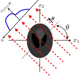
In order to specify the x-ray transform and its differential variants, we need to set the geometry of the problem. The spatial coordinates of the input function are denoted byx= [x1x2. . .xd]T and the hyperplane
projection coordinates arey= [y1. . .yd−1]T. The unit vectorθ∈Sd−1(Sd−1is the unit sphere inRd) points along the direction of integration. The projection matrixPθ⊥∈R(d−1)×d is constructed such that its rows
1A part of this chapter has been presented in [36]
✓
✓
x
1
x
2Figure 2.1:The object lies in a 2-D plane and is imaged along angleθ.
specify the normal basis of the hyperplane perpendicular to the direction of integrationθ. Then, a pointx
can be expressed in the rotated coordinate[θ,PT
θ⊥]as
x=tθ+PT
θ⊥y, (2.4)
wherePT
θ⊥is the transpose of the matrixPθ⊥.
2.1.1
Problem geometry
We now explicitly describe the geometry in the case of 2-D and 3-D input functions.
We start with two-dimensional functions. The unit vectorθ= (−sinθ,cosθ)lies along the line of
integration as depicted in Figure 2.1. The spatial coordinates of the input function are denoted by x= (x1,x2). They are also expressed in a rotated coordinate system as x=tθ+yθ⊥, wheret∈Randθ⊥= (cosθ,sinθ)is the unit vector orthogonal to the integral line (θ⊥specifies the direction of projection).
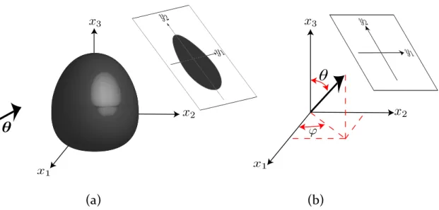
In the case of three-dimensional problem, the integral orientation can be determined by knowing two Euler anglesϕandθas depicted in Figures 2.2(a) and 2.2(b) and is given by
θ= (sinθcosϕ,sinθsinϕ,cosθ), (2.5)
In order to specify the projection matrixPθ⊥, it is necessary to determine the unit vectorsy1andy2in
the input function coordinates. The unit vectory2 is the projection of the unit vectorx3= (0,0,1)along directionθand is computed as
(a)
(b)
Figure 2.2:The object lies in a 2-D plane and is imaged along angleθ.
This yields
y2=
1 |sinθ|
−−cossinϕϕcoscosθθsinsinθθ
sin2θ
. (2.7)
The unit vectory1is the cross product ofy2andθ
y1=y2×θ (2.8)
which is in the form of
y1= 1 |sinθ| sinϕsinθ −cosϕsinθ 0 (2.9)
Then the projection matrix is
Pθ⊥= [y1,y2]T. (2.10)
2.1.2
Definition and properties
The x-ray transform is the continuous-domain operator that maps a d-dimensional function into its line integrals;P:L2(Rd)→L2(Rd−1×Sd−1)where Sd−1is the unit sphere inRd. More specifically,
P{f}(y,θ) = Z
R
f(tθ+PTθ⊥y)dt, (2.11)
Note that when f∈L2(R2), the x-ray transform is equivalent to the Radon transform. In some cases, we use
Theorem 2.1. Fourier slice theorem in the context of x-ray transformFor f∈L2(Rd), we have
Fx{f(x)}(PTθ⊥ω) =Fy{P{f(x)}(y,θ)}(ω), (2.12)
whereω∈Rd−1.
The Fourier slice theorem states that the(d−1)-dimensional Fourier transform of the x-ray transform of a function f is equal to the d-dimensional Fourier transform of the given function on the hyperplane parallel to the projection coordinate.
One can define the x-ray transform with the use of the Dirac impulse within the framework of distribution theory
P{f}(y;θ) =hf(x),δ(Pθ⊥x−y)ix∈Rd, (2.13)
whereδ(Pθ⊥x−y)is a line distribution that specifies a line that passes through the pointyin the projection
coordinate and is parallel to the orientationθ. Initially one assumes that f and its x-ray transform are in the Schwartz space, of smooth and rapidly decaying functions. One can extend the transformation toL2(Rd)by applying a standard density argument. The main properties of the x-ray transform are as follows:
• Linear:
It is a linear map by definition since it is an integral operator. Then, the x-ray transform adjoint (back-projection) is well-definedP∗:L2(Rd−1×Sd−1)→L2(Rd)and is in the form of
P∗{g(y,θ)}(x) =Z
Sd−1g(Pθ⊥x,θ)dθ. (2.14)
• Scale invariance
For anyα>0,
P{f(αx)}(y,θ) =α−1P{f}(αy,θ). (2.15) The proof is achieved with the use of the x-ray transform definition,
P{f(αx)}(y,θ) =hf(αx),δ(Pθ⊥x−y)ix∈Rd (1) =Df(x),α−dδ(α−1Pθ⊥x−y) E x∈Rd (2) =f(x),α−1δ(Pθ⊥x−αy)x∈ Rd (3) =α−1hf(x),δ(Pθ⊥x−αy)ix∈ Rd . (2.16)
The change of variablex=αx and consequently dx=|α|ddx yield the equality(1). The scaling
property of the Dirac impulse and the linearity of the inner product with respect to the second term implies the other two equalities, respectively.
• Projection translation invariance
P{f(· −x0)}(y,θ) =P{f}(y−Pθ⊥x0,θ). (2.17) Starting form the definition, we have
P{f(x−x0)}(y,θ) =hf(x−x0),δ(Pθ⊥x−y)ix
(1)
=hf(x),δ(Pθ⊥x+Pθ⊥x0−y)ix
The adjoint of a shift operator is exactly the same shift with opposite direction which implies equality (1).
• Pseudo-distributivity with respect to convolution
P{f∗g}(y,θ) = (Pf(·,θ)∗Pg(·,θ))(y,θ). (2.19)
2.2
Differential variants of the x-ray transform
Since in the parallel-beam geometry, the 3-D problem can be decomposed into a set of two dimensional problems, we focus on two dimensional function f ∈L2(R2). Thenth derivative of the x-ray transform of a function f(x)is denoted by
P(n)f(y,
θ) = ∂
n
∂ynPf(y,θ). (2.20)
The derivatives of the x-ray transform are linear operators with the following properties: • Scale invariance
P(n){f(αx)}(y,θ) =αn+1P(n)f(αy,θ), α∈
R+. (2.21)
• Pseudo-distributivity with respect to convolution
P(n)
{f∗g}(y,θ) = (P(n)f(·,θ)∗Pg(·,θ))(y,θ) = (Pf(·,θ)∗P(n)g(·,θ))(y,θ). (2.22) • Projected translation invariance
P(n)
{f(· −x0)}(y,θ) =P(n)f(y− hx0,θi,θ). (2.23)
2.3
Direct inversion formula
To derive the necessary relations, we define a new operator, the Hilbert transform along the second coordi-nate.
Definition 2.1. The Hilbert transform along the x2axis,H2:L2(R2)−→L2(R2), is defined in the Fourier domain as
\
H2{f}(ω1,ω2) =−j·sgn(ω2)bf(ω1,ω2), (2.24)
where(ω1,ω2)are spatial frequency coordinates.
Proposition 2.1. The sequential application of the x-ray transform, the nth derivative operator and the adjoint of the x-ray transform on function f∈L2(R2)is
P∗{ ∂n ∂ynP{f}(y,θ)}(x) =2π(−1) nHn 2(−4) n−1 2 {f}(x), (2.25)
where(−4)12 is the fractional Laplace operator with transfer functionkωk,(−4)n2 is n times application
of this operator, and the adjoint of x-ray transform(2.14)can be restated in two dimensional case in the form of
P∗{g}(x) =Z π
0 g(x1
Proof. Letg(y,θ) =Pf(y,θ). The Fourier Slice Theorem states that:
b
g(ω,θ) =bf(ωcosθ,ωsinθ). (2.27)
The Fourier transform of thenth derivative ofg(y,θ)with respect toyis(iω)nbg(ω,θ). Thus,
(jω)ng(bω,θ) =jn×sgnn(ω)|ω|nbf(ωcosθ,ωsinθ). (2.28)
whereω=kωk withω= (ω1,ω2) =ω(cosθ,sinθ). Forθ∈(0,π), sgn(ω) =sgn(ωsinθ) =sgn(ω2).
The space-domain equivalent is
∂n ∂ynPf(y,θ) =P{((−1) n(H 2)n(−4) n 2{f})(x)}(y,θ),∀θ∈(0,π). (2.29) Therefore we have P∗{ ∂n ∂ynP{f}(y,θ)}(x) =P ∗P{((−1)n(H 2)n(−4) n 2{f})(x)}(y,θ),∀θ∈(0,π) (2.30)
which, owing to the property thatP∗P= (−4)1/2, yields the desired results.
Equation(2.25) is a key equation. It implies that if one is interested in solving the inverse problem
P(n)f(y,
θ) =g(y,θ), (2.31)
the direct solution is to first apply the x-ray adjoint on the measured data and then apply the inverse of the operator 2π(−1)nH2n(−4)(n−1)/2. The transfer function of this inverse is21
πinsgn(ω2)nkωk−(n−1).
An equivalent form of (2.25) using the fact that(∂ ∂y)∗=− ∂ ∂yis P(n)∗ {(q∗P(n)f( ·,θ))(y)}(x) = f(x), (2.32)
whereP(n)is the adjoint of then-th derivative of the x-ray transform and the transfer function ofq(y)is b q(ωy) = 1 2π× 1 |ωy|2n−1 . (2.33)
Equation (2.32) is the basis for the generalized filtered back projection (GFBP). The full procedure is de-scribed in Algorithm 1.
Input:gθ(y) =P (n)
θ f(y)as data Output: Reconstructed image f(x) initializationNθ =The number of angles; fori=1→Nθ do
Filter the input data with the transfer function b q(ωy) = 1 2π 1 |ωy|2n−1 (2.34) end
Apply the adjoint of then-th derivative of the x-ray transform on the output of the previous stage;
return f(x).
Algorithm 1:GENERALIZED FILTERED BACK PROJECTION(GFBP) FOR THE INVERSE PROBLEM
P(n)
Chapter 3
Discretization Scheme
1
Since the mathematical model of straight-ray imaging modalities are based on the x-ray transform and its differential variants, the object can be reconstructed using direct methods such as filtered back-projection. These techniques require large number of projections with high signal-to-noise ratio in order to provide high quality reconstruction. It is equivalent to a long exposure time and high radiation dose. It is highly desirable to reduce the radiation dose in different imaging modalities. This can be achieved by either a reduction in the intensity of the photons or in the number of projection angles. The price to pay for this reduction is that the reconstruction problem becomes more ill-posed and its solution can no longer be well approximated using traditional direct methods. Instead the deployment of more sophisticated iterative schemes is needed. In order to specify such methods, one first discretizes the imaging operator, and then selects a reconstruction scheme that typically involves the choice of a cost functional to minimize.
In this chapter we concentrate on the first aspect: we use generalized sampling framework to discretize the forward operator. Therefore, the discretization problem is summarized in the choice of a suitable recon-struction space. This space is usually determined as a set of functions of the form
f(x) =
∑
k∈Zd c[k]ϕ x T −k , (3.1)whereTis the sampling step. The reconstruction space is then specified through the choice of the generating functionϕ.
In computed tomography, where the mathematical model is based on the x-ray transform and its variants, it is beneficial to use a generating function that has 1) short support and 2) good approximation properties. Isotropy is an additional property that simplifies the implementation since the footprint (x-ray transform of the function) is independent of the orientation, but typically introduces some loss of accuracy.
Two favorable candidates are box splines (in particular B-splines) and Kaiser-Bessel window func-tions. B-splines are compactly supported functions with the best cost/quality trade-off for the interpo-lation of discrete data on uniform grid. Their tensor product is used in order to extend them to higher dimension. Note that high degree B-splines are approximately isotropic. They are a special case of the box-splines investigated in this chapter. Kaiser-Bessel window functions (KBWFs) are widely used in electron microscopy [52–54] and conventional and differential phase-contrast x-ray computed tomogra-phy [34, 44, 55–57]. KBWFs involve three parameters that need to be adjusted [44, 45]. In this chapter,
1A part of this chapter has been presented in [51]
we investigate approximation-theoretic properties of the basis functions and we show how to optimize the parameters for the best performance. We also present experimental results that corroborate our theoretical prediction.
3.1
Discretization Using Shift-Invariant Functional Spaces
We first explain how the discretization of the forward model is intimately connected with the choice of a given basis function. We then recall some fundamental results from approximation theory that ensure stability and allow one to predict the expected discretization error. This will point to the importance of the partition-of-unity property which, unfortunately as we shall prove for compactly support functions, is incompatible with isotropy properties.
3.1.1
Matrix formulation
Reconstruction is usually formulated as a linear inverse problem. To solve it, it is convenient to introduce discrete representations of the object and the imaging operator. Here, we consider an object in two dimen-sions. The model of the object, from the perspective of the generalized sampling theory [58], is obtained by specifying a suitable reconstruction space. Specifically, we selectVT(ϕ)as the principal shift-invariant
space generated by the functionϕ∈L2(R2). This space is defined by
VT(ϕ) = (
∑
k∈Z2 c[k]ϕ x T −k :c∈`2(Z2) ) , (3.2)wherex∈R2. The corresponding orthogonal projection operatorPT:L2(R2)→VT(ϕ)is defined as
PTf=argmin g∈VT(ϕ)
kf−gkL
2. (3.3)
In practice, however, the values ofcin (3.2) are determined based on the solution of an inverse problem. As the derivative variants of x-ray transform are linear, pseudo shift-invariant operators, their application on a function f ∈VT is P(n) {f}(y,θ) =
∑
k∈Z2 c[k]P(n){ϕT(· −Tk)}(y,θ) =∑
k∈Z2 c[k]P(n) {ϕT}(y−Thk,θi,θ), (3.4)whereϕT(x) =ϕ(x/T),θ= (cosθ,sinθ), and
P(n)f(y,
θ) =∂
nPf
∂yn (y,θ),
withP:L2(R2)→L2(R×[0,π])being the x-ray transform operator.
The formulation of the reconstruction as a linear inverse problem is then restated as the matrix equation
g=Hc, (3.5)
wheregis the measurement vector,His the system matrix, andcis the discrete representation of the object of interest. Using (3.4), the matrix formulation can be obtained as follows: The measurement vectorg
contains values of the imaging transformP(n){f}(y,θ)at the sampled pointsy
j=j∆yandθi=i∆θ, where
i,j∈Z. The object fis represented with its coefficientscwithin the spaceVT. The system matrixHis given
by
[H](i,j),k=P(n){ϕT}(yj−Thk,θii,θi). (3.6)
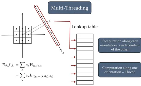
Note that, in order to compute the imaging operator, there is no need to store the whole system matrix because it is sufficient to have access to a lookup table that contains the projection of one basis function along every direction. For an isotropic basis function, storing its footprint along one orientation is enough since its footprint is independent of the orientation.
3.1.2
Fast implementation
The calculation of the x-ray transform (or its variants) involves the application of the system matrix on the coefficients of the object. However the system matrix Hcannot be stored explicitly due to its size. To circumvent this problem we exploit the translation-invariance of the x-ray transform. This property implies that all the matrix entries in (3.6) can be derived from a single derivative of the x-ray transform, namely that of the generating functionϕ:
H(i,j),k=P(n)
ϕT(yj−Thk,θii,θi).
To improve the speed, we oversampleP(n)ϕ(y,θ)on a fine gridY×Θwith for example 100 samples along each angular direction and store the values in a lookup tableL. To compute the matrix entries we define a mapping
I: R×[0,π]−→ {1,2,···,K} × {1,2, ...,P}
(y,θ)7−→(j,i), (3.7)
withKis the number of samples along each direction,Pis the number of projections and(Y(j),Θ(i))is the sample inY×Θthat is nearest to(y,θ). Therefore, we have
[H](i,j),k=LI(yj−hk,θii,θi). (3.8)
In the case of isotropic basis functions, it is sufficient to store its footprint along one orientation in the look-up table since its x-ray transform is independent of the orientation. Note that the algorithm can easily be parallelized, since projections corresponding to different angles are completely independent of each other (Figure 3.1). We designed a multithreaded implementation for an 8-core workstation which allows for 8 simultaneous projection computations. Similarly, for the adjoint of the forward model, the computation can be parallelized with respect to each object point.
In summary, our implementation is based on an accurate continuous-to-discrete model. Moreover it is fast thanks to the use of look-up tables and multi-threading. Note that our method could be also adapted to fan beam geometry by mapping it back to the parallel beam geometry. This would lead to a non-uniform sampling pattern but our method can account for this at no additional cost (thanks to our look-up-table-based implementation).
3.1.3
Desirable properties of the basis functions
Computation along each orientation is independent
of the other
Computation along one orientation = Thread d
Multi-‐Threading
y Lookup table R✓if[j] = X k ckH(i,j),k =X k ckLI(yj hk,✓ii,✓i)Figure 3.1: A simple demonstration of the implementation of the projection operator using lookup table in multithread scenario.
1)Riesz basis.Every object f∈V(ϕ)must be uniquely specified by its coefficientsc. This requires the
existence of a positive constantAsuch that
∀c∈`2, A· kck2`2 ≤ k
∑
∈Z2 c[k]ϕ x T −k L2 . (3.9)In addition, the representation should be stable. This requires the existence of a positive constantBsuch that
∀c∈`2, k
∑
∈Z2 c[k]ϕ x T −k L2 ≤B· kck2` 2. (3.10)Together, these two conditions are equivalent toϕbeing a Riesz basis ofV(ϕ).
2)Partition of unity. It is constructive for such a discretization scheme that the model approximate any input function as closely as desired by choosing a sufficiently small sampling step. More precisely, the approximation error should vanish whenever the sampling stepT tends to zero. We thus require that
lim T→0 n kf−PTfkL2 o =0. (3.11)
Theorem 3.1( [59]). Let f be a continuously defined function. The L2-approximation error of the operator
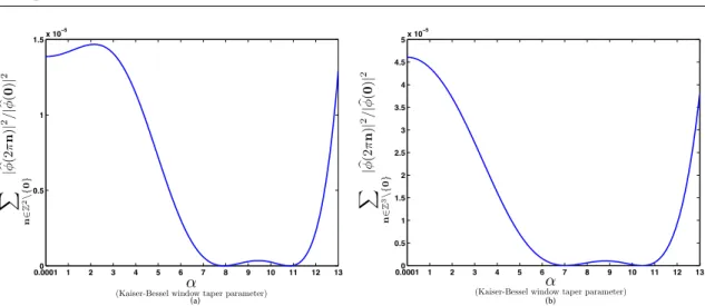
PT :L2→VT(ϕ)can be written as εf(T) =kf−PT{f}kL2 = Z R2 Eϕ(Tω)|fb(ω)| 2dω 2π 1/2 +εcorr, (3.12)
whereεcorris a correction term and Eϕis the error kernel defined in the least-squares case as Eϕ(ω) =1− | b ϕ(ω)|2 ∑k∈Z2|ϕb(ω+2kπ)|2 , (3.13)
whereϕbis the Fourier transform ofϕ. Specifically, if f ∈W2r(Sobolev space of order r) with r>1/2, then |εcorr|<γTrkf(r)kL2, whereγis some constant.
The asymptotic convergence
lim
T→0εf(T) =0 (3.14)
is achieved if and only if the basis functionϕsatisfies the partition-of-unity condition [58]
∑
k∈Z2
ϕ(x+k) =1,∀x∈R2. (3.15) The equivalent formulation of the partition of unity in the frequency domain is
b
ϕ(2πn) =δ[n],∀n∈Z2, (3.16) whereδ is the two-dimensional Kronecker delta function.
3) Compact support. The basis function ϕ should be compactly supported in order to reduce the
computational cost and also for localization in the spatial domain.
4)Isotropy. For the implementation of the imaging operator, it is required to store the values of its application on the basis function along different directions. If the basis function is isotropic, its projections do not depend on the direction, which leads to simplicity and efficiency of implementation.
3.1.4
Revisiting optimality in the projection domain
We now bound the error of approximation incurred byPTf=P{PTf}. It can be extended to any derivative
of the x-ray transform through the Fourier-slice theorem since||P(n)f||L2 =||PF−
1{|ω|nbf(ω)}||
L2. To
this end, we use the Sobolev normk·k2
W21/2 in the projection domain. Ifg∈L2(R
2), then kgk2 W21/2 = Z 2π 0 Z ∞ 0 (1+ω2) 1 2|g(bω,θ)|dθdω, (3.17)
whereg(bω,θ)is the polar form of the Fourier transform ofg.
Theorem 3.2. LetεPf(T) ==kPf−PTfk
W21/2be the Sobolev approximation error of the operatorPT.
Then, there exist positive constants r1,R1>0such that
r1εf(T)≤εPf(T)≤R1εf(T). (3.18)
Lemma 3.1. LetΩ⊂R2be a compact domain. Then, there exist positive constants r2and R2such that, for
any L2(R2)function f that is supported onΩ, it holds that
Proof. The Fourier-slice theorem implies that d
Pf(ω,θ) =bf(ω,θ), ∀ω∈[0,+∞),θ∈[0,2π). (3.20)
To show the left-hand-side inequality, we write that kfk2L 2 = bf 2 L2 = Z 2π 0 Z ∞ 0 | b f(ω,θ)|2|ω|dωdθ = Z 2π 0 Z ∞ 0 | d Pf(ω,θ)|2|ω|dωdθ ≤ Z 2π 0 Z ∞ 0 | d Pf(ω,θ)|2(1+ω2)1/2dωdθ =kPfk2 W21/2 . (3.21)
For the right-hand side, we decompose the integral into an integral over|ω| ≥1 and an integral over|ω| ≤1.
In the first one, we have that 2|ω| ≥(1+|ω|2)1/2. So, Z 2π 0 Z ∞ 1 | d Pf(ω,θ)|2(1+ω2)1/2dωdθ≤2 Z 2π 0 Z ∞ 1 | d Pf(ω,θ)|2|ω|dωdθ ≤2kfk2L 2. (3.22)
The integral over|ω| ≤1 is estimated using Z 2π 0 Z 1 0 | d Pf(ω,θ)|2(1+ω2)1/2dωdθ≤ Z 2π 0 Z 1 0 | b f(ω,θ)|2(1+ω2)1/2dωdθ ≤ sup θ∈[0,2π),ω∈[0,1)| b f(ω,θ)|2 Z 2π 0 Z 1 0 (1+ ω2)1/2dωdθ ≤C˜kfk2 L2 . (3.23)
Details concerning the last inequality can be found in [60, Section II.5]. Together, these inequalities yield the desired result.
Proof of Theorem 3.2. By letting f ←(f−PT{f})in (3.19), we obtain (3.18).
This theorem implies that the average error over all angles is small in the transform domain when the error of approximation is small in the object domain. While the theorem is an average result that involves a continuum of angles, it is still useful practically because it gives us the approximation error in the transform domain over a family of images that would correspond to all rotated versions of a given reference image.
3.1.5
Incompatible properties
There is an inconvenient result that is expressed in Theorem 3.3:
Theorem 3.3. The following properties are mutually exclusive for an isotropic basis function: 1. compact support;
2. partition of unity.
Proof. Here, we first provide a sketch of the argument. The partition-of-unity condition implies the configu-ration (3.16) of zeros of the Fourier transform of the basis function. At the same time, the Hankel transform of an even compactly supported function is an entire function of finite exponential type. Jensen’s theorem provides a contradiction between these two properties.
We prove Theorem 3.3 using a proof by contradiction. We suppose that there is a compactly supported isotropic functionφ that satisfies the partition-of-unity condition. Then, using Jensen’s theorem, we obtain
a contradiction.
Theorem 3.4( [61]). (J.L. Griffith) Letν>−1/2and1/p+1/q=1. Let f be an even entire function of exponential type1. If1<p≤2and tν+1/2f(t)∈Lp(0,∞), then f can be represented by
f(z) = Z 1 0 (xz) −νJ ν(xz)φ(x)dx(z∈C), (3.24) with x−ν−1/2
φ(x)∈Lq(0,1). Conversely, if f has this representation and x−ν−1/2φ(x)∈Lp(0,1),1<p≤2,
then f is an even entire function of exponential type1such that tν+1/2f(t)∈Lq(0,∞).
Without loss of generality, let us assume thatφ(x) =0, forkxk ≥1. We have the following:
• The functionφ is isotropic, so its Fourier transform is the Hankel transform of the functionφ(x) = φ(kxk)withx=kxk. We write that
F{φ}(ω) =2π Z ∞ 0 xφ(x)J0(kωkx)dx. (3.25) • We define f(z) =2π Z ∞ 0 xφ(x)J0(zx)dx, (3.26)
so f(kωk) =F{φ}(ω). According to Theorem 3.4 (withν=0),
f(z) =
Z ∞
0
ψ(x)J0(zx)dx, (3.27)
whereψ(x) =2πxφ(x). Sincex−21ψ(x)∈L2(0,1), f is an even entire function of exponential type 1.
• Satisfying the partition of unity is equivalent to having the equality in the Fourier domain b
φ(2πn) =δ[n], (3.28)
wheren∈Z2andδ is the two-dimensional Kronecker-delta function. It means that the set of zeros of
f(z)is{z=2πknk,∀n∈Z2\ {0}}. Therefore,
n(R)≥cR2, (3.29)
• Jensen’s theorem implies the inequality Z R 0 n(t) t dt≤max|z|=R log|f(z)|. (3.30) This inequality restricts the number of zeros inside the disc. We have that
n(R/2)log2= Z R R/2 n(R/2) t dt ≤ Z R R/2 n(t) t dt ≤max |z|=Rlog|f(z)|. (3.31)
• Since f is of exponential type 1, it implies that|f(z)| ≤Ae|z|. Therefore, max
|z|=Rlog|f(z)| ≤CR, (3.32)
whereCis a positive constant.
• Equations (3.29), (3.31), and (3.32) imply that
c(R/2)2log2≤n(R/2)log2 ≤max
|z|=R
log|f(z)|
≤CR. (3.33)
TakingRsufficiently large, we reach a contradiction.
3.2
Basis functions
Here, we investigate two favorable Basis functions, box splines and Kaise-Bessel window functions in order to discretize the projection operator. We first discuss box-splines, particularly B-spline functions which satisfy all the desirable properties of basis functions for tomographic application except the isotropy one. Note that high degree B-splines are approximately isotropic. We show that the space of these functions are close under the x-ray transform and we derive the analytical formula for their x-ray projection. We then present Kaiser-Bessel window functions which are compactly supported and isotropic. As implied by Theorem 3.3, these functions do not satisfy the partition of unity condition. Subsequently, we propose an optimal parameter selection based on approximation theory to have minimal deviation from the partition of unity condition.
The main interest of basis functions is to provide an effective and consistent way to discretize the forward model of a computed-tomography reconstruction problem. The basis for such an approach is to characterize one image by its coefficients c= (ck)k∈Ω whereΩ denotes the domain of the image and to apply (3.4)

![Figure 1.2: [25] (a) Conventional x-ray image based on attenuation. (b) Differential phase- phase-contrast image based on x-ray refraction](https://thumb-us.123doks.com/thumbv2/123dok_us/11058639.2992689/21.918.161.713.119.408/figure-conventional-image-based-attenuation-differential-contrast-refraction.webp)