comment
reviews
reports
deposited research
interactions
information
refereed research
Review
An overview of the structures of protein-DNA complexes
Nicholas M Luscombe*, Susan E Austin*, Helen M Berman
and Janet M Thornton*
Addresses: *Biomolecular Structure and Modelling Unit, Department of Biochemistry and Molecular Biology, University College, Gower Street, London WC1E 6BT, UK. Department of Chemistry, Rutgers State University, Piscataway, New Jersey 08855, USA. Department of
Crystallography, Birkbeck College, Malet Street, London WC1E 7HX, UK. E-mail: [email protected]; [email protected]; [email protected]; [email protected]
Correspondence: Janet M Thornton.
Abstract
On the basis of a structural analysis of 240 protein-DNA complexes contained in the Protein Data Bank (PDB), we have classified the DNA-binding proteins involved into eight different structural/functional groups, which are further classified into 54 structural families. Here we present this classification and review the functions, structures and binding interactions of these protein-DNA complexes.
Published: 9 June 2000
GenomeBiology2000, 1(1):reviews001.1–001.37
The electronic version of this article is the complete one and can be found online at http://genomebiology.com/2000/1/1/reviews/001 © GenomeBiology.com (Print ISSN 1465-6906; Online ISSN 1465-6914)
Introduction
DNA-binding proteins have a central role in all aspects of genetic activity within an organism, such as transcription, packaging, rearrangement, replication and repair. It is there-fore extremely important to examine the nature of com-plexes that are formed between proteins and DNA, as they form the basis of our understanding of how these processes take place. Over the past ten years, we have witnessed a great expansion in the determination of high-quality struc-tures of DNA-binding proteins. The strucstruc-tures, especially those of their complexes with DNA, have provided valuable insight into the stereochemical principles of binding, includ-ing how particular base sequences are recognized and how the DNA structure is quite often modified on binding.
A classification of protein-DNA complexes based on the structures of the DNA-binding regions in the proteins is described. The taxonomy was first proposed by Harrison [1] and later modified by Luisi [2]. Here, we build on the original classification with appropriate extensions to accommodate the new structures that have been solved. Assembling the structures in such a system simplifies comparison of the different modes of binding, allowing
identification of common themes between structurally related proteins and also highlighting unusual features that distinguish a particular protein from others. Examination of genes that are functionally assigned in PEDANT [3] show that typically 2-3% of a prokaryotic genome and 6-7% of a eukaryotic genome encodes DNA-binding proteins. Therefore, the classification of structures presented here is far from complete and many more entries are anticipated. It should be noted that the number of structures in the PDB does not necessarily reflect the relative importance of the protein in the organism. The helix-turn-helix (HTH), the ββα zinc finger, and the zipper-type motifs are, however, expected to be very common.
Constructing the classification
Dataset of protein-DNA complex structures
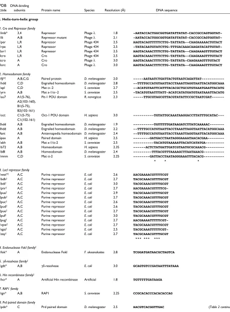
Protein-DNA complexes solved by X-ray crystallography to a resolution of higher than 3.0 Å were obtained from the January 2000 release (04/01/00) of the Protein Data Bank (PDB) [4,5] and the Nucleic Acid Database (NDB) [6]. The complexes were defined as any structure containing one or more protein chains and at least one double-stranded DNA of more than four base-pairs (bp) in length. From this set we excluded structures containing single- and quadruple-stranded DNA and non-contiguous DNA (that is, with a break in the strand). This resulted in a dataset of 240 protein-DNA complexes (Tables 1,2). Box 1 shows the selection process.
Included in the dataset were 24 homodimeric complexes whose asymmetric unit contained only half the structure. The full coordinate files were obtained from the NDB, which calculates the coordinates for the complete molecule by applying the transformation matrices provided in the PDB files to the half structure. These entries are marked accord-ingly in Table 2.
Structural taxonomy and classification of protein-DNA complexes
The PDB entries were classified according to the structures of the proteins in the complex. The classification system categorizes them in a two-tier system at the group and family levels. At the first level, proteins were sorted manu-ally into eight groups by visual inspection using RasMol [7] and from the literature. Members of the same group share a prominent structural feature used for DNA recog-nition and are related to each other in varying degrees. The eight groups are: (I) HTH (including winged HTH), (II) zinc-coordinating, (III) zipper-type, (IV) other αhelix,
(V) β sheet, (VI) β hairpin/ribbon, (VII) other, and (VIII) enzymes (Table2). The enzyme group is an exception to the structural criterion, as it contains all proteins that display enzy-matic activity when bound to DNA. Five enzymes also qualify on structural grounds for the HTH and other αhelix groups: restriction endonuclease FokI (PDB entry 1fok), γδ-resolvase (1gdt), Hin recombinase (1hcr), Tc3 transposase (1tc3) and Cre recombinase (1crx). These proteins are listed under the HTH group in Table 2 and are marked appropriately.
At the second level of classification, the DNA-recognition domains were classified into homologous families by com-paring their structures in pairs using the secondary struc-ture alignment program SSAP [8]. The program uses a dynamic programming method [9] and assesses the simi-larity between proteins by comparing the structural envi-ronments of the constituent amino acids. SSAP returns a score of 100 for identical proteins, and >80 for homolo-gous proteins; proteins are automatically assigned to the same family if they score above this cut-off. More distantly related proteins that give scores of >70 are also placed in the same family if they perform similar biological functions [10].
[image:2.609.59.558.543.723.2]Proteins were broken down into their constituent DNA-binding domains before conducting the alignments. In most dimers, each domain corresponds to a distinct subunit and the structure simply needs to be separated into the constituent chains. In proteins such as those with ββα zinc fingers, however, a chain contains several binding domains; in such cases, therefore, the subunits were separated into the appropriate segments, which are listed in Table 2. In this review, structures are identified by the standard four-digit PDB code (for example, 1aay).
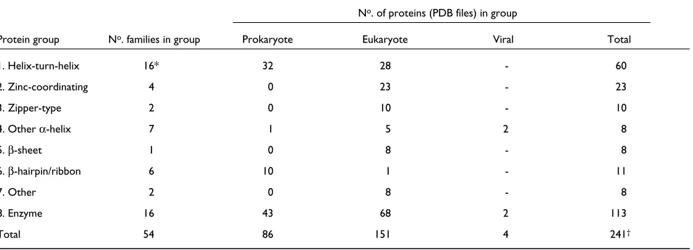
Table 1
The groups of protein structures found in the dataset, the number of families within each group and the number of PDB files each family contains.
No. of proteins (PDB files) in group
Protein group No. families in group Prokaryote Eukaryote Viral Total
1. Helix-turn-helix 16* 32 28 - 60
2. Zinc-coordinating 4 0 23 - 23
3. Zipper-type 2 0 10 - 10
4. Other α-helix 7 1 5 2 8
5. β-sheet 1 0 8 - 8
6. β-hairpin/ribbon 6 10 1 - 11
7. Other 2 0 8 - 8
8. Enzyme 16 43 68 2 113
Total 54 86 151 4 241†
*Includes the two ‘winged’ helix-turn-helix families.
comment
reviews
reports
deposited research
interactions
information
[image:3.609.55.522.117.749.2]refereed research
Table 2
List of the 240 structures of protein-DNA complexes in the dataset.
PDB DNA-binding
code subunits Protein name Species Resolution (Å) DNA sequence
I. Helix-turn-helix group
1. Cro and Repressor family
1lmb* 3,4 Repressor Phage λ 1.8
-AATACCACTGGCGGTGATATTATAT-CACCGCCAGTGGTAT-1lli A,B Repressor mutant Phage λ 2.1
-AATACCACTGGCGGTGATATTATAT-CACCGCCAGTGGTAT-1per L,R Repressor Phage 434 2.5 AAGTACAGTTTTTCTTG-TATTATA--CAAGAAAAACTGTACT
1rpe L,R Repressor Phage 434 2.5
-TATACAATGTATCTTG-TTTGACAAACAAGATACATTGTAT-2or1 L,R Repressor Phage 434 2.5 AAGTACAAACTTTCTTG-TATTATA--CAAGAAAGTTTGTACT
3cro L,R Cro Phage 434 2.5 AAGTACAAACTTTCTTG-TATTATA--CAAGAAAGTTTGTACT
6cro A Cro Phage λ 3.0 AAGTACAAACTTTCTTG-TATTATA-CAAGAAAGTTTGTACT
3orc A Cro Phage λ 3.0 AAGTACAAACTTTCTTG-TATTATA--CAAGAAAGTTTGTACT
2. Homeodomain family
1fjl*† A,B,C,G Paired protein D. melanogaster 2.0
---AATAATCTGATTACTGTAATCAGATTAT---1hdd C,D Engrailed homeodomain D. melanogaster 2.8 --TTTTGCCATGTAATTACCTAAATTAGGTAATTACATGGCAAA
1apl C,D Mat α−2 S. cerevisiae 2.7 --ACATGTAATTCATTTACACGCTGCGTGTAAATGAATTACATG
1yrn A,B Mat a-1/α−2 S. cerevisiae 2.5 -TACATGTAATTTATT-ACATCATATGATGTAATAAATTACATG
1au7 A1(5-76), Pit-1 POU domain R. norvegicus 2.3
---TTGCGTAGCGTTACGTATATTCCGCTAATCGAT---A2(103-160), B1(5-75), B2(102-161)
1oct C1(5-75) Oct-1 POU domain H. sapiens 3.0
---TGTATTGCAAATAAGGACCTTATTTGCATAC---C1(102-161)
2hdd A,B Engrailed homeodomain D. melanogaster 1.9
---TGTTTTTGATAAGATCTTATCAAAAAC---3hdd A,B Engrailed homeodomain D. melanogaster 2.2 --TTTTGCCATGTAATTACCTAAATTAGGTAATTACATGGCAAA
9ant A,B Antennapedia homeodomain D. melanogaster 2.4 --TTTTGCCATGTAATTACCTAAATTAGGTAATTACATGGCAAA
6pax A Paired domain H. sapiens 2.5
---GATGACCTAATAGGGAAAATAACACGAA---1akh A,B Mat a-1/α-2 S. cerevisiae 2.5
---TACATGTAAAAATTACATCATATGA---1b72 A,B Homeodomain H. sapiens 2.35
---ACTCTATGATTGATCGTAATGCGCAAACG---1b8I A,B Homeodomain D. melanogaster 2.4
---TTTACGTTTAAAAGCTTAATAAACG---1mnm C,D Mat α-2 S. cerevisiae 2.25 ---GATTACCTAATAGGGAAATTTACACG---* ---GATTACCTAATAGGGAAATTTACACG---* ---GATTACCTAATAGGGAAATTTACACG---*
3. LacI repressor family
1wet*† A,C Purine repressor E. coli 2.6 AACGAAAACGTTTTCGT
1bdh† A,C Purine repressor E. coli 2.7 TACGCAAACGTTTGCGT
1bdi† A,C Purine repressor E. coli 3.0 TACGCAAACGTTTGCGT
1pnr† A,C Purine repressor E. coli 2.7
AACGAAAACGTTTTCG-2pua† A,C Purine repressor E. coli 2.9 TACGCAAACGTTTGCGT
2pub† A,C Purine repressor E. coli 2.7 TACGCAAACGTTTGCGT
2puc† A,C Purine repressor E. coli 2.6 TACGCAAACGTTTGCGT
2pud† A,C Purine repressor E. coli 2.6 TACGCAAACGTTTGCGT
2pue† A,C Purine repressor E. coli 2.7 TACGCAAACGTTTGCGT
2puf† A,C Purine repressor E. coli 3.0 TACGCAAACGTTTGCGT
2pug† A,C Purine repressor E. coli 2.7
AACGAAAATTTTTCGT-1vpw† A,C Purine repressor E. coli 2.7 TACGCAAACGTTTGCGT
1qpz† A,C Purine repressor E. coli 2.5
TACGCAAATTTTTCGT-1zay† A,C Purine repressor E. coli 2.7 TACGCAAACGTTTGCGT *** *** ***
4. Endonuclease FokI family‡
1fok* A Endonuclease FokI F. okeanokoites 2.8 TCGGATGATAACGCTAGTCA
5. γδ-resolvase family‡
1gdt* A,B γδ-resolvase E. coli 3.0 GCAGTGTCCGATAATTTATAAA
6. Hin recombinase family‡
1hcr* A Artificial Hin recombinase Artificial 1.8 TGTTTTTGATAAGA
7. RAP1 family
1ign* A,B RAP1 S. cerevisiae 2.25 CCGCACACCCACACACCAG
8. Prd paired domain family
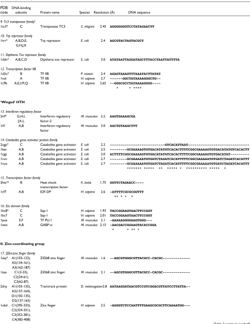
Table 2(continued)
PDB DNA-binding
code subunits Protein name Species Resolution (Å) DNA sequence 9. Tc3 transposase family‡
1tc3* C Transposase TC3 C. elegans 2.45 AGGGGGGGTCCTATAGAACTT
10. Trp repressor family
1trr* A,B,D,E, Trp repressor E. coli 2.4 AGCGTACTAGTACGCT
G,H,J,K
11. Diptheria Tox repressor family
1ddn* A,B,C,D Diptheria tox repressor E. coli 3.0 ATATAATTAGGATAGCTTTACCTAATTATTTTA
12. Transcription factor IIB
1d3u* B TF IIB P. woesei 2.4 AGAGTAAAGTTTAAATACTTATAT
1vol A TF IIB H. sapiens 2.7
---GGCTATAAAAGGGCTG--1c9b A,E,I,M,Q TF IIB H. sapiens 2.65 --GGGCGCCTATAAAAGGGG----* --GGGCGCCTATAAAAGGGG----* --GGGCGCCTATAAAAGGGG----*--GGGCGCCTATAAAAGGGG----*--GGGCGCCTATAAAAGGGG----*--GGGCGCCTATAAAAGGGG----*
‘Winged’ HTH
13. Interferon regulatory factor
2irf* G,H,I, Interferon regulatory M. musculus 2.2 AAGTGAAAGCGA
J,K,L factor-2
1if1 A,B Interferon regulatory M. musculus 3.0 AACTGTAAGCTTT
factor
14. Catabolite gene activator protein family
2cgp* C Catabolite gene activator E. coli 2.2
---GTCACATTAAT---1ber A,B Catabolite gene activator E. coli 2.5 ---GCGAAAAGTGTGACATATGTCACACTTTTCGGCGAAAAGTGTGACATATGTCACACTT
1cgp A,B Catabolite gene activator E. coli 3.0
ACTTTTCGGCGAAAAGTGTGACATATGTCACACTTTTCGGCGAAAAGTGTGACATAT---1run A,B Catabolite gene activator E. coli 2.7 ---GCGAAAAATGTGATCTAGATCACATTTTTCGGCGAAAAATGTGATCTAGATCACATTT
1ruo A,B Catabolite gene activator E. coli 2.7 ---GCGAAAAATGTGATCTAGATCACATTTTTCGGCGAAAAATGTGATCTAGATCACATTT ******* ***** ** ***** * ********** ***** *
15. Transcription factor family
3hts*† B Heat shock K. lactis 1.75
GGTTCTAGAACC----transcription factor
1cf7 A,B E2F-DP H. sapiens 2.6 -ATTTTCGCGCGGTTT ** * * *
16. Ets domain family
1bc8* C Sap-1 H. sapiens 1.93 TACCGGAAGTAACTTCCGGT
1bc7 C Sap-1 H. sapiens 2.01 TACCGGAAGTAACTTCCGGT
1pue E,F TF PU.1 M. musculus 2.1
-AAAAAGGGGAAGTGGG---1awc A,B GABP-α M. musculus 2.15 -AACGACCGGAAGTACACCGGA * * ** *
II. Zinc-coordinating group
17. ββα-zinc finger family
1aay* A1(103-133), Zif268 zinc finger M. musculus 1.6
--AGCGTGGGCGTTACGCC-CACGC---A2(134-161), A3(162-187)
1zaa C1(3-33), Zif268 zinc finger M. musculus 2.1
--AGCGTGGGCGTTACGCC-CACGC---C2(34-61), C3(62-87)
2drp A1(103-135), Tramtrack protein D. melanogaster2.8
AATAAGGATAACGTCCGTCGGACGTTATCCTTATTA--A2(137-164), D1(102-135), D2(137-165)
1ubd C1(295-323), Zinc finger H. sapiens 2.5
-AGGGTCTCCAATTTTGAAGCGCGCTTCAAAATGG---C2(324-351), C3(352-381), C4(382-408)
comment
reviews
reports
deposited research
interactions
information
refereed research
Table 2 (continued)
PDB DNA-binding
code subunits Protein name Species Resolution (Å) DNA sequence
1mey C1(1-31), Consensus zinc finger Artificial 2.2
---ATGAGGCAGAACTTAGTTCTGCCTCA---C2(32-59), C3(60-84) F1(1-31), F2(32-59), F3(60-84),
G
1a1g A1(103-132), DSNR (Zif268 variant) M. musculus 1.9 ---AGCGTGGGCGTTACGCCCACGC
A2(133-159) A3(160186)
1a1h ” QSGR (Zif268 variant) M. musculus 1.6 ---AGCGTGGGCGTTACGCCCACGC
1a1I ” RADR (Zif268 variant) M. musculus 1.9 ---AGCGTGGGCGTTACGCCCACGC
1a1j ” ” M. musculus 1.6 ---AGCGTGGGCGTTACGCCCACGC
1a1k ” ” M. musculus 2.0 ---AGCGTGGGCGTTACGCCCACGC
1a1l ” Zif268 zinc finger M. musculus 2.3 ---AGCGTGGGCGTTACGCCCACGC
2gli A1(103-135) Five-zinc finger H. sapiens 2.6 ---AGCGTGGGCGTTACGCCCACGC
A2(136-167) ” ” ” ---TTTCGTCTTGGGTGGTCCACG
A3(168-196) ” ” ”
A4(197-228) ” ” ”
A5(229-257) ” ” ”
*
18. Hormone-nuclear receptor family
2nll* A,B Retinoic acid receptor H. sapiens 1.9
---CAGGTCAT-TTCAGGTCAGCTGACCTGAAATGACCTG--1hcq A,B,E,F Estrogen receptor H. sapiens 2.4
--CCAGGTCAC-AGTGACCTGCCAGGTCACTG-TGACCTG--1glu A,B Glucococorticoid receptor R. norvegicus 2.9
--CCAGAACATCGATGTTCTGCCAGAACATCGATGTTCTG--1lat A,B Glucococorticoid receptor R. norvegicus 1.9 TTCCAGAACATGTTCTGGATTCCAGAACAT----GTTCTGGA
1by4 A,B,C,D Retinoic acid receptor H. sapiens 2.1
---CAGGACATCTAGTAAATTCCAGATCTTACGTTGTCTG--1cit A Orphan nuclear receptor H. sapiens 2.7
--CCAGAACATCGAGCCTCTGCCAGAACATCGTTGTTCTG--1a6y A,B Orphan nuclear receptor H. sapiens 2.3 -TCCAGGACATCGCTAAGCTTGCTGGTCATTGCGGTTCTG *** ** * * * ***
19. Loop-sheet-helix family
1tsr* A,B,C p53 tumor suppressor H. sapiens 2.2 TTTCCTAGACTTGCCCAATTAATAATTGGGCAAGTCTAGGAA
1tup A,B,C p53 tumor suppressor H. sapiens 2.2 TTTCCTAGACTTGCCCAATTAATAATTGGGCAAGTCTAGGAA
20. GAL4-type family:
1zme* C,D Proline utilization S. cerevisiae 2.5
-ACGGGAAGCCAACTCCG-1d66 A,B GAL4 S. cerevisiae 2.7 CCGGAGGACAGTCCTCCGG
* * * *****
III. Zipper-type group
21. Leucine zipper family
2dgc*† A,C GCN4 S. cerevisiae 2.2
---TGGAGATGACGTCATCTCC--1dgc† A,C GCN4 S. cerevisiae 3.0
---TGGAGATGACGTCATCTCC--1ysa C,D GCN4 S. cerevisiae 2.9 ---TTCCTATGAGCTCATCCAGTT
1a02 F C-Fos H. sapiens 2.7
TTGGGAAATTTCTTTCATAG----J C-Jun
* ****
22. Helix-loop-helix family
1am9* A,B,C,D Srebp-1A H. sapiens 2.3
-TTGCAAGTGGGGTGATCCATGA---1hlo A,B Max H. sapiens 2.8
-CACCACGTGGTGTGGTGCACCA---1an4 A,B USF H. sapiens 2.9
AGGCCACGTGACCGG-GGTACATCCGGTGCACT---1an2 A,C Max M. musculus 2.9 AGGTCACGTGACCTACACCACATCCAGTGCACTGGATG
1mdy A,B,C,D Myod M. musculus 3.0
TCAACAGCTGTTGA---TCAACAGCTGTTGAC---1a0a A,B Pho4 S. cerevisiae 2.8
** ** * ** **
[image:5.609.53.550.99.728.2]Table 2 (continued)
PDB DNA-binding
code subunits Protein name Species Resolution (Å) DNA sequence
IV. Other αα-helix group
23. Papillomavirus-1 E2 family
2bop*† A,C Papillomavirus-1 E2 Bovine 1.7 CCGACCGACGTCGGTCG
papillomavirus
24. Histone family
1aoi* A,B,C,D, Histone X. laevis 2.8 ATCAATATCCACCTGCAGATTCTACCAAAAGTGTATTTGGAAACTGCTC
E,F,G,H CATCAAAAGGCATGTTCAGCTGAATTCAGCTGAACATGCCTTTTGATGG
AGCAGTTTCCAAATACACTTTTGGTAGAATCTGCAGGTGGATATTGAT
25. EBNA1 nuclear protein family
1b3t* A,B Ebna-1 H. herpesvirus 4 2.2 GGGAAGCATAGCTTCCC
26. Skn-1 transcription factor
1skn* P Skn-1 C. elegans 2.5 TGACAATGTCATCCC
27. Cre recombinase family‡
1crx* A,B Cre recombinase Bacteriophage P1 2.4 TATAACTTCGTATAG
28. High mobility group family
1qrv* A,B High mobility group-1 D. melanogaster 2.2
GCGATATCGC---1ckt A High mobility group-1 R. norvegicus 2.5 CCCCTCTGGACCTTCC * *
29. MADS box family
1mnm* A,B Pheromone transcription S. cerevisiae 2.25 GATTACCTAATAGGGAAATTTACACG
factor MCM-1
V. ββ-sheet group
30. TATA box-binding family
1ytb* B TATA box-binding protein S. cerevisiae 1.8
--GTATATAAAACGGGTGGCGTTTTATATAC---1ytf A TATA box-binding protein S. cerevisiae 2.5 ----TGTATGTATATAAAACGTTTTATATACATACA
1ais A TATA box-binding protein P. woesei 2.1
AACTTACTTTIIAAAGCTTGAATGAAAAATTTCA--1cdw A TATA box-binding protein H. sapiens 1.9
CTGCTATAAAAGGCTGCAGCCTTTTATAGCAG----1tgh A TATA box-binding protein H. sapiens 2.0
---CGTATATATACGCGTATATATACG----1vol B TATA-box-BP H. sapiens 2.7
----TGATCCCTTAAACTCGCTTGTATATGA---1d3u A TATA-box-BP P. woesei 2.4
---CTTAATCGCTATATCCGTTTCTATAGCTTTCA-1c9b B,F,J,N,R TATA-box-BP H. sapiens 2.65 --GCTATAACGGTTAACGTTATTGTATAGCCAA---* --GCTATAACGGTTAACGTTATTGTATAGCCAA---*--GCTATAACGGTTAACGTTATTGTATAGCCAA---*--GCTATAACGGTTAACGTTATTGTATAGCCAA---*--GCTATAACGGTTAACGTTATTGTATAGCCAA---*
VI. ββ-hairpin/ribbon group
31. MetJ repressor protein
1cma* A,B Met repressor E. coli 2.8 TTAGACGTCTAGACGTCTA
32. Tus replication terminator family
1ecr* A Tus replication terminator E. coli 2.7 TTAGTTACAACATACT
33. Integration host factor family
1ihf* A,B Integration host factor E. coli 2.5 CGGTGCAACAAATTGATAAGCAATGCTTTTTTGGC
34. Transcription factor T-domain
1xbr* A,B T-domain X. laevis 2.5 AATTTCACACCTAGGTGTGAAAATT
35. Hyperthermophile DNA-BP.
1azp* A Sac7D S. acidocaldarius 1.6 GCGATCGC
1azq A Sac7D S. acidocaldarius 1.94 GCGATCGC
1bnz A 7A S. acidocaldarius 2.0 GTAATTAC
1bf4 A Ss07D S. acidocaldarius 1.6 GTAATTAC
* ** *
[image:6.609.59.520.101.718.2]comment
reviews
reports
deposited research
interactions
information
[image:7.609.58.513.102.719.2]refereed research
Table 2(continued)
PDB DNA-binding
code subunits Protein name Species Resolution (Å) DNA sequence 36. Arc repressor
1bdt* A,B,C,D Arc repressor Bacteriophage P22 2.5 TATAGTAGAGTGCTTCTATCAT
1bdv A,B,C,D Arc repressor Bacteriophage P22 2.8 TATAGTAGAGTGCTTCTATCAT
1par A,B,C,D Arc repressor Bacteriophage P22 2.6 TATAGTAGAGTGCTTCTATCAT
VII. Other
37. Rel homology region family
1a3q* A, B NF κB p52 H. sapiens 2.1
GATTCCCCGGGGAATTCCCC---1nfk A,B NF-κB p50 M. musculus 2.3
GAATTCCCTGGGAATTCCC---1svc P,T NF-κB p50 H. sapiens 2.6
----AGATGGGGAATCCCCTAGA---1vkx A,B NF κB p50/p65 M. musculus 2.9
---CTGGGGAATTTCCCAG----1ram A,B NF κB p65 M. musculus 2.7
GGGACTTTCCGAAATTCCCC---1bvo A Gambif1 TF A. gambiae 2.7
---CGGGCTGGAAATTTCCAGCCG--1a02 NNfat H. sapiens 2.7 ---TTGGGAAATTTCTTTCATAG
* *** *
38. Stat protein family
1bf5* A Stat-1 H. sapiens 2.9 ACAGTTTCCCGTAAATGC
VIII. Enzyme group
39. Methyltransferase family
6mht* A Hhal methyltransferase H. haemolyticus 2.05
---GATAGCGCTATCTGATAGCGCTATC---4mht A Hhal methyltransferase H. haemolyticus 2.7
---GATAG-GCTATC-GATAGCGCTATC---1mht A Hhal methyltransferase H. haemolyticus 2.8
---GATAGCGCTATCTGATAGCGCTATC---3mht A Hhal methyltransferase H. haemolyticus 2.7
---GATAGCGCTATCTGATAGCGCTATC---5mht A HhaI methyltransferase H. haemolyticus 2.7
---GATAGCGCTATCTGATAGCGCTATC---7mht A HhaI methyltransferase H. haemolyticus 2.87
---GATAGCGCTATCTGATAGCGCTATC---8mht A HhaI methyltransferase H. haemolyticus 2.76
---GATAGCGCTATCTGATAGCGCTATC---9mht A HhaI methyltransferase H. haemolyticus 2.87
---GATAG-GCTATC-GATAGCGCTATC---10mh A HhaI methyltransferase H. haemolyticus 2.55
---GATAGCGCTATCTGATAGCGCTATC---1dct A,B HaeIII methyltransferase H. aegyptus 2.8 ACCAGCAG-GCCACC-AGTGTCACTGGTGGCCTGCTGG ** ** * * * * **
40. Endonuclease PvuII family
3pvi* A,B Endonuclease PvuII P. vulgaris 1.59 TGACCAGCTGGTC
2pvi A,B Endonuclease PvuII P. vulgaris 1.76 TGACCAGCTGGTC
1pvi A,B Endonuclease PvuII P. vulgaris 2.8 TGACCAGCTGGTC
41. Endonuclease EcoRV family
1rva* A,B Endonuclease EcoRV E. coli 2.0
---AAAGATATCTTAAA---GATATCTT-1rvb A,B Endonuclease EcoRV E. coli 2.1
---AAAGATATCTTAAA---GATATCTT-1rvc A,B Endonuclease EcoRV E. coli 2.1
---AAAGATATCTTAAA---GATATCTT-2rve A,B Endonuclease EcoRV E. coli 3.0 CGAGCTCGCGAGCTCGCGAGCTCGCGAGCTCG
4rve† A,B,C,G Endonuclease EcoRV E. coli 3.0
---GGGATATCCCGG----GATATCCC-1rve A,B Endonuclease EcoRV E. coli 2.5
---AAAGATATCTTAAA---GATATCTT-1rv5 A,B Endonuclease EcoRV E. coli 2.1
---CGGGATATCCC---CGGGATATCCC-1bgb A,B Endonuclease EcoRV E. coli 2.0
---AAAGATATCTTAAA---GATATCTT-1bss A,B Endonuclease EcoRV E. coli 2.0
---AAAGATATCTTAAA---GATATCTT-1bua A,B Endonuclease EcoRV E. coli 2.15
---AAAGACATCTT---1bsu A,B Endonuclease EcoRV E. coli 2.0 ---AAAGACATCTT---** ---AAAGACATCTT---** * * *
42. Endonuclease EcoRI family
1qps*† A,M Endonuclease EcoRI E. coli 2.5 TCGCGAATTCGCG
1eri† A,C Endonuclease EcoRI E. coli 2.7 TCGCGAATTCGCG
1qrh† A Endonuclease EcoRI E. coli 2.5 TCGCGAATTCGCG
1qri† A Endonuclease EcoRI E. coli 2.6 TCGCGAATTCGCG
43. Endonuclease BamHI family
3bam* A,B Endonuclease BamHI B. amyloliquefaciens 1.8 TATGGATCCATATATG
1bhm A,B Endonuclease BamHI B. amyloliquefaciens 2.2 TATGGATCCATA----************
Table 2 (continued)
PDB DNA-binding
code subunits Protein name Species Resolution (Å) DNA sequence 44. Endonuclease V family
1vas* A Endonuclease V Phage T4 2.75 ATCGCGTTGCGCTTAGCGCAACGCCGA
45. Dnase I family
2dnj* A Deoxyribonuclease I B. taurus 2.0
GCGATCGCGCGATC--1dnk A Deoxyribonuclease I B. taurus 2.3 GGTATACGGCTATACC * ** * **
46. DNA mismatch endonuclease
1cw0* A DNA mismatch endonuclease E. coli 2.3 ACGTACCTGGCTAGCTAGGTACGT
47. DNA polymerase-βfamily
1bpy* A DNA polymerase-β H. sapiens 2.2 CCGACCACGCATCAGC
1bpx A ” ” 2.4 CCGACCACGCATCAGC
1bpz A ” ” 2.6 CCGACCACGCATCAGC
1zqa A ” ” 2.7
CATTAGAATCTAATG-1zqf A ” ” 2.9
CATTAGAATCTAATG-1zqi A ” ” 2.7
CATTAGAATCTAATG-1zqn A ” ” 3.0
CATTAGAATCTAATG-1zqp A ” ” 2.8
CATCTG--TCAGATG-7ice A ” ” 2.8
CATCTG--TCAGATG-7icg A ” ” 3.0
CATCTG--TCAGATG-7ich A ” ” 2.9
CATCTG--TCAGATG-7ici A ” ” 2.8
CATCTG--TCAGATG-7ick A ” ” 2.9
CATCTG--TCAGATG-7icm A ” ” 3.0
CATCTG--TCAGATG-7icn A ” ” 2.8
CATCTG--TCAGATG-7icp A ” ” 3.0
CATCTG--TCAGATG-7icq A ” ” 2.9
CATCTG--TCAGATG-7icr A ” ” 3.0
CATCTG--TCAGATG-7ics A ” ” 2.8
CATCTG--TCAGATG-7ict A ” ” 2.8
CATCTG--TCAGATG-7icv A ” ” 2.8
CATCTG---CAGATG-8ica A ” ” 3.0
CATTAGAATCTAATG-8icc A ” ” 2.8 CATTAGAATCTAATGA
8icf A ” ” 2.9
CATTAGAATCTAATG-8ici A ” ” 2.8 CATTAGAATCTAATGA
8ick A ” ” 2.7
CATTAGAATCTAATG-8icm A ” ” 2.9 CATTAGAATCTAATGA
8icn A ” ” 2.8
CATTAGAATCTAATG-8ico A ” ” 2.7 CATTAGAATCTAATGA
8icp A ” ” 2.9
CATTAGAATCTAATG-8icq A ” ” 3.0 CATTAGAATCTAATGA
8icr A ” ” 2.9 CATTAGAATCTAATGA
8ics A ” ” 2.9 CATTAGAATCTAATGA
8icu A ” ” 3.0
CATTAGAATCTAATG-8icx A ” ” 3.0
CATTAGAATCTAATG-9ica A ” ” 3.0
CATTAGAA-CTAATG-9icf A ” ” 3.0
CATTAGAT-CTAATG-9icg A ” ” 3.0
CATTAGAT-CTAATG-9ich A ” ” 2.9
CATTAGAT-CTAATG-9ick A ” ” 2.7
CATTAGAT-CTAATG-9icl A ” ” 2.8
CATTAGAT-CTAATG-9icm A ” ” 2.9
CATTAGAT-CTAATG-9icn A ” ” 3.0
CATTAGAATCTAATG-9ico A ” ” 2.9
CATTAGAATCTAATG-9icq A ” ” 2.9
CATTAGAATCTAATG-9icr A ” ” 3.0
CATTAGAA-CTAATG-9ics A ” ” 2.9
CATTAGAT-CTAATG-9ict A ” ” 3.0
CATTAGAT-CTAATG-9icu A ” ” 2.9
CATTAGAATCTAATG-9icv A ” ” 2.7
CATTAGAATCTAATG-9icw A ” ” 2.6
CATTAGAATCTAATG-9icx A ” ” 2.6
CATTAGAATCTAATG-9icy A ” ” 3.0
CATTAGAATCTAATG-2bpf A ” R. norvegicus 2.9
GGGCGCCGCGGCGCC-* GGGCGCCGCGGCGCC-* GGGCGCCGCGGCGCC-*
[image:8.609.63.510.102.715.2]Where a protein subunit is specified, the corresponding chain identity in the PDB file is added to the four-digit code (for example, 1aayA). For a particular segment within a subunit, an identifier number, as defined in Table 2, is added (for example, 1aayA1).
The result is a total of 54 protein families of which 33 contain more than one PDB entry. Within each family
there are structures of the same protein bound to different DNA sequences (for example, the phage 434 repressor complexes 1per and 1rpe in the Cro and Repressor family) and structures of different proteins bound to different DNA sequences, (for example, the phage 434 and λ repressor complexes, 1per and 1lli respectively, in the Cro and Repressor family). Table 2 lists all the protein-DNA complex structures in the dataset and their classifications.
comment
reviews
reports
deposited research
interactions
information
[image:9.609.59.553.96.568.2]refereed research
Table 2(continued)
PDB DNA-binding
code subunits Protein name Species Resolution (Å) DNA sequence 48. DNA polymerase I
2bdp* A DNA polymerase I B. stearothermophilus 1.8
--GCATGATGCAGCATCATGC---4bdp A DNA polymerase I B. stearothermophilus 1.8
--GCATGATGCAGCATCATGC---1qsy A DNA polymerase I T. aquaticus 2.3 GACCACGGCGCAGGGCGCCGTGGT
1qss A DNA polymerase I T. aquaticus 2.3 GACCACGGCGCAGGGCGCCGTGGT
2ktq A DNA polymerase I T. aquaticus 2.3 GACCACGGCGCGGGCGCCGTAATC
3ktq A DNA polymerase I T. aquaticus 2.3 GACCACGGCGCGGGCGCCGTAATC
4ktq A DNA polymerase I T. aquaticus 2.5 GACCACGGCGCGGGCGCCGTAATC
1tau A DNA polymerase I T. aquaticus 3.0
-GCCATGCGGCAGTCGC---1d8y A DNA polymerase I E. coli 2.08 TTTTTTTTTTTTTTTTT ** * ** *
49. DNA polymerase T7
1t7p* A DNA polymerase Bacteriophage 2.2 GCCAGTGCCAACCTTGGCACTGGC
1clq A DNA polymerase Bacteriophage 2.7 GCGGAACTACTAGTAGTTCCGAG ** * * * *
50. HIV reverse transcriptase
1hmi* A,B HIV reverse transcriptase HIV type 1 2.8 ATGGCGCCCGAACAGGAC
2hmi A,B HIV reverse transcriptase HIV type 1 2.8 ATGGCGCCCGAACAGGAC
51. Uracil-DNA glycosylase
1ssp* E Uracil-DNA glycosylase H. sapiens 1.9 CTGTATCTTAAAGATAACAG
2ssp E Uracil-DNA glycosylase H. sapiens 2.25 CTGTATCTTAAAGATAACAG
4skn A Uracil-DNA glycosylase H. sapiens 2.9 TGGGGGCTTAAAGCCGCCC-* TGGGGGCTTAAAGCCGCCC-*TGGGGGCTTAAAGCCGCCC-*TGGGGGCTTAAAGCCGCCC-*TGGGGGCTTAAAGCCGCCC-*TGGGGGCTTAAAGCCGCCC-*TGGGGGCTTAAAGCCGCCC-*TGGGGGCTTAAAGCCGCCC-* TGGGGGCTTAAAGCCGCCC-*
52. 3-Methyladenine DNA glycosylase
1bnk* A 3-Methyladenine DNA glycosylase H. H. sapiens 2.7 GACATGTTGCCTGGCAATCATGTCA
53. Homing endonuclease
1a73* A,B, Intron-encoded P. polycephalum 1.8 -TTGACTCTCTTAAGCGAGTCA
Endonuclease I-Ppoi
1a74 A,B, Intron-encoded P. polycephalum 2.2 -TTGACTCTCTTAAGCGAGTCA
Endonuclease I-Ppoi
1cyq A,B, Intron-encoded P. polycephalum 1.93 -TTGACTCTCTTAAGCGAGTCA
Endonuclease I-Ppoi
1ipp A,B, Intron-encoded P. polycephalum 2.2 -TTGACTCTCTTAAGCGAGTCA
Endonuclease I-Ppoi
1bp7 A,B,C,D Homing endlonuclease C. reinhardtii 3.0 GCAAAACGTCGTGAGACAGTTCG
I-CreI
* ** * ** ***
54. Topoisomerase I
1a31* A Topoisomerase I H. sapiens 2.1 AAAAAGACCCTGAAAAACCCCT
1a35 A Topoisomerase I H. sapiens 2.5 AAAAAGACCCTGAAAAACCCCT
1a36 A Topoisomerase I H. sapiens 2.8 AAAAAGACTTAGAAAAATTTTT
Also shown are multiple alignments of the DNA sequences that are bound in each family, as computed by ClustalX [11].
Here we review the eight groups of protein-DNA com-plexes listed above and their individual families.
Group I: helix-turn-helix proteins
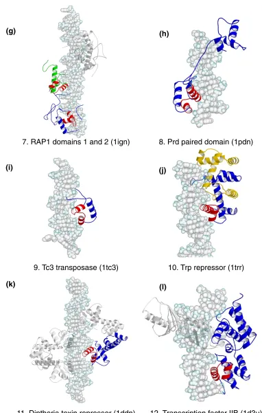
The HTH motif is a common recognition element used by transcription regulators and enzymes of prokaryotes and eukaryotes [1,2,12-15]. Although the motif is traditionally defined as a 20-amino-acid segment of two almost perpen-dicular αhelices connected by a four-residue βturn (Cro and Repressor family, 1lmb; Figure 1a) here we extend the defini-tion to those with longer linkers, such as loops, as long as the relative orientation of the α helices is maintained (for example, the RAP1 protein family, 1ign). Examples from each family within the HTH group [16,17] are shown in Figure 1. In the figure, the HTH is highlighted in red.
The motif invariably binds in the DNA major groove; the second α helix, commonly known as the recognition, or probe, helix, is inserted in the groove. In most complexes, direct contacts are made between amino-acid side chains and nucleotide bases; in a few examples, however, protein backbone atoms or bridging water molecules are used (for example, the Trp repressor family, 1trrA). Supporting con-tacts with the DNA backbone are mainly made by the linker and the first αhelix in the motif, which bridges the major groove at the amino-terminal end of the recognition helix. Further interactions with the nucleic acid can also be made by the rest of the protein and sometimes contribute to further specification of the DNA sequence. For example, the Hin recombinase protein (1hcrA) interacts with bases
in the minor grooves adjacent to the one bound by the recognition helix [18].
The HTH motif is typically found in a bundle of three to six α helices, which provides a stabilizing hydrophobic core. Although motifs from different protein families are struc-turally very similar [19] little homology is observed outside the motif. In structures such as the 434 repressor protein (1lli, Cro and Repressor family), the HTH motif is part of the main body of the protein [20]. In others, such as the purine repressor (1wet, LacI repressor family), it is placed in a small domain extending out of the main structure [21]. There is little sequence similarity between the motifs of different families and this variation allows them to recognize distinct sets of DNA sequences.
The precise positioning of the recognition helix in the DNA major groove also varies, reflecting the structural and functional requirements of each protein. The recogni-tion helices of prokaryotic transcriprecogni-tion factors (for example, those of the Cro and Repressor family, such as 1lli) are generally aligned with their axes parallel to base-pairing edges of the nucleotides, whereas those of eukary-otic proteins (for example, the homeodomain family, see 1oct) are parallel to the sugar-phosphate backbone in order to accommodate the longer α helices [22]. Binding by the Trp repressor (1trrA) is unique, with the amino-ter-minal end of the α helix practically pointing into the groove. Although this last arrangement limits the role of amino acids further down the αhelix, it is needed in order to allow a second repressor subunit into the same major groove when binding in tandem [23]. Helix binding in the major groove, which is also very common in other groups, provides a geometrically favorable framework in which components in both protein and DNA can change to allow multispecific complementarity. The protein sequences and the modes of interaction vary considerably, but the need for the helix to be presented on the surface of the protein, ready for interaction with DNA, is satisfied by the HTH motif.
In general, the prokaryotic transcription factors bind to palindromic DNA sequences as homodimers, whereas eukaryotic proteins, such as members of the home-odomain family, bind both as monomers or heterodimers to non-symmetrical target sites. The latter arrangement potentially allows recognition of a much wider range of DNA sequences. The prokaryotic enzymes in the group (for example, FokI endonuclease, 1fok) which function as monomers possess more than one motif in a single subunit.
There are 16 homologous families in the HTH group. Eight contain only one structure each, and, of the remaining six, only the Cro and Repressor and homeodomain families contain proteins with different amino-acid sequences. The Box 1
Flow diagram showing the selection of the protein-DNA complexes from the PDB (04/01/00). The protein-DNA complexes were groups into structurally related families using the secondary structure alignment program SSAP (see text).
PDB and NDB
314 Protein—DNA complexes
236 Protein—DNA complexes
240 Protein—double helix DNA complexes
54 Protein structure families 17 Single-stranded DNA
3 Quadruple DNA helix 3 Unclassified DNA Query "Protein + DNA, X-ray diffraction"
comment
reviews
reports
deposited research
interactions
information
refereed research
1. Cro and Repressor (1lmb)
2. Homeodomain (1fjl)
3. LacI repressor (1wet)
4. Endonuclease
Fok
I (1fok)
5.
γδ
-resolvase (1gdt)
6. Hin recombinase (1hcr)
(a)
(b)
(c)
(d)
[image:11.609.72.489.79.710.2](e)
(f)
Figure 1 (continues overleaf)
7. RAP1 domains 1 and 2 (1ign)
8. Prd paired domain (1pdn)
9. Tc3 transposase (1tc3)
10. Trp repressor (1trr)
11. Diptheria toxin repressor (1ddn)
12. Transcription factor IIB (1d3u)
(g)
(h)
(i)
(j)
[image:12.609.99.481.91.692.2](k)
(l)
Figure 1 (continued)
pairwise sequence identities between the subunits in the Cro and Repressor family range from 68% (1lmbA and 1perA) to 100% for identical proteins (1lliA and 1lmbA). Pairwise SSAP scores are above 85. In the homeodomain family, although POU domain proteins are often consid-ered separately, they have been included together in this study because of the high SSAP scores that are found between the proteins. For example, the Matα-2 protein (1aplA) and the POU domain protein Pit-1 (1au7A1) have an SSAP score of 88.3 in an alignment of 59 protein residues. As a result, there is greater variation in pairwise sequence identities, which are as low as 42% (1aplA and 1au7A1). The Hin recombinase, γδ-resolvase, FokI restric-tion endonuclease, Tc3 transposase and Cre recombinase families belong to both the HTH and enzyme groups.
‘Winged’ HTH proteins
The winged HTH motif is an extension of the HTH group which is characterized by the presence of a third αhelix and an adjacent βsheet (Figure 1m-p), which are considered to be components of the DNA-binding motif. The recognition helix binds as in the regular HTH motifs, and the extra sec-ondary structural elements provide additional contacts with the DNA backbone.
Group II: zinc-coordinating proteins
Zinc-coordinating proteins make up the largest single group of transcription factors in eukaryotic genomes, and the DNA-binding motif is characterized by the tetrahedral coordination of one or two zinc ions by conserved cysteine and histidine
comment
reviews
reports
deposited research
interactions
information
refereed research
13. Interferon regulatory factor (2irf)
14. Catabolic gene activator (2cgp)
15. Transcription factor (3hts)
16. Ets domain (1bc8)
(m)
(n)
[image:13.609.76.511.79.548.2](o)
(p)
Figure 1 (continued)
residues [1,2,15]. The widespread use of this arrangement is believed to be due to the structural stability the metal ions offer to domains that are not sufficiently large for a stable hydrophobic core [24]. The use of zinc-coordinating motifs is not limited to DNA binding and they are also found in domains that mediate protein-protein interactions [25]. Pro-teins in this group are more structurally diverse than those of the HTH group, and six principal families have been identi-fied so far, of which four are represented in the dataset of complexes. The representative structures are shown in Figure 2, with the zinc-coordinating motif colored red. To avoid con-fusion over the use of the term zinc finger, we reserve its use for proteins that have the Zif-268-style (1aayA1) motif with two β strands and an αhelix (Figure 2a). The name zinc-coordinating will be used as the generic term for all proteins with zinc ions in the DNA-binding motif.
The ββββααzinc-finger family
The ββαzinc-finger proteins constitute the largest individual family in the group and more than a thousand distinct sequence motifs have been identified in transcription factors [26]. The structure of the finger is characterized by a short two-stranded antiparallel β sheet followed by an αhelix (Figure 2a). Two pairs of conserved histidine and cysteine residues in the α helix and second βstrand coordinate a single zinc ion.
Protein subunits often contain multiple fingers that wrap round the DNA in a spiral manner. Fingers bind adjacent 3 bp subsites by inserting the αhelix in the major groove, and the recognition pattern between the helix and DNA is well characterized. Amino acids at positions -1, 2, 3 and 6 relative to the start of the αhelix are used to interact with the bases, -1 being the position that precedes the helix [27,28]. Although there are examples of complexes that do not follow this pattern [29], mutagenesis experiments have shown that by altering the amino acids at the key positions, different subsite sequences are recognized [30]. By adjusting the number of fingers in a protein, binding sites of varying lengths can be bound with different speci-ficities. For example, a protein with five fingers is expected to bind a long target site very selectively, whereas a protein with only a single finger could poten-tially bind a wide range of sites containing the required subsite sequence. However, the structure of the human glioblastoma protein (1gli) suggests that binding is not always straightforward; of the five fingers in the structure, one does not contact the DNA at all, and only two appear to make specific contacts with bases [31].
As described earlier, the protein subunits in this study have been split into distinct domains, each containing a single zinc-finger motif. The pairwise sequence identities of the aligned domains are all high, ranging from 73% (for example, human zinc-finger protein, 1udbA1, and Drosophila tramtrack protein, 2drpA1) to 100% (for example, mouse Zif268 protein, 1aayA1, and artificial
protein, 1mey). All domains are structurally very similar, returning SSAP scores of over 90.
Hormone receptor family
Nuclear receptors for steroid hormones, thyroid hormones and retinoids form the second family in the group (Figure 2b). On binding the appropriate ligand, these receptors translocate from the cytoplasm to the nucleus and regu-late transcription at DNA sequences called hormone response elements [2,32]. Hormone receptors normally function as homo- or hetero-dimers and each monomer typically consists of ligand-binding, DNA-binding, and transcription regulatory domains. The zinc-coordinating motif is found in the DNA-binding domain and is charac-terized by two antiparallel α helices capped by loops at their amino-terminal ends; each helix-loop pair coordi-nates a single zinc ion using four conserved cysteines. The two α helices lie approximately at right angles to each other; the first is inserted in the DNA major groove to provide interactions with bases, while the loops and the second α helix contact the DNA backbone. The DNA-binding domain alone is sufficient for dimerization, and the interface is provided by the loops leading into the second αhelix.
All receptor subunits recognize one of two half-site sequences, 5′-AGAACA-3′ or 5′-AGGTCA-3′. The identity of the full target site is determined by the two half-site sequences that are present, their relative orientation (either symmetric or palindromic) and the spacing between them (between 3 and 6 bp). Thus recognition of the target sequence depends on the read-out of the half-site sequences by each subunit and the manner in which the two subunits dimerize [33]. The sequences of all entries in the current dataset are very similar (sequence identities > 90%) except for the thyroid hormone receptor (for example, 1bsx), which has two extra helices in the carboxy-terminal tail. The structures are all very similar, with pairwise SSAP scores of over 90.
Loop-sheet-helix family
The third family of zinc-coordinating motifs is the loop-sheet-helix zinc-coordinating motif (Figure 2c). This is rep-resented by the DNA-binding region of the protein p53, a transcriptional activator implicated in tumor suppression [2,34]. As the name indicates, the DNA-binding domain consists of a loop leading out of the main body of the protein, followed by a small β sheet, an αhelix and then another loop that leads back into the protein. Three cysteines and a histidine in the two loop regions coordinate the zinc ion.
separate 5 bp recognition sequence positioned one after another. Regions outside the DNA-binding motif make the intersubunit interactions.
Gal4 family
The final zinc-coordinating family contains only the Gal4 protein [35]. It is a transcriptional regulator of galactose-induced genes and its zinc-coordinating motif has so far only been identified in yeast proteins. The motif comprises a pair of α helices that coordinate two zinc ions through six cysteine residues, where two of the cysteines are shared by both metal atoms (Figure 2d). The first αhelix is presented in the DNA major groove for binding with bases, and the second α helix makes the backbone interactions. Gal4
functions as a homodimer, and the dimerization interface is located outside the zinc-coordinating motif.
Group III: zipper-type proteins
The zipper-type group derives its name from the method of dimerization used by its members, which so far only com-prise those from eukaryotic organisms. Two families, the leucine zipper (Figure 3a) and helix-loop-helix proteins (Figure 3b), are defined; the latter must not be confused with the HTH group described earlier. While some members are reported to function as heterodimers (for example the Fos-Jun complex), all the PDB entries in the current dataset are of homodimers.
comment
reviews
reports
deposited research
interactions
information
[image:15.609.120.509.88.531.2]refereed research
Figure 2
Group II, zinc-coordinating proteins. Colors, numbers and names are used as in Figure 1.
17.
ββα
-zinc finger (1aay)
18. Hormone receptor (2nll)
19. Loop-sheet-helix (1tsr)
20. Gal4-type (1d66)
(a)
(b)
Leucine zipper family
In the leucine zipper family, the structure of the protein can be split into two parts: the dimerization region and the DNA-binding region. As shown in Figure 3a, each subunit in the leucine zipper protein consists of a single α helix about 60 amino acids long. Dimerization is mediated through the formation of a coiled coil by a 30-amino-acid section at the carboxy-terminal end of each helix. The segment, known as the zipper region, consists of leucine or a similar hydrophobic amino acid every eight residue posi-tions - roughly every two turns of the αhelix. Correspond-ing side chains from each subunit mediate hydrophobic contacts at the interface through side-by-side packing. The DNA-binding region, also known as the basic region, is found in the amino terminus and for the leucine zipper proteins, the binding segment is a direct extension of the dimerization region. The α helices of the two subunits diverge from the coiled coil and enter the DNA major groove in opposing directions, each binding to half of the target [36]. The leucine zipper family consists entirely of the yeast GCN4 proteins, which have near-identical structures and bind promoter regions of genes that encode enzymes involved in amino-acid biosynthesis.
Helix-loop-helix family
As the name suggests, helix-loop-helix proteins are a modifi-cation of the continuous α helices of the leucine zipper proteins in which the DNA-binding and dimerization regions are separated by a loop, resulting in a four-helix bundle (Figure 3b). Like those of leucine zippers, the dimerization helices interact with each other in a coiled-coil arrangement and the DNA-binding helices are inserted into the DNA major groove. By separating the two segments, more flexibil-ity is allowed in positioning the probe helices on the nucleic acid [37,38]. The helix-loop-helix family is represented by the mouse and human forms of Max, mouse MyoD, and human USF proteins. Sequence identities range from 66% (Max protein, 1an2A, and USF protein, 1an4A) to 97% (mouse Max
protein, 1an2A, and human Max protein, 1hloA) and with the exception of the MyoD (1mdyA) and USF (1an4A) protein pair (pairwise SSAP score 70), SSAP scores are above 80. Structural differences between proteins mainly arise from the variation in lengths and positioning of the loops.
Group IV: other
αα
-helix proteins
There are seven families with very different functions in the other αhelix group (Figure 4). Skn-1 (1skn; Figure 4d) and MADS (see Figure 4g for the MADS box, 1mnm) are transcription regulatory regions in eukaryotic proteins, papillomavirus-1 E2 (2bop; Figure 4a) and EBNA1 (1b3t; Figure 4c) are viral transcription regulators and replication initiators, histones (1aoi; Figure 4b) and high-mobility group (HMG) proteins (1qrv; Figure 4f) are architectural proteins for DNA packaging, and Cre (1crx; Figure 4e) is a site-specific recombinase. Although the protein structures are very different, all use αhelices (colored red in Figure 4) as the main method of DNA binding.
The Skn-1 and MADS proteins bind long probe helices in the DNA major groove in a manner similar to zipper-type pro-teins. Skn-1 (Figure 4d) is monomeric with a compact four-helix unit; the longest αhelix at the carboxy-terminal end binds the major groove, and the rest of the domain contacts the DNA backbone [39]. In MADS (Figure 4g), an anti-parallel β sheet and an adjacent coiled-coil provide the dimerization interface. The αhelices on the opposite face of the sheet diverge from the center of the binding site into adjacent major grooves, contacting base and backbone groups. The DNA is bent towards the protein [40].
[image:16.609.105.483.553.704.2]Papillomavirus-1 E2 and EBNA1 are structurally similar dimeric proteins that can be divided into two regions (Figure 4a,c). In the core region, four βstrands from each subunit combine in an eight-stranded β barrel. The flanking DNA-binding regions project single αhelices into the DNA major
Figure 3
Group III, zipper-type proteins. Colors, numbers and names are used as in Figure 1.
21. Leucine zipper (2dgc)
22. Helix-loop-helix (1am9)
groove symmetrically. As is apparent from their structures (Figure 4a,c), the binding orientations of the helices are very different in the two families [41,42].
Histone and HMG are multimeric proteins that bind DNA independent of base sequence. Histone (Figure 4b) is an octameric protein whose structure can be approximated to a cylinder. Each subunit comprises a bundles of three or four helices that pack against each other; the long DNA segment wraps around the circular edge of the protein. Neighboring α helices make extensive contacts with DNA backbone groups to stabilize the distortion, but none is inserted in the groove and there are few interactions with bases [43]. The HMG subunit comprises three αhelices that are arranged in an L shape (Figure 4f). The first and second helices bind base and backbone groups from the minor groove and cause severe distortion in the DNA structure through intercalation of amino-acid side chains [44].
Finally, Cre (Figure 4e) is a dimeric protein. Each subunit consists of two structural domains that fold into complex helical bundles. Jointly the domains form a clamp around the DNA, inserting αhelices into both the major and minor grooves [45].
Group V: the
ββ
-sheet proteins
In contrast to the proteins described so far, groups V and VI comprise proteins that use β-strand structures for DNA recognition and binding. Group V, which only contains the TATA box-binding protein family, is characterized by the use of a wide βsheet to bind the DNA (Figure 5).
TATA box-binding proteins are an essential component of multiprotein transcription initiator complexes that assem-ble on promoters of genes transcribed by RNA polymerase II. Although they are single-chain molecules, their struc-tures are generally considered to consist of two pseudo-identical domains. A ten-stranded antiparallel β sheet joining the domains covers the DNA minor groove; it creates two substantial kinks away from the main body of the protein by intercalating phenylalanine side chains from either end of the sheet [46,47]. The family is represented by proteins from the bacterium Pyrococcus woesei, yeast and humans. Both sequence and structural alignments of the various subunits yield very high SSAP scores (>90% and >90 respectively).
Group VI: the
ββ
-hairpin/ribbon proteins
The members of this group are different from the TATA box-binding proteins in that they use smaller two- or three-stranded βsheets or hairpin motifs to bind in either the DNA major or minor grooves (Figure 6). Six protein families of very diverse function are represented: the MetJ repressor (1cma; Figure 6a), Arc repressor (1bdt; Figure 6f) and T-domain families (1xbr; Figure 6d) constitute DNA-binding regions of transcriptional regulators; the integration host factor (1ihf; Figure 6c) and the hyperthermophile chromoso-mal proteins (1azp; Figure 6e) act as scaffolds to dictate the DNA structure for formation of high-order protein-DNA complexes; and the Tus protein (1ecr; Figure 6b) terminates DNA replication by helicases. Although the overall struc-tures of the proteins are different, there are common themes in the use of the βstrands.
comment
reviews
reports
deposited research
interactions
information
[image:17.609.96.518.477.707.2]refereed research
Figure 4 (continues overleaf)
Group IV, ‘other αhelix proteins’. Colors, numbers and names are used as in Figure 1.
23. Papillomavirus-1 E2 (2bop)
24. Histone (1aoi)
25. Ebna-1 nuclear protein (1b3t)
26. Skn-1 transcription factor (1skn)
27. Cre recombinase (1crx)
28. High mobility group (1qrv)
29. MADS box (1mnm)
(c)
(d)
(e)
(f)
[image:18.609.96.505.81.712.2](g)
Figure 4 (continued)
The MetJ and Arc repressors are both dimers with very similar modes of binding (Figure 6a,f). Each protein subunit comprises a helical bundle and a single βstrand; the strands from each subunit pack side by side, forming an antiparallel sheet that binds in the DNA major groove. The sheets lie flat in the groove; therefore protein side chains from just one face of the strand interact with base edges [48-50].
The Tus replication terminator and T-domain proteins use β-strand motifs to bind the DNA major groove (Figure 6b,d). In both, the strands are positioned almost perpendicular to the base edges, enabling contacts from amino acids that expose their side chains on either face of the sheet. The Tus replication terminator is a monomeric protein made of amino- and carboxy-terminal α-helical bundles that are con-nected by antiparallel βstrands. The structure forms a large cleft in which the DNA is bound with the major groove facing the strands [50]. In contrast, the T-domain binds as a dimer. Each subunit consists of a β barrel: one end of the barrel points towards the DNA and presents two βstrands, one of which extends into the major groove [51].
Both the integration host factor and chromosomal protein bind in the minor groove and distort the DNA by intercalat-ing side chains from the β sheet motifs (Figure 6c,e). The integration host factor acts as a dimer; a β-hairpin arm from each subunit extends towards the opposing face of the DNA and inserts proline side chains between distinct base-steps [52]. The minor groove is widened in the region of binding and the DNA bends toward the main body of the protein. In contrast, the hyperthermophile chromosomal protein acts as a monomer and uses a three-stranded βsheet to bind against the minor groove. Two hydrophobic side chains from neigh-boring strands intercalate at a single base-step, causing the DNA to bend away from the protein [53].
Only the chromosomal protein and Arc repressor families contain more than one structure. Pairwise sequence identi-ties and SSAP scores between subunits within families are high (>90% and >90 respectively).
Group VII: other
There are two non-enzymatic families in the current dataset that do not use a well-defined secondary structural motif for DNA binding (Figure 7). Both function as dimers and have multidomain subunits that mediate DNA-binding, dimerization and localization to the nucleus. Unlike the dimeric transcription factors encountered so far, these proteins envelop the nucleic acid and the com-plexes are symmetrical when viewed parallel to the DNA long axis. Interstrand and interdomain loops provide most of the base and backbone contacts.
The Rel homology region (Figure 7a) is a conserved amino-terminal domain of transcriptional regulators involved in
cellular defense and differentiation. Each subunit com-prises two β-sandwich domains, which are joined by up to ten interstrand loops that bind in the DNA major groove [54]. The STAT family (Figure 7b) contains transcription factors that mediate responses to cytokines and growth factors. Each protein subunit consists of four structural domains and the functional dimer resembles a pair of pliers with the DNA bound at the hinge. Surrounding loops and an α helix approach the DNA from both the major and minor grooves [55].
Group VIII: the enzymes
The enzyme group completes the classification of the dataset (Figure 8). Rather than having a common struc-tural motif for binding DNA, proteins in the enzyme group are brought together on the basis of their functions; all alter DNA structure through the catalysis of a chemical process.
Unlike the proteins met with so far, the DNA-binding regions used by enzymes are generally hard to describe in terms of simple structural motifs, and these proteins use an extensive combination of αhelices, βstrands and loops to recognize and bind DNA (Figure 8). As described in the Outline of the families section, below, many enzymes comprise three distinct domains: a DNA-recognition domain that reads the DNA sequence; a catalytic domain with the enzymatic active site; and, where applicable, a dimerization domain, although clearly there are excep-tions. The resulting structure often has a U-shaped cavity in which the DNA is bound [56] and often the DNA struc-ture is severely deformed on binding.
comment
reviews
reports
deposited research
interactions
information
[image:19.609.334.511.95.272.2]refereed research
Figure 5
Group V, β-sheet proteins. Colors, numbers and names are are used as in Figure 1.
For sequence-specific enzymes, the target sequences are typically 4-8 bp long, and binding is far more discriminat-ing than that of the transcription regulators. For example,
[image:20.609.87.508.134.713.2]in proteins such as HhaI methyltransferases and endonu-cleases, a single change in the target sequence can lead to over a million-fold reduction in binding affinity. Proteins
Figure 6
Group VI, the β-hairpin/ribbon proteins. Colors, numbers and names are used as in Figure 1.
31. Met repressor (1cma)
32. Tus replication terminator (1ecr)
33. Integration host factor (1ihf)
34. Transcription factor T-domain (1xbr)
35. Hyperthermophile protein (1azp)
36. Arc repressor (1bdt)
(a)
(b)
(c)
(d)
are thought to derive their specificity from both read-out of the base sequence and the catalytic action on the DNA, as in endonucleases BamHI (3bam; Figure 8e) and EcoRI (1qps; Figure 8d), or even primarily from the catalytic process, as in endonuclease EcoRV (1rva; Figure 8c) [57]. Other proteins, such as polymerases, must, however, provide sequence-independent interactions with their DNA substrate yet retain the specificity to distinguish correctly paired bases from mismatches. Seven endo-nucleases and four polymerases (see families 40-46 and 47-50, and Figures 8b-h and 8i-l, respectively), dominate this group of 16 families.
A protein-DNA complex website
A website that summarizes the groups and families of protein-DNA complexes can be found at [http://www.biochem. ucl.ac.uk/bsm/prot_dna/prot_dna.html]. The pages include a brief description of each family, similar to those given in the online information published with this review, as well as infor-mation on the aligned subunits of each protein, structural alignments, tables of pairwise sequence identities and SSAP scores. The proteins are linked to their respective PDB and NDB entries and a PRINTS [58] sequence motif analysis. Also available are links to PDBsum [59], our database of sum-maries and structural analyses of PDB data files. Each struc-ture has information on its CATH [60], PROCHECK [61] and PROMOTIF [62] analyses and links to SCOP [63], WHATIF [64] check and FSSP [65] structural alignments.
The classification process will be automated in the near future so that a newly solved protein structure can submitted to the website and either grouped into an existing family or identified as novel. This would facilitate the possibility of being able to predict a DNA-binding motif and its binding site given a protein sequence, or pave the way to designing proteins to bind a given DNA sequence.
Conclusions
This data collection provides the basis for improving our understanding of protein-DNA complex formation. It high-lights the diversity of protein-DNA complex geometries found in nature, but also underlines the importance of inter-actions between αhelices and the major groove, which is the main method of binding in 28 of the 54 families. In particu-lar, the HTH and zinc-coordinating motifs are used repeat-edly, and provide compact frameworks that present the α helix on the surfaces of structurally diverse proteins, ready for interaction with the DNA. These structures show many variations, both in amino-acid sequence and detailed geome-try, and have clearly evolved independently in accordance with the requirements of the contexts in which they are found. While achieving a close fit between the αhelix and major groove, there is enough flexibility to allow both the protein and DNA to adopt distinct conformations, resulting in multispecific complementarity. Even for this interaction there does not appear to be a simple code relating amino-acid sequence to the DNA sequence it binds. Given the addi-tional complexities of totally different frameworks, it is now clear that detailed rules for DNA base recognition will be family specific, but with underlying trends such as the arginine-guanine interactions.
This survey also highlights the differences between protein domains that just bind DNA and those involved in catalysis. Although there are exceptions, the former typically approach the DNA from a single face and slot into the grooves to interact with the base edges. The latter com-monly envelop the substrate using complex networks of secondary structures and loops, often causing significant distortions in the DNA - normally a requirement for the catalytic process. The ability to bend DNA is not only limited to the enzymes, however; although not as severe, DNA bending is clearly also a common feature of com-plexes formed by transcription factors. This and other
comment
reviews
reports
deposited research
interactions
information
[image:21.609.116.513.87.261.2]refereed research
Figure 7
Group VII, ‘other DNA-binding proteins’. Colors, numbers and names are used as in Figure 1.
37. Rel homology region (1a3q)
38. Stat family (1bf5)
effects such as electrostatic, water- and cation-mediated interactions assist indirect recognition of the DNA sequence, although they are not well understood yet.
Of interest is how the current dataset will aid our interpreta-tion of genome sequences. As summarized in Table 1, there are more structures of eukaryotic proteins than of prokary-otic, and very few are viral. It also demonstrates that, although the dataset is still limited, eukaryotic DNA-binding domains have greater structural diversity than others. This is unsurprising, given that these organisms have developed relatively sophisticated transcription and DNA-repair mecha-nisms, and therefore more eukaryotic proteins are likely to be found and to be structurally characterized. Although prelimi-nary studies of the available genomes show that many pro-teins will probably fall into existing families - notably those with HTH, zipper-type and ββαzinc-finger motifs - there are exciting possibilities of discovering further modes of DNA-binding. Genome analysis will not only facilitate identifica-tion of such proteins, but will allow us to determine functionally important target sites on the DNA and, in combi-nation with structural data, how higher-order oligomers are
formed within the cell. Ultimately, this will expand our understanding of the regulation of protein expression and DNA packaging, rearrangement, repair and replication, which are indispensable to the viability of organisms.
Outline of the families of DNA-binding proteins
A complete outline of the families of DNA-binding proteins and their functional, structural and binding properties follows. Box 1 shows the selection process by which the dataset was compiled. Table 1 provides a summary of the families and Table 2 lists the 240 structures of protein-DNA complexes in the database. Figures 1-8 show ribbon dia-grams of the relevant structures.Group I: helix-turn-helix group
1. Cro and Repressor family [image:22.609.92.550.356.700.2]Function.The Cro and Repressor proteins (Figure 1a) are part of the lysogenic/lytic growth switch mechanism in bacteriophages and function as transcriptional regulators at a set of six related operons.
Figure 8 (continues overleaf)
Group VIII, the enzymes. Colors, numbers and names are used as in Figure 1.
39. Methyltransferase family (6mht)
40. Endonuclease PvuII family (3pvi)
(a)
Structure. Both protein types function as homodimers. Each Repressor subunit has two domains: an amino-terminal five-helix bundle whose second and third α helices comprise a HTH motif; and a carboxy-terminal domain that mediates dimerization (Figure 1a). Cro is a single-domain protein with a structure homologous to the amino-terminal region of Repres-sor. The fourth and fifth αhelices mediate dimerization [66].
Binding. Cro and Repressor bind six related operons with varying affinities. Each operon is 14 bp long and pseudo-symmetrical; four bases at either end are conserved between
sites and the variation in the sequence of the central 6 bp are thought to modulate the binding affinity of the protein. The recognition helix of the HTH motif contacts base edges in the DNA major groove.
2. Homeodomain family
Function. These are transcription regulators for a wide range of genes; in particular many have a vital role in devel-opment and cell differentiation (for example, Mat α-2; 1apl). Some are expressed broadly whereas others are tissue-specific.
comment
reviews
reports
deposited research
interactions
information
refereed research
41. Endonuclease EcoRV family (1rva)
42. Endonuclease EcoRI family (1qps)
43. Endonuclease BamHI family (3bam)
(c)
(d)
[image:23.609.106.504.227.705.2](e)
Figure 8 (continued)
Structure. The proteins are small (just over 100 amino-acid residues in length) and consist of four helices.
Binding.The protein binds DNA either as a monomer or a dimer, depending on the protein and many are capable of both.
Typical HTH binding is displayed in Figure 1b, with the second helix of the motif inserted in the DNA major groove.
3. LacI repressor family
Function. Lac repressor regulates the lac operon, which
44. Endonuclease V family (1vas)
45. Dnase I family (2dnj)
46. DNA mismatch repair endonuclease (1cw0)
47. DNA polymerase
β
(1bpy)
48. DNA polymerase I (2bdp)
49. DNA polymerase T7 (1t7p)
(f)
(g)
(h)
(i)
[image:24.609.99.495.161.724.2](j)
(k)
Figure 8 (continued)