Open Access
Research
Effects of deep sedation or general anesthesia on cardiac function in
mice undergoing cardiovascular magnetic resonance
Christopher J Berry
1, Daniel R Thedens
2, KellyAnn Light-McGroary
1,
Jordan D Miller
1, William Kutschke
1, Kathy A Zimmerman
3and
Robert M Weiss*
1,3Address: 1Division of Cardiovascular Medicine, Roy J. and Lucille A. Carver College of Medicine, University of Iowa, Iowa, USA, 2Department of
Radiology, Roy J. and Lucille A. Carver College of Medicine, University of Iowa, Iowa, USA and 3Iowa City Veterans Affairs Medical Center, Iowa
City, Iowa USA
Email: Christopher J Berry - [email protected]; Daniel R Thedens - [email protected]; KellyAnn Light-McGroary - [email protected]; Jordan D Miller - [email protected]; William Kutschke - [email protected]; Kathy A Zimmerman - [email protected]; Robert M Weiss* - [email protected] * Corresponding author
Abstract
Background: Genetically engineered mouse models of human cardiovascular disease provide an opportunity to understand critical pathophysiological mechanisms. Cardiovascular magnetic resonance (CMR) provides precise reproducible assessment of cardiac structure and function, but, in contrast to echocardiography, requires that the animal be immobilized during image acquisition. General anesthetic regimens yield satisfactory images, but have the potential to significantly perturb cardiac function. The purpose of this study was to assess the effects of general anesthesia and a new deep sedation regimen, respectively, on cardiac function in mice as determined by CMR, and to compare them to results obtained in mildly sedated conscious mice by echocardiography.
Results: In 6 mildly sedated normal conscious mice assessed by echo, heart rate was 615 ± 25 min-1 (mean ± SE) and left ventricular ejection fraction (LVEF) was 0.94 ± 0.01. In the CMR studies of normal mice, heart rate was slightly lower during deep sedation with morphine/midazolam (583 ± 30 min-1), but the difference was not statistically significant. General anesthesia with 1% inhaled isoflurane significantly depressed heart rate (468 ± 7 min-1, p < 0.05 vs. conscious sedation). In 6 additional mice with ischemic LV failure, trends in heart rate were similar, but not statistically significant. In normal mice, deep sedation depressed LVEF (0.79 ± 0.04, p < 0.05 compared to light sedation), but to a significantly lesser extent than general anesthesia (0.60 ± 0.04, p < 0.05 vs. deep sedation).
In mice with ischemic LV failure, ejection fraction measurements were comparable when performed during light sedation, deep sedation, and general anesthesia, respectively. Contrast-to-noise ratios were similar during deep sedation and during general anesthesia, indicating comparable image quality. Left ventricular mass measurements made by CMR during deep sedation were nearly identical to those made during general anesthesia (r2 = 0.99, mean absolute difference < 4%), indicating equivalent quantitative accuracy obtained with the two methods. The imaging procedures were well-tolerated in all mice.
Conclusion: In mice with normal cardiac function, CMR during deep sedation causes significantly less depression of heart rate and ejection fraction than imaging during general anesthesia with isoflurane. In mice with heart failure, the sedation/anesthesia regimen had no clear impact on cardiac function. Deep sedation and general anesthesia produced CMR with comparable image quality and quantitative accuracy.
Published: 19 May 2009
Journal of Cardiovascular Magnetic Resonance 2009, 11:16 doi:10.1186/1532-429X-11-16
Received: 22 December 2008 Accepted: 19 May 2009
This article is available from: http://www.jcmr-online.com/content/11/1/16
© 2009 Berry et al; licensee BioMed Central Ltd.
Background
Genetically engineered mice are useful for mechanistic studies of cardiovascular disease states, which depend, in part, on quantitative phenotypic characterization. Cardio-vascular magnetic resonance (CMR) provides quantita-tively precise characterization of cardiac structure and function noninvasively in vivo, but can be somewhat hin-dered by the need to immobilize the animal during image acquisition. This is often accomplished by administration of general anesthetic agents, all of which are known to perturb cardiac function.[1,2] Echocardiography can be performed in unsedated or minimally sedated conscious mice, avoiding anesthesia-related functional artifacts, but only provides images in a limited number of planes and is quantitatively less accurate than CMR, especially in eccen-trically remodeled ventricles.[3]
Here we introduce a new regimen of deep sedation with morphine and midazolam for mice undergoing CMR, and compare its effects on cardiac function to those observed during general anesthesia with isoflurane, using echocar-diographic methods during mild conscious sedation as a reference standard for a "physiological preparation". We report that deep sedation caused no statistically signifi-cant depression of heart rate in normal mice, compared to conscious sedation, produced significantly less depression of systolic function, and yielded comparable image qual-ity, compared to general anesthesia with isoflurane. In mice with heart failure, there was a non-significant trend toward preservation of physiologic heart rate during deep sedation, but no discernable differential effect on LV systolic function.
Methods
All procedures were approved by the Office of Animal Resources at the University of Iowa.
Mice
A group of 6 mice (3 male, age 6 ± 1 month) without known cardiovascular abnormalities, bred on a C57Bl/6 background and heterozygous for a null allele of inter-leukin-10, were identified from a breeding colony and entered this study as "normal" mice". An additional 6 mice (4 male, age 5 ± 2 months), heterozygous for defi-ciency of copper-zinc superoxide dismutase underwent surgical ligation of the proximal left coronary artery (see below), and entered the study as a group with "ischemic left ventricular failure".
Surgical Preparation
Mice in the ischemic left ventricular failure group were anesthetized with ketamine/xylazine (87.5/12.5 mg/kg, respectively), and the anterior descending branch of the left coronary artery was ligated near the left atrial append-age, using 8-0 Ethilon suture (Ethicon). Successful
liga-tion of the artery was confirmed by blanching of the myocardium. All wounds were closed with 6-0 silk, and animals were allowed to recover for 3 weeks.
Echocardiography
Echocardiography was performed as previously described [4]. Briefly, mice were minimally sedated with mida-zolam, 0.15 mg by subcutaneous injection. This produced a mouse that was conscious, mildly curious, but docile. The anterior chest was shaved and warmed gel was applied, to improve acoustic interface. The mouse was cra-dled in the investigator's left hand and images were obtained in real-time using a 15 MHz linear-array trans-ducer, coupled to a Sonos 5500R ultrasonograph (Philips,
Bothell, WA USA). Two-dimensional images were acquired at ~200 sec-1 in parasternal long- and short-axis
planes, and stored off-line for subsequent analysis. Pulse-wave Doppler interrogation of mitral inflow was used to measure heart rate.
Echocardiography image analysis
Echocardiography image analysis was performed as previ-ously described and validated.[5] Briefly, endo- and epi-cardial borders were traced electronically at end-diastole and end-systole using custom software developed for this purpose (Freeland Medical Systems, Indianapolis, IN). Left ventricular end-diastolic volume, end-systolic vol-ume, stroke volvol-ume, ejection fraction, and mass were cal-culated using the bi-plane area-length method, previously validated in our laboratory.
Preparation for CMR
Deep sedation was achieved by subcutaneous injection of morphine, 4.5 mg/kg, and midazolam, 9 mg/kg. After confirmation of loss of response to noxious stimuli, cuta-neous CMR-compatible ECG leads and a rectal tempera-ture probe were applied, the mouse was wrapped lightly in gauze, and inserted head-first into an imaging cone. Core temperature was monitored with a rectal probe, and supplemented with an external forced-air heat source so as to maintain core temperature ≥ 35°C. In 3 mice with ischemic heart failure, probe insertion was not possible due to perineal edema or other factors. In those cases, forced air settings were selected to coincide with those used in mice with temperature probe in place.
During a separate imaging session, general anesthesia was induced by inhalation of 3% isoflurane for 3 minutes, fol-lowed by steady-state inhalation of 1% isoflurane and room air via nose cone. The remainder of animal prepara-tion was the same as for deep sedaprepara-tion.
CMR
small animal receiver coil (Doty Scientific, Columbia, South Carolina, USA). After acquisition of axial and oblique localizers, image data were acquired using an ECG-gated short-axis multi-slice cine spoiled gradient echo pulse sequence covering the entire LV throughout the cardiac cycle with parameters TR/TE = 6.0/3.4 ms, flip angle = 15°, using 1 mm slices with a 256 × 128 pixel matrix covering a 56 mm × 28 mm field of view in each slice, yielding 15–20 cine frames per cardiac cycle depend-ing on the heart rate. Imagdepend-ing for the purpose of ejection fraction determination commenced within 10 minutes after confirmation of sedation or anesthesia. Heart rates were measured simultaneously with image acquisition. Respiratory monitoring was not performed. Images were batched and archived off-line for later analysis.
CMR image analysis
Images were viewed in cine mode to confirm complete-ness of the data set, spanning at least one slice below the left ventricular apex, and one slice above the aortic valve, and for a complete cardiac cycle. End-diastolic and end-systolic frames were identified, and endocardial and epi-cardial borders were planimetered electronically using standard software (MedisR, Rotterdam). Cardiac volumes
and mass were calculated using modified Simpson's Rule, and tabulated for each mouse under each experimental condition.
In order to objectively evaluate the effect of sedation vs. general anesthesia on image quality, contrast-to-noise ratio (CNR) was measured in two contiguous mid-ven-tricular slices from each mouse under both experimental conditions, using the following formula:
where S.I. = signal intensity, myo = anterior left ventricular myocardium, and S.D. = standard deviation.
Experimental protocol
On Day 1, 6 mice underwent surgical ligation of the left coronary artery; 6 additional normal mice had no surgical procedure. On Day 22 of the study, each mouse under-went echocardiography during mild conscious sedation. On Day 25, each mouse underwent CMR during deep sedation. On Day 40 ± 7, each mouse underwent CMR during general anesthesia. Because some of the mice were participants in long-term molecular and genetic studies, none were euthanized for the purpose of postmortem val-idation of image findings.
Statistics
Data are reported as mean ± SE. Group data obtained dur-ing conscious sedation, deep sedation, and general anesthesia, respectively, were compared using paired
ANOVA. Results were considered statistically significant if p ≤ 0.05.
Results
All 12 mice completed each of 3 imaging procedures, and resumed normal in-cage activity thereafter.
Image Quality
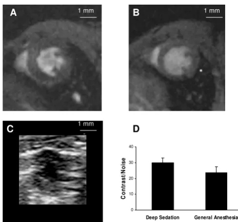
Figure 1 shows representative short-axis end-diastolic images from a single mouse, acquired by each of the 3 imaging methods. Panel D shows group-averaged con-trast-to-noise ratios for CMR images during deep sedation and general anesthesia, respectively. The two CMR tech-niques provided similar levels of image quality.
Effects of sedation or anesthesia on heart rate
During mild conscious sedation, normal mice had a heart rate of 615 ± 25 min-1. The effects of deep sedation and
general anesthesia, respectively, are shown in the Table. Deep sedation caused approximately 5% decrease in heart rate (p = NS). However, general anesthesia caused a highly significant 24% drop in heart rate, compared to mild con-scious sedation. In mice with heart failure, there was a sta-tistically non-significant trend toward lower heart rate during general anesthesia, compared to either light or deep sedation, respectively.
CNR=(Mean S.I.blood pool−Mean S.I.myo) /(S.D. of S.I.backgroound)
[image:3.612.314.546.406.623.2]Image Quality
Figure 1
Image Quality. Mid-ventricular short-axis end-diastolic images acquired (A) with CMR during deep sedation, (B) with CMR during general anesthesia, and (C) with echocardiogra-phy, all from the same mouse. Images are shown to scale, white bar = 1 mm. (D) Comparison of contrast-to-noise ratio for CMR obtained during deep sedation vs. general anesthesia, p = 0.17.
D
A B
C
1 mm 1 mm
1 mm
0 10 20 30 40
Deep Sedation General Anesthesia
C
ont
ra
s
t/
N
oi
s
Effects of sedation or anesthesia on left ventricular ejection fraction (Table 1)
During mild conscious sedation, normal mice had LVEF = 0.94 ± 0.01. Deep sedation caused a statistically signifi-cant decrease in LVEF of 16%, compared to mild con-scious sedation. However, general anesthesia caused a more profound 36% decrease in LVEF (p < 0.05 vs. deep sedation). In mice with ischemic left ventricular dysfunc-tion, ejection fractions were proportionally more variable than in normal mice. When analyzed separately, heart failure mice demonstrated no discernable effect of seda-tion vs. anesthesia upon the measured ejecseda-tion fracseda-tion.
Comparison of ejection fractions obtained during deep sedation to those obtained during general anesthesia is shown graphically in Figure 2. The data indicate a system-atic trend toward lower LVEF during general anesthesia, when all mice are considered.
Effect of deep sedation or anesthesia on core temperature
In normal mice, there was no difference in core body tem-perature during deep sedation vs. general anesthesia (Table). Furthermore, the setting on the forced air heat source required to maintain this temperature was the same for both conditions. In mice with ischemic heart failure, insertion of the temperature probe was impossible or poorly tolerated in 3 mice, due to perineal edema or other factors. For those animals, forced air settings were empirically set to match those for normal mice (data not shown).
Effect of deep sedation or anesthesia on quantitative accuracy
As noted, contrast-to-noise ratio was similar during imag-ing durimag-ing deep sedation vs. imagimag-ing durimag-ing general anesthesia. Whereas the sedation and anesthetic regimens caused disparate effects on heart rate and cardiac function, left ventricular mass should have remained nearly con-stant during the brief interval between studies in mature mice. Figure 3 shows the correlation between mass meas-urements made during each sedation or anesthesia regi-men. Indeed the measurements are nearly identical over a broad range of LV masses.
Discussion
The most important finding of this study is that it intro-duces a new deep sedation regimen for CMR of genetically engineered mice, which causes insignificant reduction in heart rate and only mild reduction in left ventricular systo-lic function in normal mice and in mice with ischemic left ventricular failure.
For these determinations, the reference standard was echocardiography performed during mild conscious seda-tion. The reported heart rate, 615 ± 25 min-1, and left
[image:4.612.53.561.463.711.2]ven-tricular ejection fraction, 0.94 ± 0.01, in normal mice, are nearly identical to previous studies performed on unse-dated mice in our laboratory.[6] Whereas all general anes-thetic preparations are known to depress cardiac function [1,2], deep sedation with morphine and midazolam pro-duces no significant decrement in heart rate, and propro-duces
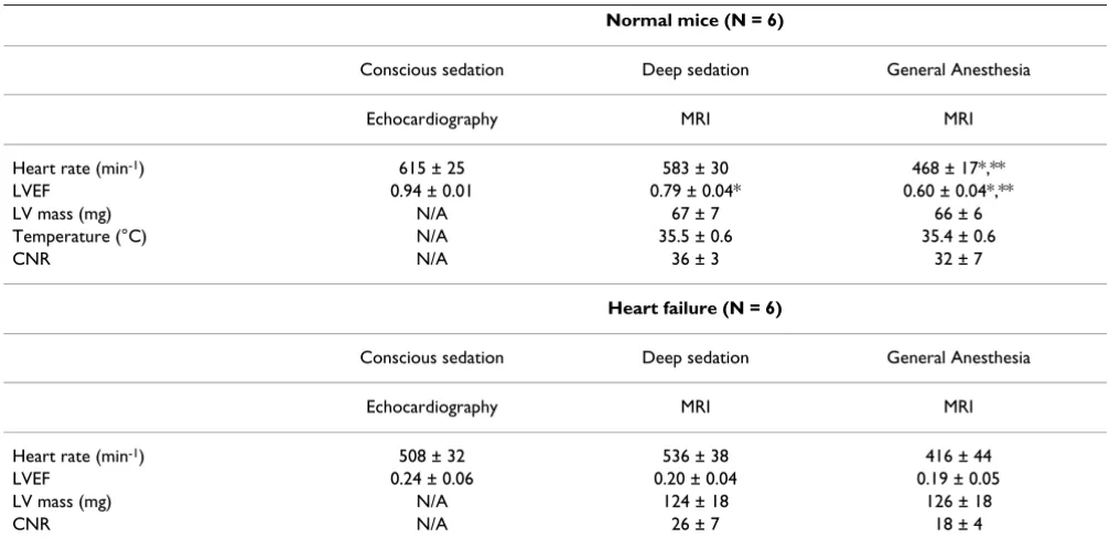
Table 1: Image-derived LV parameters.
Normal mice (N = 6)
Conscious sedation Deep sedation General Anesthesia
Echocardiography MRI MRI
Heart rate (min-1) 615 ± 25 583 ± 30 468 ± 17*,**
LVEF 0.94 ± 0.01 0.79 ± 0.04* 0.60 ± 0.04*,**
LV mass (mg) N/A 67 ± 7 66 ± 6
Temperature (°C) N/A 35.5 ± 0.6 35.4 ± 0.6
CNR N/A 36 ± 3 32 ± 7
Heart failure (N = 6)
Conscious sedation Deep sedation General Anesthesia
Echocardiography MRI MRI
Heart rate (min-1) 508 ± 32 536 ± 38 416 ± 44
LVEF 0.24 ± 0.06 0.20 ± 0.04 0.19 ± 0.05
LV mass (mg) N/A 124 ± 18 126 ± 18
CNR N/A 26 ± 7 18 ± 4
very significant improvement in left ventricular systolic function, when compared to general anesthesia with iso-flurane. These attributes accrue without sacrificing image quality or the quantitative accuracy of CMR studies, despite higher heart rates during deep sedation.
Isoflurane itself has become a preferred general anesthetic agent for cardiovascular phenotyping studies, because it causes less depression of cardiac function than injectable general anesthetics.[2] In the present study, LVEF meas-urements made during general anesthesia, while signifi-cantly depressed, maintained a consistent linear
relationship to measurements made during deep seda-tion. Thus, carefully controlled studies using isoflurane continue to report mechanistically valid results in geneti-cally engineered models of cardiovascular disease in mice [7-14].
Clinically, however, patients with decompensated heart failure or severe valvular heart disease are considered at "high risk" for procedures requiring general anesthesia. Similar concerns can arise in the study of mice with, for example, severe aortic stenosis [4]. Thus, concerns about the risks of general anesthesia in potentially valuable mice may limit the ability to assess cardiovascular function with methods such as CMR. Our finding that deep seda-tion results in less depression of heart rate and systolic function may broaden the applicability of CMR methods in "sick" mice.
In the present study, we report LVEF for normal mice dur-ing isoflurane = 0.60, which is consistent with a number of previous reports [8,14], but significantly lower than others [7]. Yang et al employed high-dose diazepam for sedation during CMR, and reported normal mean heart rate and ejection fraction of 494 and 0.65, respectively [15], which are similar to reported values for isoflurane, but significantly lower than results reported here during deep sedation with morphine/midazolam. It cannot be determined whether these differences related to differ-ences in the respective groups of "normal" mice, as opposed to differential effects of the sedation regime. A feature of the present study is that it compares cardiac function in the same mice, subjected to 3 different seda-tion/anesthesia regimens.
This study has some limitations. It was not possible to measure core temperature during echocardiography in conscious mice. Because the mice remained capable of ambulation and maintained normal heart rates, it could be inferred that core temperature was normal, i.e. higher than during CMR. It is possible that temperature differ-ences accounted for some of the observed effects on car-diac function. However, it is noted that there was no significant difference in heart rates during deep vs. light sedation. Furthermore, temperatures during deep seda-tion were similar to temperatures during general anesthe-sia, whereas cardiac effects were significantly different. Taken together, these findings argue for differential direct effects of the sedation/anesthesia agents, independent of their effects on core temperature.
[image:5.612.58.287.89.249.2]Paired CMR studies were performed 15 days apart, after healed myocardial infarction, during which cardiac func-tion may have changed. The mice strains selected for these studies were from only two genotypes – heterozygous for deletion of IL-10 ("normal mice") or copper-zinc superox-Ejection Fraction
Figure 2
Ejection Fraction. General anesthesia caused a systematic 30% decrease in left ventricular ejection fraction, compared to measurements made during deep sedation. The relation-ship was relatively consistent over the whole range repre-sented by normal (n = 6) and LV failure (n = 6) mice.
0 0.2 0.4 0.6 0.8 1
0 0.2 0.4 0.6 0.8 1
Ejection Fraction - Sedation
E
ject
io
n
F
ract
io
n
A
n
est
h
esi
a
SE = 0.08 R2= 0.91
y = 0.70x + 0.05
Left ventricular mass
Figure 3
Left ventricular mass. Mass measurements made during deep sedation and general anesthesia, respectively, were nearly identical across the entire range.
0 50 100 150 200
0 50 100 150 200
LV Mass - Sedation (mg)
L
V
M
ass
A
n
est
h
esi
a (
m
g
)
R2= 0.99
[image:5.612.57.290.521.666.2]ide dismutase (heart failure mice). While those hetero-zygous mice are not known to exhibit impaired cardiac function at the ages studied, it is possible that there were genotype-specific responses to sedation or anesthesia that would not extrapolate to other strains.
The superior safety profile of the deep sedation regimen was not specifically addressed. Indeed, all mice survived the entire experimental protocol, including imaging dur-ing general anesthesia. However, the perception of increased risk of general anesthesia in sick mice is widely held, and this continues to limit the access of those mice to CMR at our center, and others.
The imaging protocols employed for these studies were relatively simple, requiring less than 20 minutes. More complex protocols might outlast the effects of a single subcutaneous injection of morphine/midazolam, whereas inhalation of isoflurane would be expected to yield a consistent effect over time. This consideration is offset by the simplicity of the deep sedation regimen, which avoids the necessity for ambient air-handling pro-cedures and other safeguards necessary for handling vola-tile agents.
A priori, it is difficult to determine "normal" cardiovascu-lar function in mice. We have previously observed that mild conscious sedation produces no significant perturba-tion of heart rate or systolic funcperturba-tion in mice, compared to the unsedated state, and we report relatively narrow ranges of normal.[4,6] However, confrontation by investi-gators may introduce artifactual cardiostimulatory or depressant influences which are difficult or impossible to account for. It will therefore remain important to include carefully configured "control" groups in all mechanistic studies.
Conclusion
Deep anesthesia with morphine and midazolam causes less cardiac depression during magnetic imaging studies than general anesthesia with isoflurane, without sacrific-ing image quality. The new method of preparsacrific-ing geneti-cally engineered mice for quantitative cardiovascular assessment may broaden applications for CMR in this rap-idly growing and evolving field.
Abbreviations
LV: left ventricle; EF: ejection fraction; CMR: cardiovascu-lar magnetic resonance; CNR: contrast-to-noise ratio
Competing interests
The authors declare that they have no competing interests.
Authors' contributions
CB was involved in experimental design, CMR data acqui-sition, and manuscript writing. DT designed CMR image-acquisition and image-quality assessment methods. KL-M acquired and analyzed some echo and CMR images. JM participated in echocardiographic image acquisition. WK performed animal surgeries and participated in drafting and critically reviewing the manuscript. KZ analyzed and tabulated echocardiographic image data. RW designed the study and analyzed echocardiographic and CMR data, and critically revised the manuscript.
Acknowledgements
This work was supported by grant # RR017369 and # RR017838 from the National Institutes of Health, Bethesda, MD USA.
References
1. Lukasik VM, Gillies RJ: Animal anesthesia for in vivo magnetic resonance. NMR in Biomedicine 2003, 16:459-467.
2. Kober F, Iltis I, Cozzone PJ, Bernard M: Cine-MRI assessment of cardiac function in mice anesthetized with ketamine/xyla-zine and isoflurane. MAGMA 2004, 17:157-161.
3. Siri M, Frigstad S, Sagber E, Larsson H, Skjaerpe T: Accurate and reproducible measurement of left ventricular volume and ejection fraction by contrast echocardiography: A compari-son with magnetic recompari-sonance imaging. J Am Coll Cardiol 2004,
44:1030-1035.
4. Weiss RM, Ohashi M, Miller JD, Young SG, Heistad DD: Calcific aortic valve stenosis in old hypercholesterolemic mice. Circu-lation 2006, 114:2065-2069.
5. Hill JA, Karimi M, Kutschke W, Davisson RL, Zimmerman K, Wang Z, Kerber RE, Weiss RM: Cardiac hypertrophy is not a required compensatory response to short-term pressure overload. Circulation 2000, 101:2854-2862.
6. Han R, Bansal D, Miyake K, Muniz VP, Weiss RM, NcNeil PL, Camp-bell KP: Dysferlin-mediated membrane repair protects the heart from stress-induced left ventricular injury. J Clin Invest
2007, 117:1805-1813.
7. Zhou R, Pickup S, Glickson JD, Scott CH, Ferrari VA: Assessment of global and regional myocardial function in the mouse using cine and tagged MRI. Magn Reson Med 2003, 49:760-764. 8. Hove MT, Lygate CA, Fischer A, Schneider JE, Sang AE, Hulbert K,
Sebag-Montefiore L, Watkins H, Clarke K, Isbrandt D, Wallis J, Neu-bauer S: Reduced inotropic reserve and increased susceptibil-ity to cardiac ischemia/reperfusion injury in phosphocreatine-deficient guanidinoacetate-N-methyl-transferase-knockout mice. Circulation 2005, 111:2477-2485. 9. Petretto E, Sarwar R, Grieve I, Lu H, Kumaran MK, Muckett PJ,
Man-gion J, Schroen B, Benson M, Punjabi PP, Prasad SK, Pennell DJ, Kiesewetter C, Tasheva ES, Corpuz LM, Webb MD, Conrad GW, Kurtz TW, Kren V, Fisher J, Hubner N, Pinto YM, Pravenec M, Aitman TJ, Cook SA: Integrated genomic approaches implicate oste-oglycin (Ogn) in the regulation of left ventricular mass. Nature Genetics 2008, 40:546-52.
10. Jia L-G, Donnett C, Bogaev RC, Blatt RJ, McKinney CE, Day KH, Berr SS, Jones LR, Moorman JR, Sweadner KJ, Tucker AL: Hypertrophy, increased ejection fraction, and reduced Na-K-ATPase activ-ity in phospholemman-deficient mice. Am J Physiol Heart and Circ
2005, 288:H1982-H1988.
11. Voros S, Yang Z, Bove M, Gilson WD, Epstein FH, French BA, Berr SS, Bishop SP, Conaway MR, Matsubara H, Carey RM, Kramer CM:
Interaction between AT1 and AT2 receptors during postinf-arction left ventricular remodeling. Am J Physiol Heart and Circ
2006, 290:H1004-H1010.
12. Trivedi CM, Luo Y, Yin Z, Zhang M, Zhu W, Wang T, Floss T, Goet-tlicher M, Noppinger PR, Wurst W, Ferrari VA, Abrams CS, Gruber PJ, Epstein JA: Hdac2 regulates the cardiac hypertrophic response by modulating Gsk3-β activity. Nature Medicine 2007,
Publish with BioMed Central and every scientist can read your work free of charge "BioMed Central will be the most significant development for disseminating the results of biomedical researc h in our lifetime."
Sir Paul Nurse, Cancer Research UK
Your research papers will be:
available free of charge to the entire biomedical community
peer reviewed and published immediately upon acceptance
cited in PubMed and archived on PubMed Central
yours — you keep the copyright
Submit your manuscript here:
http://www.biomedcentral.com/info/publishing_adv.asp
BioMedcentral 13. Weinreb DB, Aguinaldo JG, Feig JE, Fisher EA, Favad ZA:
Non-inva-sive MRI of mouse models of atherosclerosis. NMR in Biomedi-cine 2007, 20:256-64.
14. Maslov MY, Chacko VP, Stuber M, Moens AL, Kass DA, Champion HC, Weiss RG: Altered high-energy phosphate metabolism predicts contractile dysfunction and subsequent ventricular remodeling in pressure-overload hypertrophy mice. Am J Physiol Heart Circ Physiol. 2007, 292(1):H387-H391.
15. Yang Z, Berr SS, Gilson WD, Toufektsian MC, French BA: Simulta-neous evaluation of infarct size and cardiac function in intact mice by contrast-enhanced Cardiac magnetic resonance imaging reveals contractile dysfunction in noninfarcted regions early after myocardial Infarction. Circulation 2004,