JOURNAL OFCLINICALMICROBIOLOGY, July 2002, p. 2452–2458 Vol. 40, No. 7 0095-1137/02/$04.00⫹0 DOI: 10.1128/JCM.40.7.2452–2458.2002
Copyright © 2002, American Society for Microbiology. All Rights Reserved.
Clostridium difficile
Genotyping Based on
slpA
Variable
Region in S-Layer Gene Sequence:
an Alternative to Serotyping
Tuomo Karjalainen,
1Nicolas Saumier,
1Marie-Claude Barc,
1Michel Delme´e,
2and Anne Collignon
1*
Universite´ de Paris-Sud, Faculte´ de Pharmacie, De´partement de Microbiologie, 92296 Chaˆtenay-Malabry Cedex, France,1and Universite´ Catholique de Louvain, Faculte´ de Me´decine, Unite´ de Microbiologie,
Brussels 1200, Belgium2
Received 10 January 2002/Returned for modification 16 February 2002/Accepted 13 April 2002
Recent investigations ofClostridium difficilecell wall components have revealed the presence of an S-layer
encoded by theslpAgene. The aim of this study was to determine whetherslpAgenotyping can be used as an
alternative to serotyping. The variable regions ofslpAwere amplified by PCR from serogroup reference strains
and various clinical isolates chosen randomly. Amplified products were analyzed after restriction enzyme digestion and DNA sequencing. The sequences of the variable region of the SlpA protein were found to be
strictly identical within a given serogroup but divergent between serogroups.These preliminary results suggest
that PCR-restriction fragment length polymorphism, in conjunction with DNA sequencing of theslpAvariable
region, could constitute an alternative typing method for determiningC. difficileserotypes.
Clostridium difficileis the etiological agent of antibiotic-associ-ated pseudomembranous colitis and is considered the major cause of nosocomial diarrhea. Its pathogenicity is mediated by two toxins, A and B, both of which damage the human colonic mucosa and are potent tissue-damaging enzymes (6). Con-firmed and putative accessory virulence factors that could play a role in intestinal colonization have been identified, including a capsule (11), proteolytic enzymes (22, 24), and adhesins involved in mucus and cell association (15–17, 26–29, 33).
Many bacteria express a surface-exposed proteinaceous layer, termed the S-layer, that forms a regular two-dimensional array visible by electron microscopy. Previous studies have shown that a high degree of variability exists in the molecular
masses of the two proteins composing theC. difficileS-layer (8,
10, 19). Each strain carries a higher-molecular-mass protein of 48 to 56 kDa, encoded by the C-terminal, conserved part of the
slpA gene, and a lower-molecular-mass protein of 36 to 45
kDa, coded for by the variable N-terminal part of the gene (10, 17, 19). The lower-molecular-mass S-layer protein, referred to as the P36 protein, appears to be located on the exterior surface of the bacteria and has adhesive properties (10, 17). Interestingly, the gene encoding the S-layer precursor is present in a genetic cluster locus carrying 17 open reading frames (ORFs), 11 of which carry a similar two-domain archi-tecture, likely to encode surface-anchored proteins (17).
Most epidemiological studies ofC. difficile have been
per-formed by using several typing systems. Serogrouping by slide agglutination or enzyme-linked immunosorbent assay with rab-bit antisera enables the differentiation of 10 major serogroups, which are represented by capital letters (A, B, C, D, F, G, H,
I, K, and X) (14, 31). In serogroup A, another 20 subgroups (subgroups A1 to A20) can be distinguished by polyacrylamide gel electrophoresis (14); these subgroups possess serogroup-specific somatic antigens but have a flagellar antigen in com-mon that is responsible for cross-agglutination on slides (12). Recently, new molecular techniques have been developed for
C. difficiletyping (7, 18).
The aim of this work was to study the genotypic variability of
theslpAgene encoding the outwardly exposed domain of the
majorC. difficilesurface protein.Amplicons obtained by PCR
from serogroup reference strains and various clinical isolates were analyzed by restriction fragment length polymorphism (RFLP) analysis and nucleotide sequencing.
MATERIALS AND METHODS
Bacterial strains, media, and growth conditions.Thirty-twoC. difficileisolates belonging to 10 different serogroups (A, B, C, D, F, G, H, I, K, and X) were examined. The strains included the 10 serogroup reference strains and clinical isolates (Table 1). Serogroups had been previously determined by slide aggluti-nation with rabbit antisera (13) or enzyme-linked immunosorbent assay and confirmed by typing by sodium dodecyl sulfate-polyacrylamide gel electrophore-sis (14). Toxin A production was determined by theC. difficiletoxin A test (Oxoid). In vitro cytotoxin (toxin B) determination was performed by using tissue culture cells as previously described (28).
Bacteria were grown under anaerobic conditions in Tryptone-glucose-yeast infusion broth (Difco Laboratories, Detroit, Mich.) for 48 h.
Genomic DNA isolation.DNA was extracted from 10 ml of an overnight anaerobic culture ofC. difficilein accordance with the protocol provided in the Puregene DNA gram-positive bacterium and yeast DNA extraction kit (Gentra Systems, Minneapolis, Minn.; www.gentra.com).
PCR amplification ofslpA.For amplification of the variable domain of theslpA
gene (Fig. 1) from variousC. difficileisolates, the primers used were slpAV3 (5⬘-ATGAATAAGAAAAAYWTAGCAATRGC-3⬘) and slpAV5 (5⬘-TCTATT CTATCTT CTCCWGCTAC-3⬘), where Y⫽CT, W⫽AT, and R⫽AG. For amplification of the entire gene, primers slpAV3 and slpAC5 (5⬘-AGCKATAC CTTTACCWACTTG-3⬘), where K⫽TG, were used. DNA amplification by PCR was performed in a reaction volume of 50l consisting of 1l of purified genomic DNA (1g/l), 1l each of the 5⬘and 3⬘primers at 20 pmol/l, 25l of water, and 25l of Ready-MixTaqPCR Reaction Mix with MgCl2(Sigma).
* Corresponding author. Mailing address: Universite´ de Paris-Sud, Faculte´ de Pharmacie, De´partement de Microbiologie, 5 rue JB Clem-ent, 92296 Chaˆtenay-Malabry Cedex, France. Phone: (33) 1 46 83 55 49. Fax: (33) 1 46 83 55 37. E-mail: [email protected].
on May 15, 2020 by guest
http://jcm.asm.org/
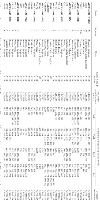
TABLE 1. C. dif ficile isolates studied and their slpA RFLP patterns a Strain Serogroup Origin Toxins A and B in vitro Size of slpA variable domain (nt/a.a.) Size(s) (nt) of fragment(s) obtained with: GenBank accession no. Dra I Hin II Pvu II Rsa I ATCC 43594 (W1194) A1 Brussels, Belgium A ⫹ B ⫹ 1,182/394 282, 366, 456 126, 136, 191, 729 391, 791 1,130 AF458877 Kohn A b Nottingham, United Kingdom A ⫺ B ⫺ 1,065/355 102, 216, 723 248, 346, 471 100, 154, 794 189, 798 AF448119 9354 A b Bondy, France A ⫹ B ⫹ 1,053/351 945 227, 826 274, 779 367, 686 AF448120 TO005 A10 Toronto, Canada A ⫹ B ⫹ 1,185/395 288, 363, 456 111, 126, 923 391, 794 1,133 AF458878 ATCC 43593 (1351) B Brussels, Belgium A ⫺ B ⫺ 1,059/353 189, 216, 327, 327 200, 201, 274, 312 1,059 144, 260, 583 AF458879 EX560 B Mauscron, Belgium A ⫺ B ⫺ 1,059/353 189, 216, 327, 327 200, 201, 274, 312 1,059 144, 260, 583 AF448121 CO109 B Jouy-en-Josas, France A ⫺ B ⫺ 1,059/353 189, 216, 327, 327 200, 201, 274, 312 1,059 144, 260, 583 AF448122 ATCC 43596 (545) C Namur, Belgium A ⫹ B ⫹ 1,077/359 402, 651 126, 175, 776 200, 265, 612 144, 335, 568 AF448123 630 C London, United Kingdom A ⫹ B ⫹ 1,077/359 402, 651 126, 175, 776 200, 265, 612 144, 335, 568 AF448124 C253 C Rome, Italy A ⫹ B ⫹ 1,077/359 402, 651 126, 175, 776 200, 265, 612 144, 335, 568 AJ291709 ATCC 43597 (3232) D Brussels, Belgium A ⫺ B ⫺ 1,167/389 141, 201, 753 171, 230, 681 1,167 155, 370, 642 AF458880 90-111 D Bondy, France A ⫺ B ⫺ 1,167/389 141, 201, 753 171, 230, 681 1,167 155, 370, 642 AF458881 93-136 D Bondy, France A ⫺ B ⫺ 1,167/389 141, 201, 753 171, 230, 681 1,167 155, 370, 642 AF458882 ATCC 43598 (1470) F Brussels, Belgium A ⫺ B ⫹ 1,062/354 1,038 111, 172, 273, 491 200, 862 113, 159, 173, 190, 332 AF448125 R7404 F London, United Kingdom ND 1,062/354 1,038 111, 172, 273, 491 200, 862 113, 159, 173, 190, 332 AJ300677 GAI95600 F Kanazawa, Japan A ⫺ B ⫹ 1,062/354 1,038 111, 172, 273, 491 200, 862 113, 159, 173, 190, 332 AF448126 GAI95601 F Kanazawa, Japan A ⫺ B ⫹ 1,062/354 1,038 111, 172, 273, 491 200, 862 113, 159, 173, 190, 332 AF448127 ATCC 43599 (2022) G Brussels, Belgium A ⫹ B ⫹ 1,065/355 846, 219 485, 580 1,065 1,065 AF448128 R8366 G London, United Kingdom ND 1,065/355 846, 219 485, 580 1,065 1,065 AJ300676 96-392 G Paris, France A ⫹ B ⫹ 1,065/355 846, 219 485, 580 1,065 1,065 AF448129 ATCC 43600 (2149) H Brussels, Belgium A ⫹ B ⫹ 1,089/363 265, 824 410, 679 222, 299, 562 407, 628 AF448365 89-638 H Bondy, France A ⫹ B ⫹ 1,089/363 265, 824 410, 679 222, 299, 562 407, 628 AF448366 90-204 H Bondy, France A ⫹ B ⫹ 1,089/363 265, 824 410, 679 222, 299, 562 407, 628 AF448367 ATCC 43601 (7322) I Brussels, Belgium A ⫺ B ⫺ 1,017/339 993 171, 701 920 136, 222, 538 AF458883 56026 I Brussels, Belgium A ⫺ B ⫺ 1,017/339 993 171, 701 920 136, 222, 538 AF458884 54823 I Brussels, Belgium A ⫺ B ⫺ 1,017/339 993 171, 701 920 136, 222, 538 AF458885 ATCC 43602 (4811) K Brussels, Belgium A ⫺ B ⫺ 1,065/355 102, 159, 233, 568 286, 699 185, 265, 522 196, 265, 409 AF448368 94-416 K Paris, France A ⫹ B ⫹ 1,065/355 102, 159, 233, 568 286, 699 185, 265, 522 196, 265, 409 AF448369 48-515 K Brussels, Belgium A ⫹ B ⫹ 1,065/355 102, 159, 233, 568 286, 699 185, 265, 522 196, 265, 409 AF448370 ATCC 43603 (5036) X Brussels, Belgium A ⫺ B ⫺ 1,038/346 507, 531 162, 268, 525 231, 380, 427 101, 153, 778 AF448371 12-934 X Verviers, Belgium A ⫺ B ⫺ 1,038/346 507, 531 162, 268, 525 231, 380, 427 101, 153, 778 AF448372 36-678 X Brussels, Belgium A ⫺ B ⫹ 1,038/346 507, 531 162, 268, 525 231, 380, 427 101, 153, 778 AF448373 a Serogroup reference strains are in bold. Classi fication is according to Delme ´e et al. (13, 14); Fragment sizes of ⬍ 100 bp are not indicated. nt, nucleotides; a.a., amino acids; ND, not determined. b Serogroup subgroup unknown.
on May 15, 2020 by guest
http://jcm.asm.org/
[image:2.587.126.456.65.724.2]Initial denaturation was carried out at 95°C for 5 min. Thirty-five cycles of amplification were performed in a Perkin-Elmer GeneAmp PCR system 2400 thermocycler. Each cycle consisted of three steps: denaturation at 95°C (30 s), annealing at 45°C (1 min), and extension at 72°C (1 min to amplify the conserved or variable domain and 2 min to amplify the entire gene). An additional step of extension for 10 min at 72°C was performed at the end of the amplification to complete the extension of the primers. Samples (3l) of amplified products were analyzed by electrophoresis in a 1.0% (wt/vol) agarose gel with a 100-bp ladder (Amersham-Pharmacia Biotech) as the molecular size marker.
RFLP analysis.Five microliters of PCR-amplified products, purified with the Wizard PCR Preps DNA purification system (Promega), were digested with the restriction enzymesDraI,HinfI,PvuII, andRsaI in accordance with the vendor’s (Life Technologies) recommendations. The digested amplified products were analyzed by electrophoresis in a 1.0% (wt/vol) agarose gel.
Sequence analysis. PCR products were purified with the QIAquick PCR purification kit (Qiagen, Hilden, Germany). DNA sequencing was carried out with the BigDye terminator DNA sequencing kit (PE Applied Biosystems, War-rington, England). The samples were analyzed with an automated DNA se-quencer, the ABI PRISM 310 genetic analyzer (Perkin-Elmer). Initial sequenc-ing was carried out with the same primers as used for PCR. More sequence was acquired by using primers the sequences of which were derived from the internal sequence data obtained for the variable region.
Computer analyses.Nucleotide and protein sequence alignments were per-formed with the DNA CLUSTAL W program (30).
Nucleotide sequence accession numbers.The GenBank nucleotide sequence accession numbers of theslpAvariable region from theC. difficileisolates studied are given in Table 1.
RESULTS
PCR amplification and sequence analysis of theslpA
vari-able region from serogroup reference strains.It has been
pre-viously demonstrated that theC. difficile slpA gene carries a
conserved 3⬘half and a variable 5⬘half in the strains studied
(Fig. 1) (17). The gene encodes a larger precursor protein that is probably cleaved into distinct S-layer proteins. The protein corresponding to the N-terminal half (referred to as P36) is exposed to the external milieu and thus could play a role in determining serogroup specificity. To investigate this
possibil-ity, the variable region of theslpA gene from 10 serogroup
reference strains was amplified by PCR with the primers indi-cated in Fig. 1 (Table 1). A PCR product of approximately 1 kb was obtained from all of the strains (Table 1).
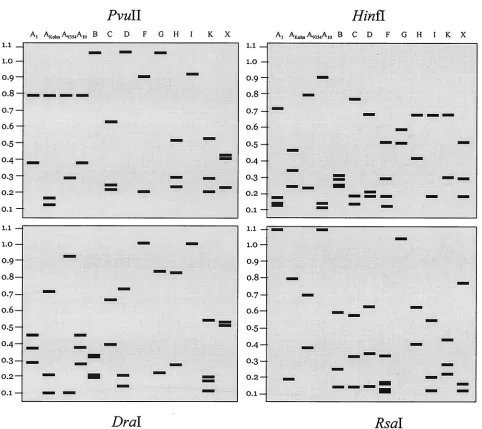
The PCR product was digested with the restriction enzymes
DraI,HinfI,PvuII, andRsaI. Each serogroup revealed a
dif-ferent RFLP profile after electrophoresis in an agarose gel of
the restriction products (not shown), suggesting that theslpA
variable-region sequence is different in each serogroup. For
this reason, theslpAgene variable region from the reference
strains was sequenced. The length of the variable region varied from 1,017 to 1,185 nucleotides (339 to 395 amino acids) (Ta-ble 1). As shown in Fig. 2, alignment of the amino acid se-quences of the variable region revealed that each sequence was unique, with interserogroup sequence homology ranging from 11% (between serogroups A and I) to 64% (between sero-groups C and F) (Table 2).
The presence of nucleotide sequence differences in theslpA
genes ofC. difficilereference strains allowed the establishment
of a precise computer-generated RFLP profile for each sero-group. As shown in Fig. 3 and Table 1, with the exception of serogroup A, unique restriction patterns were produced for
each serogroup by digestion with the restriction enzymesHinfI
andRsaI.PvuII digestion could not distinguish serogroups B,
D, and G, and DraI digestion could not differentiate
sero-groups F and I.
Sequence analysis of theslpAgene variable region fromC.
difficileclinical isolates.The uniqueslpAsequence and RFLP
patterns of each serogroup suggested that nucleotide sequence analysis and/or PCR-RFLP of the variable region of this gene could be employed to differentiate between serogroups. To test
this hypothesis, we sequenced the variable region of theslpA
genes from 22 clinical isolates whose serogroups were known (at least two sequences per serogroup) and which were chosen fortuitously and represented diverse geographic locations (Ta-ble 1). Analysis of the sequences obtained revealed that, with the exception of serogroup A, the nucleotide and deduced protein sequences were 100% identical within a given group (Fig. 2). In contrast, the four strains belonging to
sero-group A each had a uniqueslpAvariable region, although the
SlpA N-terminal regions in A1 and A10 were 93% identical.
DISCUSSION
Different methods have been developed to typeC. difficile
[image:3.587.49.281.74.150.2]strains. A correlation has been observed between the electro-phoretic patterns of proteins extracted from clinical isolates
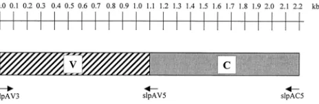
FIG. 1. Structural organization of theslpAgene ofC. difficile. The conserved (C) domain is in gray, and the variable (V) domain is represented by the hatched bar. Positions of the primers used for amplification of the domains are indicated below the gene structures. Gene length is indicated in kilobases above the gene structures.
FIG. 2. Multiple alignments of the predicted amino acid sequences encoded by variants of the variable region of theslpAgene from Delme´e’s serogroup reference strains A1, B, C, D, F, G, H, I, K, and X. Furthermore, three additional SlpA sequences from serogroup A (Kohn, 9354, and TO005) are included in the alignment. Additional sequence data were obtained for at least two isolates in each serogroup (Table 1). Analysis of the data showed that theslpAgene sequence from strains EX560 and CO109 was 100% identical to that of the serogroup B reference strain; that of strains 630 and C253 was 100% identical to that of the serogroup C reference strain; that of strains 90-111 and 93-136 was 100% identical to that of the serogroup D reference strain; that of strains R7404, GAI95600, and GAI95601 was 100% identical to that of the serogroup F reference strain; that of strains R8366 and 93-392 was 100% identical to that of the serogroup G reference strain; that of strains 89-638 and 90-204 was 100% identical to that of the serogroup H reference strain; that of strains 56026 and 54823 was 100% identical to that of the serogroup I reference strain; that of strains 94-416 and 48-515 was 100% identical to that of the serogroup K reference strain; and that of strains 12-934 and 36-678 was 100% identical to that of the serogroup X reference strain. Identical residues are indicated by asterisks below the alignment; functionally identical
2454 KARJALAINEN ET AL. J. CLIN. MICROBIOL.
on May 15, 2020 by guest
http://jcm.asm.org/
2455
on May 15, 2020 by guest
and their virulence, strains isolated during hospital outbreaks and symptomatic disease having a different profile from that observed in isolates from healthy carriers or infants (21).
Delme´e et al. (14) compared serogrouping of C. difficile by
slide agglutination with rabbit antisera and polyacrylamide gel electrophoresis of whole-cell proteins, permitting correlation between the two typing systems and establishment of a single classification. This serogrouping is also carried out by the en-zyme-linked immunosorbent assay technique.
Recently, new molecular techniques have been developed to typeC. difficilestrains based on DNA polymorphism, such as arbitrary primed PCR, a genotypic method permitting detec-tion of polymorphisms within the target genome without prior knowledge of the target nucleotide sequence. This method has been used as an efficient discriminative method for
investiga-tion of nosocomial outbreaks ofC. difficile-associated diarrhea
[image:5.587.43.286.100.228.2]but has certain drawbacks, e.g., lack of reproducibility (1, 2, 5,
[image:5.587.51.533.277.706.2]FIG. 3. Computer-generated restriction enzyme digestion profiles of the 1-kbslpAvariable region from the serogrouped strains indicated. A TABLE 2. Pairwise comparisons of amino acid sequence homology
of the variable region of the SlpA protein between
C. difficileserogroups
Serogroup % Identity
A B C D F G H I K X
A 100 30 28 14 28 35 25 11 29 31
B 30 100 32 20 33 32 28 36 26 47
C 28 32 100 23 64 36 33 38 37 32
D 14 20 23 100 21 22 23 25 20 14
F 28 33 64 21 100 35 34 40 37 32
G 35 32 36 22 35 100 28 38 31 34
H 25 28 33 23 34 28 100 32 30 26
I 11 36 38 25 40 38 32 100 33 34
K 29 26 37 20 37 31 30 33 100 28
X 31 47 32 14 32 34 26 34 28 100
2456 KARJALAINEN ET AL. J. CLIN. MICROBIOL.
on May 15, 2020 by guest
http://jcm.asm.org/
7). Pulsed-field gel electrophoresis has been applied to C. difficile, but this method is costly and technically complex and some strains are untypeable (7). PCR ribotyping is being used routinely by the United Kingdom Anaerobe Reference Unit in
Cardiff, Wales, to typeC. difficilestrains (7). This method relies
on DNA pattern profiles, obtained by PCR amplification of a specific chromosomal region such as the rRNA gene (4) or the 16S-23S rRNA gene intergenic spacer region (25). Each of the strains belonging to one of Delme´e’s serogroups gives a dif-ferent banding pattern. PCR ribotyping appears to be more discriminatory than the arbitrary primed PCR or pulsed-field
gel electrophoresis method in epidemiological studies of C.
difficileoutbreaks (7). However, most of the aforementioned typing techniques are discriminating but labor intensive. Sero-typing with specific antisera gives adequate levels of discrimi-nation for epidemiological purposes and is not technically dif-ficult. However, the reagents required for serotyping are not readily available to most diagnostic laboratories and some strains are untypeable or coagglutinable with this technique.
The epitopes for the serotypes are probably parts of
bacte-rial surface proteins. In group A streptococci,sof(serum
opac-ity factor gene) or emm (M protein gene) sequence-based
analysis has been used more successfully than serological anal-ysis for strain subtyping (3). These proteins are surface asso-ciated. Until now, no investigations have explored the useful-ness of S-layer gene sequencing for subtyping of strains. A
study ofCampylobacter fetus (9) and Lactobacillus helveticus
(32) demonstrated that amplification of the S-layer gene by PCR can be used for identification of strains. Furthermore, for
C. fetus, each strain exhibited a different Southern blotting pattern when hybridized with the PCR product. This suggested
that genotyping based onslpAgene structure could be useful
for typing of strains. Since the P36 S-layer protein ofC. difficile
is located on the surface of the bacteria and because S-layer proteins are the most abundant bacterial proteins (23), it would be logical to conclude that they play a major role in determining serogroup specificity. This was confirmed by PCR-RFLP analysis and nucleotide sequencing: DNA and deduced
amino acid sequences of theslpAvariable region were 100%
identical within a given serogroup, whereas interserogroup identity was, in general, fairly low. However, further
sequenc-ing of more strains is necessary to confirm these data. The
exception is serogroup A, which is known to carry 20 sub-groups. It is evident that these subserogroups may not be
completely specified by the S-layer, since theslpAsequences
were quite similar in serogroups A1 and A10. The subsero-group specificity of highly flagellated serosubsero-group A could be attributable to other surface proteins, most likely the flagella.
These data indicate that theslpAgene constitutes a reliable
target for differentiation of C. difficileisolates and could be
used as an alternative method to serotyping, at least in an
outbreak setting until more diagnostic data become available.
The gene could be easily amplified from various strains by PCR by using primers described here and then sequenced. Alterna-tively, the amplified DNA could be digested with restriction enzymes and profiles could be compared. However, sequence-based methods have advantages over the more commonly used RFLP methods, which are difficult to standardize. Further-more, the relatively small size of certain restriction fragments obtained after digestion may render interpretation of the
pro-files difficult in clinical laboratories. Sequence data give more reliable results, thus eliminating interlaboratory variation due to, for example, gel mobility differences (20). The technology for generating DNA sequence data has become readily avail-able for on-site or commercial service companies. Finally, this methodology should have a relatively low cost; the overall cost for generating one sequence can be estimated to be less than $10 U.S. However, because of country-to-country variations in labor and reagent costs, this estimate may not be valid for some countries. Sequencing can be expected to produce the same results in different laboratories, even with the use of different methods. This, combined with the ease of generation of tem-plate DNA for sequencing by PCR, makes sequence-based molecular typing a promising alternative to the more tradi-tional methods.
ACKNOWLEDGMENT
We thank H. Kato, Kanazawa University, Kanazawa City, Japan, for the gift ofC. difficilestrains GAI95600 and GAI95601.
REFERENCES
1.Barbut, F., N. Mario, M. Delmee, J. Gozian, and J. C. Petit.1993. Genomic fingerprinting ofClostridium difficileisolates by using a random amplified polymorphic DNA (RAPD) assay. FEMS Microbiol. Lett.114:161–166. 2.Barbut, F., N. Mario, M. C. Meyohas, D. Binet, J. Frottier, and J. C. Petit.
1994. Investigation of a nosocomial outbreak ofClostridium difficile -associ-ated diarrhoea among AIDS patients by random amplified polymorphic DNA (RAPD) assay. J. Hosp. Infect.26:181–189.
3.Beall, B., G. Gherardi, M. Lovgren, R. R. Facklam, B. A. Forwick, and G. J. Tyrrell.2000.emmandsofgene sequence variation in relation to serological typing of opacity-factor-positive group A streptococci. Microbiology146:
1195–1209.
4.Bidet, P., F. Barbut, V. Lalande, B. Burghoffer, and J. C. Petit.1999. De-velopment of a new PCR-ribotyping method forClostridium difficilebased on ribosomal RNA gene sequencing. FEMS Microbiol. Lett.175:261–266. 5.Blair, D. F., and S. K. Dutcher.1992. Flagella in prokaryotes and lower
eukaryotes. Curr. Opin. Genet. Dev.2:756–767.
6.Borriello, S. P., H. A. Davies, S. Kamiya, P. J. Reed, and S. Seddon.1990. Virulence factors ofClostridium difficile. Rev. Infect. Dis.12(Suppl. 2):S185– S191.
7.Brazier, J. S.2001. Typing ofC. difficile. Clin. Microbiol. Infect.7:428–431. 8.Calabi, E., S. Ward, B. Wren, T. Paxton, M. Panico, H. Morris, A. Dell, G. Dougan, and N. Fairweather.2001. Molecular characterization of the surface layer proteins fromClostridium difficile. Mol. Microbiol.40:1187–1199. 9.Casademont, I., D. Chevrier, and J. L. Guesdon.1998. Cloning of asapB
homologue (sapB2) encoding a putative 112-kDaCampylobacter fetusS-layer protein and its use for identification and molecular genotyping. FEMS Im-munol. Med. Microbiol.21:269–281.
10.Cerquetti, M., A. Molinari, A. Sebastianelli, M. Diociaiuti, R. Petruzzelli, C. Capo, and P. Mastrantonio.2000. Characterization of surface layer proteins from differentClostridium difficileclinical isolates. Microb. Pathog.28:363– 372.
11.Davies, H. A., and S. P. Borriello.1990. Detection of capsule in strains of
Clostridium difficileof varying virulence and toxigenicity. Microb. Pathog.
9:141–146.
12.Delme´e, M., V. Avesani, N. Delferrie`re, and G. Burtonboy.1990. Character-ization of flagella ofClostridium difficileand their role in serogrouping reactions J. Clin. Microbiol.28:2210–2214.
13.Delme´e, M., M. Homel, and G. Wauters.1985. Serogrouping ofClostridium difficilestrains by slide agglutination. J. Clin. Microbiol.21:323–327. 14.Delme´e, M., Y. Laroche, V. Avesani, and G. Cornelis.1986. Comparison of
serogrouping and polyacrylamide gel electrophoresis for typing of Clostrid-ium difficile. J. Clin. Microbiol.24:991–994.
15.Hennequin, C., A. Collignon, and T. Karjalainen.2001. Analysis of expres-sion of GroEL (Hsp60) ofClostridium difficilein response to stress. Microb. Pathog.31:255–260.
16.Hennequin, C., F. Porcheray, A.-J. Waligora, A. Collignon, P. Bourlioux, and T. Karjalainen.2001. GroEL (Hsp60) ofClostridium difficileis involved in cell adherence. Microbiology147:87–96.
17.Karjalainen, T., A.-J. Waligora-Dupriet, M. Cerquetti, P. Spigagli, P. Mauri, and P. Mastrantonio.2001. Molecular and genomic analysis of two genes encoding surface-anchored proteins fromClostridium difficile.Infect. Im-mun.69:3442–3446.
18.Kuijper, E. J., J. H. Oudbier, W. N. Stuifbergen, A. Jansz, and H. C. Zanen.
on May 15, 2020 by guest
http://jcm.asm.org/
1987. Application of whole-cell DNA restriction endonuclease profiles to the epidemiology of Clostridium difficile-induced diarrhea. J. Clin. Microbiol.
25:751–753.
19.McCoubrey, J., and I. R. Poxton.2001. Variation in the surface layer proteins ofClostridium difficile. FEMS Immunol. Med. Microbiol.31:131–135. 20.Meinersmann, R. J., L. O. Helsel, P. I. Fields, and K. L. Hiett.1997.
Discrimination of Campylobacter jejuni isolates by fla gene sequencing. J. Clin. Microbiol.35:2810–2814.
21.Pantosti, A., M. Cerquetti, and P. M. Gianfrilli.1988. Electrophoretic char-acterization ofClostridium difficilestrains isolated from antibiotic-associated colitis and other conditions. J. Clin. Microbiol.26:540–543.
22.Poilane, I., T. Karjalainen, M. C. Barc, P. Bourlioux, and A. Collignon.1998. Protease activity ofClostridium difficilestrains. Can. J. Microbiol.44:157– 161.
23.Sa`ra, M., and U. B. Sleytr.2000. S-layer proteins. J. Bacteriol.182:859–868. 24.Seddon, S. V., and S. P. Borriello.1992. Proteolytic activity ofClostridium
difficile. J. Med. Microbiol.36:307–311.
25.Stubbs, S. L., J. S. Brazier, G. L. O’Neill, and B. I. Duerden.1999. PCR targeted to the 16S-23S rRNA gene intergenic spacer region ofClostridium difficileand construction of a library consisting of 116 different PCR ri-botypes. J. Clin. Microbiol.37:461–463.
26.Tasteyre, A., M.-C. Barc, A. Collignon, and T. Karjalainen.2001. Role of flagellar proteins ofClostridium difficilein adherence and gut colonization. Infect. Immun.69:7937–7940.
27.Tasteyre, A., M.-C. Barc, T. Karjalainen, P. Dodson, S. Hyde, P. Bourlioux, and P. Borriello.2000. AClostridium difficilegene encoding flagellin. Mi-crobiology146:957–966.
28.Tasteyre, A., T. Karjalainen, V. Avesani, M. Delme´e, A. Collignon, P. Bour-lioux, and M.-C. Barc.2000. Phenotypic and genotypic diversity of the flagellin gene (fliC) amongClostridium difficileisolates from different sero-groups. J. Clin. Microbiol.38:3179–3186.
29.Tasteyre, A., T. Karjalainen, V. Avesani, M. Delme´e, A. Collignon, P. Bour-lioux, and M.-C. Barc.2001. Molecular characterization offliDgene encod-ing flagellar cap and its expression amongClostridium difficileisolates from different serogroups. J. Clin. Microbiol.39:1178–1183.
30.Thompson, J. D., D. G. Higgins, and T. J. Gibson.1994. CLUSTAL W: improving the sensitivity of progressive multiple sequence alignment through sequence weighting, position-specific gap penalties and weight matrix choice. Nucleic Acids Res.22:4673–4680.
31.Toma, S., G. Lesiak, M. Magus, H.-L. Lo, and M. Delme´e.1988. Serotyping ofClostridium difficile. J. Clin. Microbiol.26:426–428.
32.Ventura, M., M. L. Callegari, and L. Morelli, L.2000. S-layer gene as a molecular marker for identification ofLactobacillus helveticus. FEMS Mi-crobiol. Lett.189:275–279.
33.Waligora, A.-J., C. Hennequin, P. Mullany, A. Collignon, P. Bourlioux, and T. Karjalainen.2001. Characterization of a cell surface protein ofC. difficile
with adhesive properties. Infect. Immun.69:2144–2153.
2458 KARJALAINEN ET AL. J. CLIN. MICROBIOL.