0095-1137/00/$04.00⫹0
Copyright © 2000, American Society for Microbiology. All Rights Reserved.
Development of a TT Virus DNA Quantification System Using
Real-Time Detection PCR
TAKANOBU KATO,1MASASHI MIZOKAMI,1* MOTOKAZU MUKAIDE,2ETSURO ORITO,1 TOMOYOSHI OHNO,1TATSUNORI NAKANO,1YASUHITO TANAKA,1HIDEAKI KATO,1 FUMINAKA SUGAUCHI,1RYUZO UEDA,1NOBORU HIRASHIMA,3KAZUHIDE SHIMAMATSU,4
MASAYOSHI KAGE,4ANDMASAMICHI KOJIRO4
Second Department of Medicine, Nagoya City University Medical School,1and Department of Gastroenterology,
Chyukyo Hospital,3Nagoya, Center for Molecular Biology and Cytogenetics, SRL, Inc., Tokyo,2
and Department of Pathology, Kurume University School of Medicine, Kurume,4Japan
Received 13 July 1999/Returned for modification 31 August 1999/Accepted 8 October 1999
Although TT virus (TTV) was isolated from a cryptogenic posttransfusion hepatitis patient, its pathogenic role remains unclear. It has been reported that the majority of the healthy population is infected with TTV. To elucidate the differences between TTV infection in patients with liver diseases and TTV infection in the healthy population, a quantification system was developed. TTV DNA was quantified by a real-time detection PCR (RTD-PCR) assay on an ABI Prism 7700 sequence detector. With this system, TTV DNA was quantified in 78 hepatitis C virus (HCV)-infected patients (63 with elevated serum alanine aminotransferase [ALT] levels and 15 with normal ALT levels) and in 70 voluntary blood donors (BDs). The quantification range was 2.08 to 7.35 log copies/ml. The intra-assay and interassay coefficients of variation were 0.37 to 6.33% and 0.60 to 7.07%, respectively. The mean serum TTV DNA levels in the HCV-infected patients with both elevated and normal
ALT levels and BDs were 3.69ⴞ0.89, 3.45ⴞ0.76, and 3.45ⴞ0.67 log copies/ml, respectively. Comparison of
the serum TTV DNA levels among the HCV-infected patients revealed that they were not related to the serum ALT and HCV core protein levels or to the histopathological score on liver biopsy. This study showed that (i) the RTD-PCR assay for the detection of TTV was accurate and had a high degree of sensitivity, (ii) the mean serum TTV DNA level was similar among HCV-infected patients, irrespective of their ALT level, and also among BDs, and (iii) a high serum TTV DNA level does not affect the serum ALT and HCV levels or liver damage in HCV-infected patients.
A novel DNA virus, the TT virus (TTV), named after the initials of the patient from whom it was first isolated, was cloned from the acute-phase serum of a patient with crypto-genic posttransfusion hepatitis (10). Although this virus had been believed to have a linear single-stranded DNA genome similar to that of members of the familyParvoviridae, the latest report on the full genomic structure of this virus has indicated that it has a circular genome similar to that of members of the familyCircoviridae(8, 9, 13). Since the virus was found more often in patients with various liver diseases than in voluntary blood donors, some pathogenic role is conceivable (1, 15). Recently, tests with a new highly sensitive primer set with about 10- to 100-fold greater sensitivities than the conventional primer sets used for TTV detection has revealed that the majority of the general population in Japan is infected with TTV (16). In our previous study with this primer set, we had determined that the prevalence was not significantly different between hepatitis C virus (HCV)-infected patients with chronic hepatitis and hepatocellular carcinoma or between pa-tients with hepatocellular carcinoma associated with HCV in-fection and those with neither HCV nor hepatitis B virus (HBV) in the serum. Moreover, comparison of patients with and without TTV infection revealed no significant differences in their backgrounds, biochemical findings, or histopathologi-cal scores (4). Therefore, to evaluate the differences in serum TTV DNA levels between healthy individuals and patients with
liver diseases, an accurate and highly sensitive system for the quantification of TTV has been developed.
Recently, the real-time detection PCR (RTD-PCR) assay has been described as a new system for the quantification and detection of PCR product accumulation. This system exploits the 5⬘-3⬘ exonuclease activity ofTaqDNA polymerase which hydrolyzes an internal dually labeled (a fluorescent reporter dye and quencher dye) probe (6). In the intact probe, the quencher dye suppresses the fluorescence emission of the re-porter dye. During the annealing and extension phase of PCR, the probe anneals to the target sequence and is subsequently hydrolyzed and cleaved byTaqDNA polymerase. The cleavage reduces the quenching effect and allows an increase in the emission of the reporter dye fluorescence, and continuous measurement of the increments in the fluorescence released during PCR provides an accurate estimate of the initial copy number in the sample. This report describes the use of this system for the quantification of the serum TTV DNA levels in patients with HCV infection and voluntary blood donors in-fected with TTV and determination of the correlation between the amount of TTV and the severity of hepatic damage.
MATERIALS AND METHODS
Materials.Serum samples from 78 patients infected with HCV but without hepatitis B surface antigen (HBsAg) (63 had elevated serum alanine aminotrans-ferase [ALT] levels and 15 had normal ALT levels) were analyzed. The subjects included previously reported patients who were known to be infected with TTV (4). As controls, 70 blood samples from voluntary blood donors supplied by the Japan Red Cross Blood Center, which were positive for TTV but negative for human immunodeficiency virus, HBV, and HCV and which had been confirmed to have normal ALT levels, were also obtained from the Department of Blood Transfusion, Nagoya City University, as the attached tube for a cross-matching test. All samples were stored at⫺80°C until use.
* Corresponding author. Mailing address: Second Department of Medicine, Nagoya City University Medical School, Kawasumi, Mizuho, Nagoya 467-8601, Japan. Phone: 853-8216. Fax: 81-52-852-0849. E-mail: [email protected].
94
on May 15, 2020 by guest
http://jcm.asm.org/
Anti-HCV was detected with a second-generation enzyme immunoassay kit (Ortho Diagnostics, Tokyo, Japan). The samples were tested for HBsAg and anti-HBsAg by the particle agglutination method (Fujirebio, Tokyo, Japan), and the samples were tested for the anti-hepatitis B core antigen by the passive hemagglutination method (International Regent Co., Kobe, Japan). Serum HCV core protein levels were measured by fluorescent enzyme immunoassay (14, 18). To determine the reproducibility of the tests with the two sets of primers and the probe and the correlation between the results of tests with the two sets of primers and the probe, serum samples from selected HCV patients with elevated ALT levels were used.
Detection of TTV.A total of 20l of DNA was extracted from 100-l serum samples by using the SMITEST EX-R&D (Sumitomo Metal Industries, Tokyo, Japan). TTV DNA was detected by PCR with the primers and under the con-ditions described by Takahashi et al. (16). The expected sizes of the products were confirmed by electrophoresis on 3% agarose gels with ethidium bromide staining.
Construction of primers and probe.For the RTD-PCR assay, two sets of primers and probe located in 5⬘untranslated open reading frame 2 were pre-pared. Set A primers were those reported by Takahashi et al. (16), with slight modification, and consisted of forward primer M801 (5⬘-CTACGTCACTAAC CACGTG-3⬘), reverse primer M935 (5⬘-TTCGGTGTGTAAACTCACC-3⬘), and probe TP117 (5⬘-AAACTCAGCCATTCGGAAGT-3⬘). The sequences of set B primers were designed so that they were near the sequences of set A primers with reference to the alignment of the TTV sequence on the basis of data from our studies and from the database of the DDBJ/EMBL/GenBank (data not shown) and consisted of forward primer K117 (5⬘-CACTTCCGAATGGCTGA GTT-3⬘), reverse primer K233 (5⬘-GCCTTGCCCATAGCCCGG-3⬘), and probe TP208 (5⬘-TCCCGAGCCCGAATTGCCCCT-3⬘). The probe contains a fluores-cent reporter dye (6-carboxyfluorescein) at the 5⬘end and a fluorescent quencher dye (6-carboxy-tetramethyl-rhodamine) at the 3⬘end.
RTD-PCR.Ten microliters of the extracted DNA was detected with the se-quence detector system (ABI Prism 7700; Applied Biosystems, Foster City, Calif.) in 50l of a PCR mixture containing TaqMan PCR Core Reagents with AmpliTaq Gold and AmpErase uracil-N-glycosylase (Applied Biosystems), 20 pmol of each primer, and 15 pmol of the probe. After initial activation of uracil-N-glycosylase at 50°C for 2 min, AmpliTaq Gold was activated at 95°C for 9 min. The subsequent PCR conditions consisted of 50 cycles of denaturation at 95°C for 20 s and annealing and extension at 60°C for 1 min per cycle. During the PCR amplification, the amplified products were measured continuously by de-termination of the fluorescence emission. After real-time data acquisition, the cycle threshold value was calculated by determining the point at which the fluorescence exceeds an arbitrary threshold limit. The threshold limit was man-ually set to cross the fluorescent signal of all standards in the exponential phase. The cycle threshold value is predictive of the quantity of target copies in the samples.
For the standard curve, a standard DNA was constructed. The PCR products obtained with primers M801 and K233 were cloned into the TA cloning vector (Invitrogen, San Diego, Calif.) and were sequenced with M13 primers to confirm the sequences of the primers and the probe region. This construct was used as the primary standard after quantification by optical density determination and was prepared by use of a series of 10-fold dilutions. For dilution of the standards, TE (Tris-EDTA) buffer was used, and the standards were stored at⫺20°C.
Histopathological analysis.Histopathological examinations were performed for the HCV-infected patients with elevated ALT levels. All liver tissue speci-mens were obtained by needle biopsy. Specispeci-mens were fixed in 10% neutral formalin, embedded in paraffin, cut into 4-m-thick sections, and stained with hematoxylin-eosin. Each specimen was assessed independently by three liver pathologists (M. Kage, M. Kojiro, and K. Shimamatsu) by use of a scoring system which included a grading of necroinflammation and staging of fibrosis on the basis of the recommendations of Ichida et al. (3). In the grading, inflammation in both the portal area and the lobular area was divided into four levels on the
basis of the degrees of infiltration of lymphocytes and necrosis of the hepato-cytes. If the interpretations of the pathologists differed, the score was determined to be that which two pathologists agreed on.
Statistical analysis.For statistical analysis of categorical data, Mann-Whit-ney’s U test, the Kruskal-Wallis test, and analysis of variance (ANOVA) were used. For the liner regression, Spearman’s correlation test was used. Data anal-ysis was carried out with the StatView J, version 4.5, software package (Abacus Concepts Inc., Berkeley, Calif.).
RESULTS
Sensitivity and reproducibility of RTD-PCR assay.By using
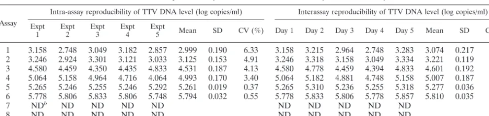
standard DNA at a series of 10-fold dilutions, the detection range was determined to be from 2.08 to 7.35 log copies/ml. To evaluate the reproducibility of the RTD-PCR assay, a total of eight samples, including six TTV-positive samples and two TTV-negative samples, were tested five times on the same day or for 5 days. The intra-assay and interassay coefficients of variation (CVs) were calculated to be within the ranges of 0.37 to 6.33% and 0.60 to 7.07%, respectively (Table 1). Identical sensitivities and reproducibilities were obtained with each of the primer and probe sets.
Correlation between two sets of primers and probes. The
[image:2.612.57.553.83.201.2]serum TTV DNA levels of 50 HCV-infected patients were measured by using primer and probe sets A and B, and these levels were almost identical and showed a strong correlation (r⫽0.871;P⬍0.0001) (Fig. 1).
TABLE 1. Reproducibility of serum TTV DNA levels by RTD-PCR assaya
Assay
Intra-assay reproducibility of TTV DNA level (log copies/ml) Interassay reproducibility of TTV DNA level (log copies/ml) Expt
1 Expt2 Expt3 Expt4 Expt5 Mean SD CV (%) Day 1 Day 2 Day 3 Day 4 Day 5 Mean SD CV (%)
1 3.158 2.748 3.049 3.182 2.857 2.999 0.190 6.33 3.158 3.215 2.964 2.748 3.283 3.074 0.217 7.07 2 3.246 2.924 3.301 3.121 3.033 3.125 0.153 4.91 3.246 3.318 3.158 3.049 3.334 3.221 0.119 3.68 3 4.580 4.459 4.350 4.435 4.833 4.531 0.187 4.13 4.580 4.778 4.459 4.394 4.833 4.601 0.192 4.17 4 5.064 5.158 4.964 4.716 4.064 4.993 0.170 3.40 5.064 5.182 4.881 4.748 5.158 5.007 0.187 3.73 5 5.265 5.246 5.255 5.246 5.292 5.261 0.019 0.37 5.265 5.310 5.236 5.255 5.318 5.277 0.036 0.68 6 5.778 5.806 5.833 5.806 5.748 5.794 0.032 0.55 5.778 5.833 5.806 5.778 5.857 5.810 0.035 0.60
7 NDb ND ND ND ND ND ND ND ND ND
8 ND ND ND ND ND ND ND ND ND ND
aPrimers and probe set B were used. bND, not detected.
FIG. 1. Correlation between serum TTV DNA levels determined with primer sets A and B.
on May 15, 2020 by guest
http://jcm.asm.org/
[image:2.612.314.548.522.707.2]Serum TTV DNA levels among HCV-infected patients and
blood donors.Among the samples from HCV-infected patients
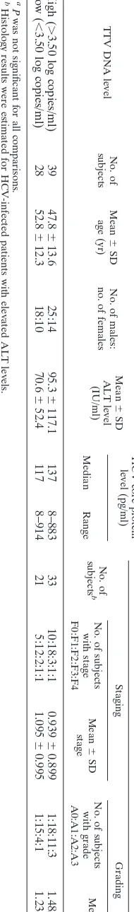
with elevated ALT levels and those with normal ALT levels and voluntary blood donors, the serum TTV DNA levels could be quantified for 54 of 63 (85.7%), 13 of 15 (86.7%), and 62 of 70 (88.6%) samples, respectively. For the remaining samples for which virus was not quantified, the serum TTV DNA level was considered to be below the detection range of this assay. The mean serum TTV DNA levels in the HCV-infected patients with elevated ALT levels and normal ALT levels and blood donors were 3.69⫾0.89, 3.45⫾0.76, and 3.45⫾0.67 log copies/ml, respectively, and these data were similar and were not significantly different (Table 2). Comparison of HCV-infected patients with high (⬎3.50 log copies/ml) and low (⬍3.50 log copies/ml) serum TTV DNA levels indicated no significant differences in terms of mean age, sex, serum ALT level, and serum HCV core protein level (Table 3). His-topathological examination was performed for HCV-infected patients with elevated ALT levels, and the results were ex-pressed as the fibrosis score, classified into five stages (stages F0 to F4), and the activity score, classified into four grades (grades A0 to A3), according to the New Inuyama Classifica-tion. The mean score for each parameter was also represented as an index. Among patients with high serum TTV DNA levels, 10 were classified into stage F0, 18 were classified into stage F1, 3 were classified into stage F2, 1 was classified into stage F3, and 1 was classified into stage F4; the mean score was 0.939 ⫾ 0.899. One was classified into grade A0, 18 were classified into grade A1, 11 were classified into grade A2, and 3 were classified into grade A3; the mean score was 1.485⫾
0.712. Among the patients with low serum TTV DNA levels, 5 were classified into stage F0, 12 were classified into stage F1, 2 were classified into stage F2, 1 was classified into stage F3, and 1 was classified into stage F4; the mean score was 1.095⫾
0.995. One was classified into grade A0, 15 were classified into grade A1, 4 were classified into grade A2, and 1 was classified into grade A3; mean score was 1.238⫾0.625. No significant difference in the ratios or the mean scores were observed (Table 3).
DISCUSSION
At first, TTV was believed to be a novel hepatitis virus because it could be isolated from three of five patients with cryptogenic posttransfusion hepatitis. However, our previous study revealed that TTV is prevalent among both patients with liver diseases and voluntary blood donors (4). Thus, to clarify the correlation between serum TTV DNA levels and liver diseases, we developed a quantification assay with high degrees of sensitivity, accuracy, specificity, and reproducibility. Some of the findings from this quantification assay are that (i) the mean serum TTV DNA level was similar among HCV-infected
pa-tients irrespective of the serum ALT level and also among voluntary blood donors, and (ii) the serum TTV DNA level is not related to the serum ALT and HCV core protein levels or the histopathological score of fibrosis and inflammation among HCV patients.
The RTD-PCR system enables the accurate and highly sen-sitive estimation of the level of TTV infection. This system is now available for quantification of HCV andEscherichia coli and even for the detection of cancer cells (2, 5, 7, 12, 17). It has a wide dynamic range (2.08 to 7.35 log copies/ml), and the intra-assay and interassay CVs ranged from 0.37 to 6.33% and 0.60 to 7.07%, respectively. In regard to specificity, the fluo-rescence signal due to cleavage of the probe is generated only if the target sequence for the probe is amplified by PCR, and no signal is observed under the condition of nonspecific am-plification. To confirm the accuracy and to negate the influence of primers and probe affinity, two sets of primers and probe were used to obtain estimates of TTV DNA levels in our study, and the quantification data obtained with these two sets were identical and showed a strong correlation. Besides these ad-vantages, it must also be mentioned that other useful systems for the quantification of TTV are not available. Therefore, this quantification system will be useful for estimation of the se-verity of TTV infection.
The results of recent studies raise a question regarding the association between TTV infection and hepatic disorders. Mushahwar et al. (9) reported that inoculation of TTV into chimpanzees could not induce hepatitis. In regard to this point, our investigation of the quantification of TTV among HCV-infected patients might be relevant. Serum TTV DNA levels were similar, irrespective of the severity of liver injury, and were even similar in HCV-infected patients and blood donors. Comparison of HCV-infected patients with both high and low serum TTV DNA levels revealed no significant differences in serum ALT and HCV core protein levels or the histopatho-logical score. These data imply that TTV infection and repli-cation occur in many healthy people and do not affect the HCV infection or the liver damage due to HCV infection. This observation does not totally negate the association between TTV infection and hepatitis. In the case of the other hepatitis viruses, such as HCV, a dissociation between the serum viral titer and the severity of hepatitis is observed. To estimate the pathogenicity of TTV, the serial monitoring of serum TTV DNA levels in patients with hepatitis of unknown etiology will be needed.
Another important use of this system is to identify the or-gans for which TTV has an affinity. The findings of the simi-larity of TTV to members of the familyCircoviridaerather than to members of the familyParvoviridaecould suggest the pos-sibility of an association of TTV with diseases other than liver diseases. For example, the chicken anemia virus, a represen-TABLE 2. Serum TTV DNA levels in HCV-infected patients and voluntary blood donorsa
Group subjectsNo. of Meanage (yr)⫾SD no. of femalesNo. of males: MeanALT level⫾SD (IU/liter)
HCV core protein
level (pg/ml) TTV DNA level (log copies/ml) Median Range
HCV-infected patients
Elevated ALT levels 54 48.6⫾12.1 35:19 100.4⫾100.8 117 8–914 3.69⫾0.89b
Normal ALT levels 13 54.7⫾15.3 8:5 20.9⫾7.6 32 8–883 3.45⫾0.76b
Blood donors 62 NA NA NA NT NT 3.45⫾0.67
aAbbreviations: NA, not available; NT, not tested. bPwas not significant.
on May 15, 2020 by guest
http://jcm.asm.org/
[image:3.612.52.551.85.172.2]tative of the familyCircoviridae, induces severe anemia, sub-cutaneous and intramuscular hemorrhage, destruction of erythroblastoid cells in the bone marrow, and depletion of certain lymphoid organs in young chickens (11). By using the quantification system described here, further studies on the quantification of TTV in various organs such as the bone marrow and estimation of the pathogenicity of this virus will be needed.
In summary, an accurate and highly sensitive system for the quantification of TTV has been developed. By using this sys-tem, it was revealed that the titer of TTV DNA in serum does not affect HCV infection or the liver damage due to HCV.
ACKNOWLEDGMENTS
We express our gratitude to Osamu Yanagihara for quantification of the HCV core protein level.
This work was supported in part by a grant from the Japanese Ministry of Health and Welfare, Health Science Research Grants (Non-A, Non-B Hepatitis Research Grants), and by a Grant-in Aid for Scientific Research (B) from the Ministry of Education, Science, Sports and Culture (grant 11691222).
REFERENCES
1.Charlton, M., P. Adjei, J. Poterucha, N. Zein, B. Moore, T. Therneau, R. Krom, and R. Wiesner.1998. TT-virus infection in North American blood donors, patients with fulminant hepatic failure, and cryptogenic cirrhosis. Hepatology28:839–842.
2.Gerard, C. J., K. Olsson, R. Ramanathan, C. Reading, and E. G. Hanania.
1998. Improved quantitation of minimal residual disease in multiple my-eloma using real-time polymerase chain reaction and plasmid-DNA comple-mentarity determining region III strand. Cancer Res.58:3957–3964. 3.Ichida, F., T. Tsuji, M. Omata, T. Ichida, K. Inoue, T. Kamimura, G.
Yamada, K. Hino, O. Yokosuka, and H. Suzuki.1996. New Inuyama classi-fication; new criteria for histological assessment of chronic hepatitis. Int. Hepatol. Commun.6:112–119.
4.Kato, T., M. Mizokami, E. Orito, T. Nakano, Y. Tanaka, R. Ueda, N. Hirashima, Y. Iijima, T. Kato, F. Sugauchi, M. Mukaide, K. Shimamatsu, M. Kage, and M. Kojiro.1999. High prevalence of TT virus infection in Japanese patients with liver diseases and blood donors. J. Hepatol.31:221– 227.
5.Kawai, S., O. Yokosuka, T. Kanda, F. Imazeki, Y. Maru, and H. Saisho.
1999. Quantification of hepatitis C virus by TaqMan PCR: comparison with HCV amplicor monitor assay. J. Med. Virol.58:121–126.
6.Livak, K., S. Flood, J. Marmoro, W. Giusti, and K. Deetz.1995. Oligonu-cleotides with fluorescent dyes at opposite ends provide a quenched probe system useful for detecting PCR product and nucleic and hybridization. PCR Methods Applications4:357–362.
7.Martell, M., J. Gomez, J. I. Esteban, S. Sauleda, J. Quer, B. Cabot, R. Esteban, and J. Guardia.1999. High-throughput real-time reverse transcrip-tion-PCR quantitation of hepatitis C virus RNA. J. Clin. Microbiol.37:327– 332.
8.Miyata, H., H. Tsunoda, A. Kazi, A. Yamada, M. A. Kahn, J. Murakami, T. Kamahora, K. Shiraki, and S. Hino.1999. Identification of an novel GC-rich 113-nucleotide region to complete the circular, single-stranded DNA ge-nome of TT virus, the first human circovirus. J. Virol73:3582–3586. 9.Mushahwar, I. K., J. C. Erker, A. S. Muerhoff, P. T. Leary, N. Simons, L. G.
Birkenmeyer, M. L. Chalmers, T. J. Pilot-Matias, and S. M. Dexai.1999. Molecular and biophysical characterization of TT virus: evidence for a new virus family infecting humans. Proc. Natl. Acad. Sci. USA96:3177–3182. 10. Nishizawa, T., H. Okamoto, K. Konishi, H. Yoshizawa, Y. Miyakawa, and M.
Mayumi.1997. A novel DNA virus (TTV) associated with elevated transam-inase levels in posttransfusion hepatitis of unknown etiology. Biochem. Bio-phys. Res. Commun.241:92–97.
11. Noteborn, M. H. M., G. F. de Boer, D. J. van Roozelaar, C. Karreman, O. Kranenburg, J. Vos, S. H. M. Jeurissen, R. C. Hoeben, A. Zantema, G. Koch, H. van Ormondt, and A. J. van der Eb.1991. Characterization of cloned chicken anemia virus DNA that contains all elements for the infectious replication cycle. J. Virol.65:3131–3139.
12. Oberst, R., D. M. P. Hays, L. K. Bohra, R. K. Phebus, C. T. Yamashiro, C. Paszko-Kolva, S. J. A. Flood, J. M. Sargeant, and J. R. Gillespie.1998. PCR-based DNA amplification and presumptive detection ofEscherichia coliO157:H7 with an internal fluorogenic probe and the 5⬘nuclease (Taq-Man) assay. Appl. Environ. Microbiol.64:3389–3396.
13. Okamoto, H., T. Nishizawa, N. Kato, M. Ukita, H. Ikeda, H. Iizuka, Y. Miyakawa, and M. Mayumi.1998. Molecular cloning and characterization of a novel DNA virus (TTV) associated with posttransfusion hepatitis of un-known etiology. Hepatol. Res.10:1–16.
TABLE 3. Comparison of HCV-infected patients with high and low serum TTV DNA levels a TTV DNA level No. of subjects Mean ⫾ SD age (yr) No. of males: no. of females Mean ⫾ SD ALT level (IU/ml) HCV core protein level (pg/ml) Histology results No. of subjects b Staging Grading Median Range No. of subjects with stage F0:F1:F2:F3:F4 Mean ⫾ SD stage No. of subjects with grade A0:A1:A2:A3 Mean ⫾ SD grade High ( ⬎ 3.50 log copies/ml) 39 47.8 ⫾ 13.6 25:14 95.3 ⫾ 117.1 137 8–883 33 10:18:3:1:1 0.939 ⫾ 0.899 1:18:11:3 1.485 ⫾ 0.712 Low ( ⬍ 3.50 log copies/ml) 28 52.8 ⫾ 12.3 18:10 70.6 ⫾ 52.4 117 8–914 21 5:12:2:1:1 1.095 ⫾ 0.995 1:15:4:1 1.238 ⫾ 0.625 a P was not significant for all comparisons. b Histology results were estimated for HCV-infected patients with elevated ALT levels.
on May 15, 2020 by guest
http://jcm.asm.org/
[image:4.612.121.216.81.726.2]14.Orito, E., M. Mizokami, T. Tanaka, J. Y. N. Lau, K. Suzuki, M. Yamauchi, Y. Ohta, A. Hasegawa, S. Tanaka, and M. Kohara.1996. Quantification of serum hepatitis C virus core protein level in patients chronically infected with different hepatitis C virus genotypes. Gut39:876–880.
15. Simmonds, P., F. Davidson, C. Lysett, L. E. Prescott, D. M. MacDonald, J. Ellender, P. L. Yap, C. A. Ludlam, G. H. Haydon, J. Gillon, and L. M. Jarvis.
1998. Detection of novel DNA virus (TTV) in blood donors and blood products. Lancet352:191–195.
16. Takahashi, K., H. Hoshino, Y. Ohta, N. Yoshida, and S. Mishiro.1998. Very
high prevalence of TT virus (TTV) infection in general population of Japan revealed by a new set of PCR primers. Hepatol. Res.12:233–239. 17. Takeuchi, T., A. Katsume, T. Tanaka, A. Abe, K. Inoue, K.
Tsukiyama-Kohara, R. Kawaguchi, S. Tanaka, and M. Kohara.1999. Highly sensitive and accurate system for quantification of hepatitis C virus genomic RNA using real-time detection PCR. Gastroenterology116:636–642.
18. Tanaka, T., J. Y. N. Lau, M. Mizokami, E. Orito, E. Tanaka, and K. Kiyo-sawa.1995. Simple fluorescent enzyme immunoassay for detection and quantification of hepatitis C viremia. J. Hepatol.23:742–745.