secreting Tregs by 1
aa
,25-dihydroxyvitamin D3
abrogates regulatory function
Zoë Urry, … , Zarin Brown, Catherine M. Hawrylowicz
J Clin Invest.
2009;
119(2)
:387-398.
https://doi.org/10.1172/JCI32354
.
Signaling through the TLR family of molecular pattern recognition receptors has been
implicated in the induction of innate and adaptive immune responses. A role for TLR
signaling in the maintenance and/or regulation of Treg function has been proposed,
however its functional relevance remains unclear. Here we have shown that TLR9 is highly
expressed by human Treg secreting the antiinflammatory cytokine IL-10 induced following
stimulation of blood and tissue CD3
+T cells in the presence of 1
a
,25-dihydroxyvitamin D3
(1
a
25VitD3), the active form of Vitamin D, with or without the glucocorticoid
dexamethasone. By contrast, TLR9 was not highly expressed by naturally occurring
CD4
+CD25
+Treg or by Th1 and Th2 effector cells. Induction of TLR9, but not other TLRs,
was IL-10 dependent and primarily regulated by 1
a
25VitD3 in vitro. Furthermore, ingestion
of calcitriol (1
a
25VitD3) by human volunteers led to an increase of both IL-10 and TLR9
expression by CD3
+CD4
+T cells analyzed directly ex vivo. Stimulation of 1
a
25VitD3-induced IL-10–secreting Treg with TLR9 agonists, CpG oligonucleotides, resulted in
decreased IL-10 and IFN-
g
synthesis and a concurrent loss of regulatory function, but,
unexpectedly, increased IL-4 synthesis. We therefore suggest that TLR9 could be used to
monitor and potentially modulate the function of 1
a
25VitD3-induced IL-10–secreting Treg in
vivo, and that this has implications in cancer therapy and vaccine design.
Research Article
Immunology
Find the latest version:
Ligation of TLR9 induced on human IL-10–
secreting Tregs by 1
α
,25-dihydroxyvitamin D3
abrogates regulatory function
Zoë Urry,1 Emmanuel Xystrakis,1 David F. Richards,1 Joanne McDonald,1 Zahid Sattar,1
David J. Cousins,1 Christopher J. Corrigan,1 Emma Hickman,2
Zarin Brown,2 and Catherine M. Hawrylowicz1,3
1MRC and Asthma UK Centre in Allergic Mechanisms of Asthma, King’s College London, London, United Kingdom.
2Novartis Institute for Biomedical Research, Horsham, West Sussex, United Kingdom. 3National Institute for Health Research Biomedical Research Centre,
Guy’s and St. Thomas’ NHS Foundation Trust/King’s College London, London, United Kingdom.
Signaling through the TLR family of molecular pattern recognition receptors has been implicated in the
induc-tion of innate and adaptive immune responses. A role for TLR signaling in the maintenance and/or regulainduc-tion
of Treg function has been proposed, however its functional relevance remains unclear. Here we have shown
that TLR9 is highly expressed by human Treg secreting the antiinflammatory cytokine IL-10 induced
follow-ing stimulation of blood and tissue CD3
+T cells in the presence of 1
α
,25-dihydroxyvitamin D3 (1
α
25VitD3),
the active form of Vitamin D, with or without the glucocorticoid dexamethasone. By contrast, TLR9 was not
highly expressed by naturally occurring CD4
+CD25
+Treg or by Th1 and Th2 effector cells. Induction of TLR9,
but not other TLRs, was IL-10 dependent and primarily regulated by 1
α
25VitD3 in vitro. Furthermore,
inges-tion of calcitriol (1
α
25VitD3) by human volunteers led to an increase of both IL-10 and TLR9 expression by
CD3
+CD4
+T cells analyzed directly ex vivo. Stimulation of 1
α
25VitD3-induced IL-10–secreting Treg with TLR9
agonists, CpG oligonucleotides, resulted in decreased IL-10 and IFN-
γ
synthesis and a concurrent loss of
regu-latory function, but, unexpectedly, increased IL-4 synthesis. We therefore suggest that TLR9 could be used to
monitor and potentially modulate the function of 1
α
25VitD3-induced IL-10–secreting Treg in vivo, and that
this has implications in cancer therapy and vaccine design.
Introduction
The TLRs represent a family of evolutionarily conserved receptors, which recognize pathogen-associated molecular patterns (PAMPs) and certain host molecules. Ten TLRs (TLR1–10) have been identi-fied in humans to date. These are proposed to play central roles in the induction of innate immune responses and in triggering host immunity to infection (1–3). The capacity of TLRs to control adaptive immunity was thought to critically involve APCs, which activate naive T cells or modulate effector T cells following ligation of one or more TLRs (4).
Recent evidence has highlighted a role for direct stimulation of T cells by PAMPs. mRNA specific for TLRs in human T cell popu-lations has been reported (5, 6). Signaling through TLR2, TLR5, and TLR7/8 has been shown to be a costimulator of highly puri-fied human T cells, enhancing cytokine production, survival, and proliferation in the absence of APCs (7–10).
A significant indication that TLR signaling may play a role in the maintenance and/or function of Tregs was an observed reduc-tion in the frequency of natural CD4+CD25+Tregs, but not CD25–
T cells, in mice lacking MyD88, a key adaptor molecule involved in signaling through the majority of TLRs (11). mRNA for a range of TLRs has now been detected in rat, mouse, and human CD4+CD25+ Tregs (9, 11–15). An early study reported that the TLR
agonist LPS potently enhanced proliferation and regulatory activ-ity of murine CD4+CD25+
Tregs (12). However, more recent stud-ies have indicated that ligation of other TLRs, for example TLR2, on effector T cells or CD4+CD25+
Tregs may actually alleviate sup-pression by enhancing effector T cell proliferation and, in some cases, diminishing FoxP3 expression in Tregs (9, 11, 13–15).
The binding of PAMP to TLRs on APCs results in the production of an array of proinflammatory cytokines, including IL-12, IFN-α, IL-6, IFN-γ, and IL-8, in order to mount an effective innate response. TLR signal transduction can, under certain circumstances, also elicit a counterregulatory response in the form of IL-10 secre-tion, as demonstrated in Tlr2–/– mice, which have an impaired
capacity to synthesize IL-10 (16). Human natural CD4+CD25+
Tregs have also been shown to secrete IL-10 in response to TLR2 stimulation and subsequently to induce IL-10 synthesis in cocul-tured CD25– T cells (17). However, the presence of TLRs on IL-10–
secreting T cells themselves and the functional consequences of TLR signaling in these Tregs have not been reported.
IL-10 is a potent antiinflammatory cytokine and inhibits Th1 and Th2 immune responses, which has led to considerable inter- est in its therapeutic potential to treat a wide range of immune-mediated pathologies, including allergy, transplantation, and autoimmune disease (18, 19). We have shown that human IL-10– secreting Tregs (IL-10–Tregs), which express low levels of the CD4+CD25+ Treg-associated transcription factor FoxP3, can be
induced following activation, through either polyclonal stimuli or antigen presented by APCs in the presence of the glucocor-ticoid dexamethasone (Dex) and the active form of vitamin D, 1α,25-dihydroxyvitamin D3 (1α25VitD3) (20, 21). In our search
Conflict of interest: The authors have declared that no conflict of interest exists.
Nonstandard abbreviations used: Dex, dexamethasone; IL-10–Treg, IL-10–secreting Treg; CpG-ODN, CpG oligonucleotide; PAMP, pathogen-associated molecular pattern; VDR, vitamin D receptor; 1α25VitD3, 1α,25-dihydroxyvitamin D3.
to identify molecules uniquely expressed by this population, the profile of TLR expression on 1α25VitD3 and Dex-induced IL-10– Tregs was examined and compared with other relevant peripheral cell populations. High TLR9 transcript abundance was detected in human drug-induced IL-10–Tregs but not in other human effector cell or Treg populations. The functional consequences of signaling via TLR9 on IL-10–Tregs were therefore examined and shown to impair regulatory function. These findings have implications for the use of TLR9 ligands in cancer therapy and as adjuvants in vaccine design.
Results
TLR9 expression is increased in human drug-induced IL-10–Tregs. Human peripheral blood derived CD3+CD4+
T cells were stimu-lated with CD3-specific antibody, IL-2, and IL-4 in the absence or presence of 10–7 M 1α25VitD3 and 10–7 M Dex for 7 days and then
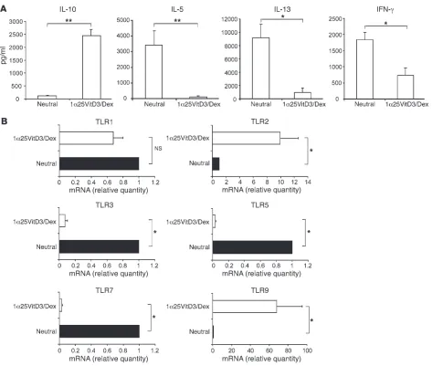
restimulated for a further 7 days under identical conditions. This protocol induces T cells producing high levels of IL-10 but low levels of Th1- and Th2-associated cytokine mRNA and protein, referred to as “drug-induced IL-10–Tregs” (20, 21) (Figure 1A). The profile of TLR gene expression by drug-induced IL-10–Tregs was compared with that of cells in control cultures lacking any drugs (“neutral”). Expression of TLR2 and TLR9 mRNA was clearly and significantly elevated in drug-induced IL-10–Tregs in comparison with cells from control cultures and freshly isolated CD4+ T cells
[image:3.585.55.528.79.480.2](Figure 1B and Supplemental Figure 1; supplemental material available online with this article; doi:10.1172/JCI32354). In con-trast, TLR1 transcript abundance was not significantly different, Figure 1
Profile of TLR gene expression by human drug-induced IL-10–Tregs. CD4+ T cells were cultured for two 7-day cycles with anti-CD3, IL-2, and
while TLR3, TLR5, and TLR7 mRNA were significantly decreased in IL-10–Tregs compared with control cultures (Figure 1B). TLR6,
TLR8, and TLR10 mRNA expression was undetectable on both T cell populations, while that of TLR4 was barely detectable (Sup-plemental Figure 1 and data not shown).
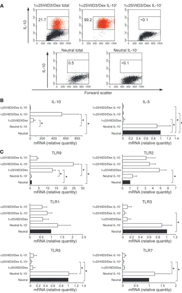
[image:4.585.44.410.77.665.2]TLR9 expression correlates with that of IL-10. To examine whether the expression of TLR2 and TLR9 correlated with IL-10 expression, live IL-10–Tregs were enriched from the 1α25VitD3/Dex-treated cul- tures using an established antibody capture technique and cell sort-ing. This resulted in an enrichment in bulk drug-treated cultures
Figure 2
TLR9 expression is selectively enhanced in human drug-induced IL-10–Tregs. (A)At day 14, neutral and 1α25VitD3/Dex-treated cells were restimulated for 16 hours with anti-CD3 and IL-2. IL-10+
and IL-10– cells were detected
and isolated using a commercially available IL-10 secretion assay and a cell sorter. FACS profiles of IL-10 expression by control T cell lines (neutral total), drug-induced IL-10–Tregs (1α25VitD3/ Dex total) and the isolated IL-10+
(1α25VitD3/Dex IL-10+) and IL-10–
(1α25VitD3/Dex IL-10– and neutral
IL-10–) T cell fractions are shown.
Values represent the percentage of gated IL-10+ cells. Data are
routinely containing 15%–30% IL-10+ T cells, to greater than 98%
viable IL-10+ T cells (Figure 2A). TLR profiles were compared with
those of the IL-10–depleted cell fraction (<0.1% IL-10+ T cells) and
control (neutral) cultures (<1% IL-10+ T cells). The predicted profile
of cytokine mRNA expression in these populations was confirmed by real-time RT-PCR (Figure 2B). TLR9 transcript abundance was elevated in the IL-10+ enriched cell fraction in comparison with
both the bulk 1α25VitD3/Dex and IL-10–depleted cultures (Fig-ure 2C). However, no difference in TLR2 expression was observed between the IL-10+ and IL-10–
fractions, suggesting TLR2 expres- sion did not directly correlate with that of IL-10, and therefore sub-sequent studies focused exclusively on TLR9. TLR1, TLR3, TLR5, and TLR7 gene expression were also examined for comparative pur-poses and were not increased in IL-10+ T cell fractions, compared
with the IL-10– T cell population (Figure 2C). These data confirm
elevated expression of TLR9 mRNA by drug-induced IL-10–Tregs. In order to identify whether TLR9 reflects a specific marker for IL-10–Tregs, expression was measured in other human peripheral blood–derived CD3+CD4+ Treg and effector T cell populations.
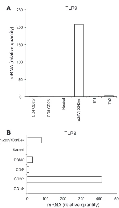
Expression of TLR9 mRNA was detectable in Th1 and Th2 effector cell lines differentiated using previously described methodology (22), as well as in naturally occurring Tregs isolated on the basis of high levels of expression of the CD25 antigen by flow cytometry (Figure 3A). However, in all cases, TLR9 was expressed at much lower levels than in the drug-induced IL-10–Tregs. For compara-tive purposes, the complete TLR profile of CD4+CD25+ Tregs is
shown in Supplemental Figure 2.
Human B lymphocytes are reported to express high levels of TLR9 (23). Expression of TLR9 in bulk 1α25VitD3/Dex-treated cells, which routinely contain 15%–30% IL-10+
T cells, was there-fore compared with that of highly purified B cell populations (>99% CD19+). 1α25VitD3/Dex cultures expressed approximately
25% of TLR9 mRNA levels detected in the B cell population (Fig-ure 3B), implying comparable levels of expression between IL-10+
T cells and B cells.
TLR9 is expressed on both human peripheral blood– and respiratory tissue–derived IL-10–Tregs. The clinical symptoms of disease occur at tissue sites. We therefore addressed whether IL-10–Tregs could be induced from human respiratory tissue–derived T cells and whether TLR9 represents a marker of these cells. As seen in periph-eral blood–derived cultures, CD3+CD4+
T cells derived from lung-draining lymph nodes and CD3+
T cells from nasal polyps, stimu-lated with anti-CD3 in the presence of 1α25VitD3 and Dex for 14 days, expressed elevated levels of IL-10 but low amounts of Th1- and Th2-associated cytokines in comparison with cells stimulated under neutral conditions (Supplemental Figure 3 and data not shown). Drug-induced IL-10–Tregs derived from both lung-drain-ing lymph nodes and from nasal polyps expressed elevated levels of TLR9 compared with the same cells cultured in the absence of drugs (Supplemental Figure 3).
TLR9 expression is induced by 1α25VitD3 in vitro. To investigate whether TLR9 expression was primarily regulated by 1α25VitD3 or Dex, peripheral blood–derived CD3+CD4+
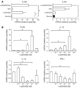
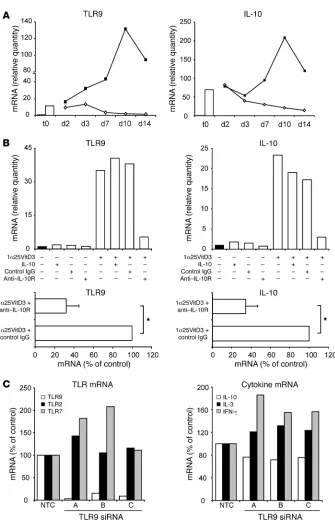
T cells were stimu-lated in the presence of either drug alone or in combination. TLR9 induction was predominantly regulated by 1α25VitD3, with little or no effect of Dex alone. TLR9 was maintained or slightly enhanced in cultures containing both drugs, although this did not reach statistical significance (Figure 4A). In contrast to effects on TLR9, 1α25VitD3 profoundly downregulated TLR3, TLR5, and TLR7 but had little effect on TLR1 (Supplemental Figure 4). The effect of 1α25VitD3 upon TLR and cytokine transcript abun- dance was dose dependent, and a highly comparable concentra-tion dependency for the induction of both IL-10 and TLR9 was observed (Figure 4B). Concentrations of 10–9–10–7 M 1α25VitD3
enhanced TLR9 and IL10 mRNA but inhibited the expression of the Th1- and Th2-associated cytokines IL-13 and IFN-γ. However, the highest concentration of 1α25VitD3 (10–6 M), likely to represent
[image:5.585.59.257.79.423.2]supraphysiological levels (24), failed to significantly induce IL-10 or TLR9 and resulted in less profound reduction in the expression of the effector cytokines. These data highlight the close associa-tion and regulation of IL-10 and TLR9 expression.
Figure 3
Comparison of TLR9 expression by IL-10–Tregs with other human peripheral blood–derived populations. (A and B) TLR9 transcript abundance as assessed by real-time RT-PCR in day 14 drug-induced IL-10–Tregs (1α25VitD3/Dex) and control cultures (neutral) was com-pared with expression in other human T cell populations including naive (CD4+CD25–), CD4+CD25hi, and day 28 highly differentiated Th1
and Th2 cell lines (A) and in non–T cell populations (B). CD4+CD25+
and CD4+CD25– populations were isolated by cell sorting and were
routinely more than 99% pure. Th1 and Th2 cells were generated from naive T cells according to a previously published protocol (22). B cells were isolated on the basis of CD20 antigen expression and were more than 99% CD19+. Monocytes (CD14+) were more than
96% pure. Data are shown normalized to an endogenous control (18S rRNA) and expressed relative to CD25– T cells. One representative
Ingestion of calcitriol by humans induces a parallel increase of IL-10 and TLR9 in CD3+CD4+ T cells
. We previously demonstrated that admin- istration of oral calcitriol (1α25VitD3) to 3 steroid-insensitive asth- ma patients restores IL-10 synthesis by their T cells (21). To deter-mine whether TLR9 represents a potential marker of drug-induced IL-10–Tregs in vivo, we assessed how ingestion of calcitriol for 1 week by these individuals influenced expression of TLR9 and IL-10. CD3+CD4+
T cells were purified from freshly derived periph-eral blood before and after calcitriol ingestion and analyzed directly ex vivo in the absence of any in vitro manipulation. In all individu- als tested, IL-10 and TLR9 expression were increased following cal-citriol ingestion, with the greatest increase being observed on day 3 (Figure 5). No increase or reduction in the Th2 cytokines IL-5 or IL-13 and Th1 cytokine IFN-γ or TLR1, TLR2, TLR3, or TLR5 was observed in the same T cell population.
Induction of IL-10 and TLR9 by 1α25VitD3 is IL-10 dependent and requires continued presence of 1α25VitD3 to maintain expression. The relationship between TLR9 and IL-10 expression was examined fur-ther. Kinetic studies indicated that the induction of TLR9 and IL-10 expression by 1α25VitD3 alone was not significantly increased above neutral cultures at day 7 (data not shown) and was most marked following 14 days or more of culture (Figure 6A). Removal of 1α25VitD3 at day 14 resulted in a gradual decline of both IL10 and TLR9 mRNA levels. TLR9 transcript abundance was compara-ble with neutral cultures 7 days after 1α25VitD3 withdrawal, while IL-10 showed a more gradual but steady loss of gene expression
(Figure 6A), suggesting that the continued presence of 1α25VitD3 is required to maintain optimal expression of both molecules.
As shown in Figure 6B, blocking of IL-10 action throughout the 14-day culture period inhibited the 1α25VitD3-mediated increase in TLR9 and IL10 gene expression, indicating that the capacity of 1α25VitD3 to modulate both molecules was IL-10 dependent (n = 4;
P < 0.05 for both TLR9 and IL10). However, addition of exogenous IL-10 to the control cultures did not result in significant induction of either molecule, demonstrating that IL-10 was necessary but not sufficient to increase TLR9 and IL-10 expression. Supplementing 1α25VitD3 cultures with recombinant IL-10 increased TLR9 and IL-10 expression in 2 of the 4 donors tested, but overall this did not reach statistical significance (Figure 6B and data not shown).
In an attempt to dissect the association between TLR9 and IL-10 expression, knockdown of TLR9 was performed using 3 gene-specific lentiviral shRNA constructs containing 3 different sequences of TLR9 siRNA. CD4+ T cells were cultured for 7 days
[image:6.585.47.368.82.437.2]with 1α25VitD3 and then transduced with either a non-targeting control siRNA or each of the 3 TLR9 shRNA lentiviral constructs (siRNA A, B, or C; Figure 6C). Knockdown of TLR9 of 85% or more compared with the control siRNA was achieved with all 3 lentiviral constructs in the 2 experiments performed. This effect appeared to be specific for TLR9, since mRNA for control TLRs (TLR2, TLR5 and TLR7) were not reduced by this treatment. TLR9 knockdown was associated with a slight reduction (maximally 20%) of IL10 mRNA, but no decline in IL-13 or IFN-γ effector cytokine transcript
Figure 4
TLR9 expression is primarily induced by 1α25VitD3 in vitro. (A) The effects of 2 rounds of 7-day cultures with 1α25VitD3 and Dex, singly or in combination, on the expression of
TLR2 and TLR9 mRNA by CD4+ T cells were
abundance was observed (Figure 6C). At this level, it seems prob-able that the loss of IL-10 is due to the non-specific loss of IL-10+
T cells rather than specific downregulation of the IL10 gene.
Pretreatment of drug-induced IL-10–Tregs with CpG oligonucleotides leads to loss of regulatory activity and reduced IL-10 synthesis . The func-tional relevance of TLR9 expression upon the suppressive activity of IL-10–Tregs was examined using the TLR9 agonist CpG oligonu-cleotide (CpG-ODN) 2006. CD4+ T cells that had been stimulated
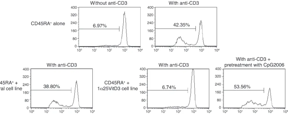
for 14 days in the presence of 1α25VitD3 were pretreated over-night in medium or with CpG-ODNs and then washed extensively. The capacity of these cells to inhibit the proliferative response of freshly isolated, autologous CFSE-labeled naive CD4+CD45RA+
T cells was then assessed. The CFSE-labeled naive T cells prolifer-ated following stimulation with anti-CD3 in culture, with around 42% of cells entering cell cycle at day 5 (Figure 7). Addition of the control (neutral) cell line did not alter their proliferative response. In contrast, the cell line generated in the presence of 1α25VitD3 inhibited naive T cell proliferation to a level comparable with that of unstimulated cells. Pretreatment of IL-10–Tregs with CpG-ODNs prevented their capacity to block the proliferation of the naive T cells, implying that ligation of TLR9 on drug-induced IL-10–Tregs impairs regulatory function.
Parallel experiments demonstrate that when CD4+
T cells stimu-lated for 14 days in the presence of 1α25VitD3 were restimulated in the presence of CpG-ODNs for 48 hours, downregulation of IL-10 synthesis was observed in response to 2 separate CpG-ODN sequences 2216 and 2006 (Figure 8A). Inhibition of IL-10 synthe-sis by CpG-ODNs was concentration dependent, with statistically significant effects seen with concentrations of 5 μM or higher. An unexpected and reproducible observation was the dose-dependent upregulation of the Th2 cytokine IL-4 with CpG-ODN 2006, and to a lesser extent with CpG-ODN 2216. Although cytokine analysis
by FACS demonstrated a 50% or greater reduction in IL-10 positiv-ity upon CpG exposure, staining for IL-4 was consistently at less than 1%, and therefore the change in frequency of these cells could not be assessed (data not shown). Regulation of the Th2 cytokine gene cluster is generally thought to occur in parallel. However, the low levels of synthesis of the Th2 cytokines IL-5 and IL-13 were unaffected by either class of CpG-ODNs (IL-5 data not shown). CpG-ODN 2006, but not CpG-ODN 2216, inhibited IFN-γ synthe-sis. No evidence for the induction of either cell death or expansion of the CpG-ODNs exposed IL-10–Tregs was observed (data not shown). These data imply that CpG-ODNs actively modulate the function of IL-10–Tregs.
In order to confirm the specificity of TLR9 agonist effects on IL-10– Treg cytokine production, control TLR ligands specific for TLR3 (poly [I:C]), TLR4 (LPS), and TLR7 (imiquimod) were also assessed in an identical manner to CpG-ODNs (Figure 8B). All of these con-trol TLR ligands failed to inhibit IL-10 or increase IL-4 expression.
Discussion
The present study demonstrates that 1α25VitD3 (calcitriol) increased IL-10 and TLR9 expression by human CD3+CD4+
[image:7.585.76.520.79.316.2]T cells both in vitro and following ingestion by patients. Ligation of TLR9 by specific agonist CpG-ODNs inhibited IL-10 synthesis and the regulatory activity of 1α25VitD3-induced IL-10–Tregs. These data suggest that modulation of IL-10–Treg function by TLR9 ligands may occur during infection and natural expo- sure to ligands and might also influence the outcome of clini-cal strategies using TLR9 agonists for immune intervention. We propose a model whereby 1α25VitD3 plays a role in maintaining IL-10–Tregs in the host. Coexpression of TLR9 with IL-10 pro-vides a mechanism whereby inappropriate actions of Tregs can be temporarily disabled to enhance the host immune response Figure 5
1α25VitD3 elevates IL-10 and TLR9 expression in vivo. CD3+CD4+ T cells were isolated from 3 steroid-insensitive asthma patients before
to infection. IL-10 is known to impair the clearance of both viral and bacterial pathogens (25, 26).
There is increasing evidence to support the role of the vitamin D pathway in the regulation of immune function (27–29), in addi- tion to its well-established role in the homeostatic control of cal- cium and bone metabolism. The prevalence of vitamin D insuffi-ciency (defined by serum 25-hydroxyvitamin D3 levels of less than 75 nmol/l) is remarkably common. A recent study in the white British population suggested that up to 87% of individuals exhibit vitamin D insufficiency/deficiency in winter and spring (30). Epidemiological studies have assessed the effects of vitamin D
[image:8.585.45.380.74.601.2]availability on a range of immune pathologies, concluding that vitamin D sufficiency is associated with reduced risk of numer-ous cancers (31), while deficiency is linked with a higher risk of autoimmune conditions (32). In respiratory disease, a recent study showed a positive correlation between serum 25-hydroxyvitamin D3 levels and predicted lung function in a large sample of the USA population (approximately 14,000 subjects) (33, 34). In parallel, a high rate of vitamin D deficiency (<50 nmol/l serum 25-hydroxyvi-tamin D3) was reported among individuals of South Asian ethnic origin in London, a population that also exhibited high levels of severe and poorly controlled asthma (35). 25-hydroxyvitamin D3
Figure 6
1α25VitD3-induced IL-10 and TLR9 are IL-10 dependent and require continued presence of 1α25VitD3 to maintain expres-sion. (A)CD4+ T cells were cultured in
neutral conditions or with 1α25VitD3 (open bar) until day 14 (t0). Cells cultured with 1α25VitD3 for 14 days were then restimu-lated with (filled squares) or without (open diamonds) 1α25VitD3 for up 2 weeks.
IL10 and TLR9 mRNA was assessed at the indicated time points. A representa-tive experiment from 2 different healthy donors is shown. (B)Top: CD4+ T cells
levels have also been positively associated with protective immune responses to infection in human populations, including infection with mycobacteria and influenza (reviewed in refs. 28 and 36). Indeed, one of the most striking effects of the vitamin D pathway is on the innate immune system. 1α25VitD3 and its analogs induce antimicrobial gene expression, including the human cathelicidin antimicrobial peptide and defensin β2 genes, in human keratino-cytes, monocytes, epithelial cells, and neutrophils (36–40).
Our study was originally designed to identify biomarkers of IL-10–Tregs. However, our data support the conclusion that TLR9 is not specific to all IL-10–secreting T cells per se (e.g., steroid-induced IL-10+ T cells; CD46-induced IL-10+
T cells; our unpub-lished observations), which represent cells that are rapidly but transiently induced to express IL-10 (41). In contrast, induction by 1α25VitD3 occurs more slowly but is stable in the presence of 1α25VitD3, and under these conditions TLR9 and IL-10 expression is tightly linked. We therefore propose that vitamin D status of the host controls IL-10 (and TLR9) expression. There is emerging lit- erature on the high prevalence of vitamin D deficiency and its asso-ciation with immune disorders (e.g., Crohn disease, type 1 diabetes mellitus, rheumatoid arthritis) and, as we have emphasized, poor respiratory health (33, 42). We and others (e.g., ref. 43) propose a model whereby vitamin D sufficiency is essential to maintain appropriate regulatory pathways, specifically IL-10, to help prevent immune disorders and to maintain respiratory health.
A close association between the capacity of 1α25VitD3 to increase the expression of IL-10 and TLR9 on human T cells was observed not only in vitro, but also in vivo. We previously reported that ingestion of calcitriol by steroid-insensitive asth-matic patients, who respond poorly to steroids for the induction of IL-10 in vitro, restored their capacity to synthesize IL-10 in response to steroids, suggesting a potential role for calcitriol as a steroid-enhancing agent in chronic inflammation (18). Here we show the direct capacity of ingestion of calcitriol to increase IL-10
and that this correlates with increased TLR9 expression by their CD3+CD4+ T cells analyzed directly ex vivo. In vitro, the kinetics
and 1α25VitD3 concentration dependency of increased TLR9 and IL-10 expression by human T cells were highly comparable. The sustained expression of both IL-10 and TLR9 by human T cells was dependent on the continuing presence of 1α25VitD3 in culture, suggesting that the expression of both of these molecules is likely to be influenced by vitamin D status. Inhibition of IL-10 signaling profoundly downregulated (by 75%–90%) both IL-10 and TLR9, implying that induction of both molecules is IL-10 dependent. In contrast, knockdown of TLR9 is highly effective, but only a slight (<20%) reduction in IL10 mRNA is observed, likely due to the non-specific loss of IL-10+ T cells rather than specific regulation of the
IL10 gene. Furthermore, the 1α25VitD3 withdrawal experiments demonstrate that TLR9 expression is lost more rapidly than IL-10. Together these data imply that expression of IL-10 is more stable and can occur independently of TLR9, but that TLR9 (and IL-10) expression is highly IL-10 dependent.
The effects of 1α25VitD3 to increase TLR9 expression may be cell specific, as an independent study has shown that short-term culture of murine islet cells with a vitamin D receptor (VDR) agonist does not alter TLR transcript abundance, including TLR9 (44). However, its capacity to regulate IL-10 synthesis and tolerance appears to be more widely applicable. In vitro studies indicate that 1α25VitD3 enhances IL-10 secretion by human dendritic cells (45), in addition to the effects on CD4+ T cells described here, while in vivo 1α25VitD3
has been shown to promote tolerance, presumably at least in part via its effects on APCs (46–48). In support of the present study in asthma patients, a double-blind, randomized placebo-controlled trial demonstrated that vitamin D supplementation in patients with congestive heart failure improved cytokine profiles by enhanc-ing IL-10 and improving TNF/IL-10 ratios (49). Furthermore, in an animal model of allergic experimental encephalomyelitis, IL-10 signaling was essential for 1α25VitD3-mediated inhibition of dis-Figure 7
CpG-ODNs abrogate regulatory activity of drug-induced IL-10–Tregs. CD4+ T cells were cultured for two 7-day cycles with anti-CD3, IL-2, IL-4
alone (neutral) or with 1α25VitD3, then harvested, washed, and pretreated for 24 hours with anti-CD3 and IL-2 alone or together with 10 μM CpG-ODN 2006. Autologous CD45RA+ T cells were isolated, CFSE labeled, and cocultured with the cell lines at a ratio of 2:1 responder/cell
[image:9.585.63.536.83.275.2]ease (50). Together these data further suggest a role for vitamin D in controlling IL-10 production as well as playing a protective role in the host response to infection and inflammation.
Interactions between other TLRs and the vitamin D pathway on other cell lineages have been reported. For example, ligation of TLR2/1 on human monocytes or macrophages enhances their gene expression of VDR and Cyp27B1 (1α-hydroxylase; the enzyme that catalyses conversion of 25-hydroxyvitamin D3 into the active form 1α25VitD3), thereby increasing 1α25VitD3 synthesis, leading to activation of downstream VDR target genes (40). In keratinocytes, a similar pathway of TLR2-induced enhancement of 1α25VitD3 synthesis has been observed, but with an additional positive feed-back loop whereby 1α25VitD3-VDR interaction then upregulates TLR2 expression (39). These studies, together with those of infec-tion, support a link between TLRs and the vitamin D pathway and their capacity to control innate immunity.
The profile of TLR expression on dendritic cell popu-lations is widely acknowledged to differ between mice and humans, and our data suggest differences also exist on T cells (10). TLR9 is expressed on murine effec-tor T cell populations, and ligation with CpG-ODNs directly enhanced IL-2 production, proliferation, and survival via MyD88 and PI3K-dependent pathways (51). In rodent T cells, CpG-ODNs increased proliferation of CD4+CD25– T cells, enabling these cells to overcome
suppression mediated by natural CD4+CD25+ Tregs (14,
15). However, the effects of CpG-ODNs on human CD4+
T cell function are conflicting. One study describing the costimulatory capacity of TLR2 ligands also briefly mentioned that human memory and naive T cells failed to respond to CpG-ODNs (8). Conversely, type A CpG- ODNs (such as CpG-ODN 2216), but not type B CpG- ODNs, has been shown to inhibit the suppressive activ-ity of human natural CD4+CD25+ Tregs through direct
stimulation of TLR8 (13). It is unlikely that CpG-ODNs acted through TLR8 in the current study, considering the complete absence of TLR8 mRNA in 1α25VitD3-induced IL-10–Tregs. In a series of experiments designed to demonstrate the specificity of CpG-ODNs on IL-10– Tregs, we first demonstrated a loss of IL-10 synthesis when CpG-ODNs were added to cultures of IL-10–Tregs. Only the TLR9 agonist CpG-ODNs, and not control TLR ligands (LPS, poly [I:C], and imiquimod), modulated cytokine production by drug-induced IL-10–Tregs. Secondly, in functional inhibition assays, loss of regulatory function was observed when IL-10–Tregs were pre-pulsed with CpG-ODNs and then washed thoroughly prior to the addition to CFSE-labeled naive responder T cells. Based on these data, we conclude that the inhibition seen here is most likely due to the direct action of CpG-ODNs on human IL-10–Tregs.
[image:10.585.42.318.84.537.2]Natural exposure to CpG motifs is likely to occur following infection with bacteria or viruses, which are known to exacerbate established asthmatic disease. We propose that our data, which show that stimulation of IL-10–Tregs with TLR9 agonist leads to a reduction in IL-10 synthesis and loss of regulatory activity, support a model in which loss of Treg function during infection promotes resolution of infection by cells of the innate and adaptive immune
Figure 8
The TLR9 agonist CpG-ODN inhibit IL-10 production by drug-induced IL-10–Tregs. (A) CD4+ T cells were cultured
system. Our demonstration that T cells derived directly from human lung-draining lymph nodes and nasal tissue respond to 1α25VitD3 by increased expression of IL-10 and TLR9 highlights that this pathway is present and likely to function at the active site of disease. TLR2 is expressed at increased levels by both human and murine naturally occurring (Foxp3+) CD4+CD25+ Tregs. In
most but not all studies, ligation of TLR2 results in expansion but temporary loss of suppressive activity (11, 17, 52, 53). These data have been used to support a similar model in which, during early inflammatory responses, Treg activity is impaired directly and/or indirectly through TLR-induced signals from dendritic cells and/ or by enhanced IL-2 secretion by effector cells, allowing effector T cell populations to resolve the inflammatory insult. Upon reso-lution of the inflammation, the expanded Treg population would regain its functional competence in order to maintain immune homeostasis and prevent host damage and autoimmunity that might arise from overzealous effector cells (52). The evidence that TLR5-stimulated CD4+
effector T cells are refractory to suppres-sion (9), and that TLR8 ligation abrogates CD25+ Treg function
(13), suggests that the direct actions of other TLRs on both effec-tor and regulatory populations fit this model. The data reported here suggest that adaptive IL-10–Tregs equally lose suppressive function during active infection as a result of TLR9 stimulation. Interestingly, TLR9 ligation on non–T cell populations has recently been shown to impair conversion of naive T cells into FoxP3+ Tregs
in the periphery (54). We have also become aware of studies dem-onstrating associations of polymorphisms in the TLR9 gene with asthma (55, 56) and Crohn disease (57). Both conditions are influ-enced by vitamin D deficiency (42), and it would be of interest to determine whether control of Treg function by TLR9 is implicated in these associations.
Synthetic CpG-ODNs are currently of interest as immunomodu- latory agents. Preclinical studies and human clinical trials docu-ment their capacity to improve vaccines and treat cancer, infectious disease, and allergy and/or asthma (58). One important mechanism of action proposed for CpG-ODNs is to activate endogenous plas-macytoid dendritic cells to enhance protective host T cell responses (58). For example, signaling via TLR9 enhances the antigen-pre-senting capacity of plasmacytoid dendritic cells isolated from the lung-draining lymph nodes of patients with non–small cell lung carcinoma for both CD4 and CD8 type I immune responses (59). Additional add-on strategies being considered include depletion of Treg populations that are proposed to impair the development of protective antitumor immune responses. The evidence in the pres-ent study that CpG-ODN signaling of inducible IL-10–Tregs leads to loss of regulatory activity is likely to be beneficial and desirable in conditions such as cancer or infection in which the aim is to boost immunological responses and competence.
Our findings that drug-induced IL-10–Tregs are susceptible to modulation by CpG-ODNs also has implications for the treatment of allergy. Conjugation of CpG-ODNs to specific allergens has been tested in human clinical trials and been shown to improve the safety of allergen immunotherapy by reducing allergenicity (60–63). A proposed mechanism is the deviation of the disease-promoting Th2 responses toward a Th1 response not associated with the allergic phenotype (60, 61, 64). We previously observed that drug-induced IL-10–Tregs are as effective, if not more so, in inhibiting Th1 responses than Th2 responses (21), suggesting that IL-10–Tregs might impair immune deviation. On this basis, possible recognition of CpG-allergen conjugates by IL-10–Tregs
would result in loss of Treg inhibitory function and might facili-tate deviation. Alternatively the pathway described here may limit the effectiveness of CpG-allergen immunotherapy, providing the opportunity for further optimization of CpG conjugate design to maximize benefit in treating allergic disease.
In summary, we have observed the expression of TLR9 by human adaptive IL-10–Treg populations is regulated by 1α25VitD3, and that ligation results in loss of Treg function, further highlighting the role of the vitamin D pathway in regulating immune function. Vitamin D deficiency is surprisingly widespread in populations within the northern hemisphere. It will therefore be important to fully identify the impact of the vitamin D pathway on immune func-tion, including effects of endogenous and therapeutic vitamin D on IL-10, Tregs, and TLRs and more widely with respect to their influence in both allergy and immunity to infection.
Methods
Patient details. With the exception of calcitriol ingestion experiments, PBMCs were obtained from normal healthy individuals and used in all experiments. For calcitriol ingestion experiments, PBMCs were obtained from patients attending the Asthma Clinic at Guy’s Hospital, London, United Kingdom. “Asthma” was defined by American Thoracic Society cri-teria as reversible obstruction (>15%) of the airways (65). “Glucocorticoid resistance” was defined as the failure of forced expired volume in 1 second (FEV1) to improve by greater than 15% from a baseline of less than 75% after
14 days of 40 mg/day oral prednisolone. PBMCs were obtained from 3 glu-cocorticoid-resistant patients (all male), mean (SD) age 54 (15) years, mean (SD) basal FEV1
55% (20%) predicted, and range of prednisolone reversibil-ity 0% to 14% as previously described (18). Human lung-draining lymph nodes were obtained following resection from patients with early stage non–small cell lung cancer. Only non-involved lung-draining lymph nodes, as determined by histopathology, were used for this study (59). Nasal pol-yps were acquired from patients undergoing surgery for nasal polyposis. All donors signed a consent form, and all studies were fully approved by the Ethics Committee at Guy’s Hospital, London, United Kingdom.
Cell purification and culture. PBMCs were isolated as previously described (21). Human lung-draining lymph nodes and nasal polyps were dissected and digested in HBSS with endotoxin-free collagenase (2 mg/ml; Liberase C1; Roche) for 1 hour at 37°C (59). CD4+ T cells or CD3+
T cells were puri-fied by positive selection using Dynabeads (Dynal; typical purity 98.5%) or cell sorting (typical purity 99.5%) (21). CD4+CD25hi (purity >99%) and
CD4+CD25– (purity >99.5%) T cells were isolated by cell sorting from Buffy
coats obtained from the National Blood Service. CD20+ B cells (purity
>99%) and CD14+ monocytes (purity >98%) were also purified from PBMCs
by cell sorting. Highly differentiated human Th1 and Th2 cells were gener-ated from naive T cells as previously described (22). Isolation of live IL-10– secreting cells (purity >98%) was performed using an IL-10 Secretion Assay Detection Kit (Miltenyi Biotec).
CD4+ T cells (1 × 10⁶ cells/ml) were stimulated with 1 μg/ml plate-bound
anti-CD3 (OKT-3), 50 U/ml IL-2 (Eurocetus), 10 ng/ml IL-4 (NBS), Dex (Sigma-Aldrich), and/or calcitriol (1α25VitD3; BIOMOL Research Labs), for 7 day cycles. In some experiments, 5 ng/ml recombinant human IL-10 (R&D Systems), 5 μg/ml anti–IL-10 receptor (clone 3F9-2; BD Biosciences — Pharmingen), or isotype control rat IgG2a (clone R35-9S; BD Biosciences — Pharmingen) was added. At the end of each cycle, cells were recultured with cross-linked anti-CD3 and IL-2 alone, and supernatants were harvested at 48 hours for cytokine analysis.
-TCGTCGTTTTGTCGTTTTGTC-GTT) and CpG-ODN type A 2216 (5′-GGGGGACGATCGTCGGGGGG). LPS was purchased from Sigma-Aldrich, and the remaining TLR agonists imiquimod and poly (I:C) were from Invivogen. On day 14, following 2 rounds of culture with anti-CD3, IL-2, IL-4, and 1α25VitD3, cells were harvested, washed, and stimulated for a further 48 hours with anti-CD3, IL-2, and various concentrations of the relevant TLR agonist as indicated in Figure 8. Supernatants were harvested for cytokine analysis.
Functional assays of regulatory function. Cell lines were generated by culture of isolated CD4+ T cells under neutral conditions (anti-CD3, IL-2, and
IL-4) or additionally with 1α25VitD3. On day 14, cell lines were harvested, washed, and pretreated for 24 hours with anti-CD3 and IL-2 alone or with 10 μM CpG-ODN 2006. Fresh autologous CD4+CD45RA+ naive T cells
were purified and labeled with 2 μM CFSE (Invitrogen), and 2 × 10⁵ cells
were cocultured with 1 × 10⁵
cells of the relevant line as indicated in Fig-ure 7. Cultures were stimulated with 0.1 μg/ml plate-bound anti-CD3 and 2 μg/ml anti-CD28 (clone 15E8; Sanquin) for 5 days. Propidium iodide (Sigma-Aldrich) was then added to exclude dead cells, and 30,000 CFSE-positive responder cells were analyzed by FACS.
Cytokine analysis. IL-5, IL-10, IL-13, and IFN-γ were measured using ELISA and matched antibody pairs (BD Biosciences — Pharmingen), with refer- ence to commercial standards (R&D Systems). The lower limits of detec-tion were 50 pg/ml for IFN-γ and IL-10, 100 pg/ml for IL-5, and 100 pg/ml for IL-13. When less than 200 μl of supernatant was available, the Luminex (Luminex Corp.) or Meso Scale Discovery systems were used.
1α25VitD3 ingestion by asthmatic volunteers. This study was approved by the Research Ethics Committee of Guy’s Hospital, and informed consent was obtained from volunteers. Three glucocorticoid-resistant asthma patients were given 0.5 μg/day (2 × 0.25 μg) oral calcitriol for 7 days, and PBMCs were obtained before treatment and on days 1, 3, and 7 after ingestion of calcitriol as previously described (21). CD3+CD4+
T lymphocytes were iso-lated by cell sorting and cell pellets kept for ex vivo mRNA extraction.
Real time RT-PCR. RNA was extracted from cell pellets using the RNeasy Mini kit (Qiagen) and quantified using Ribogreen RNA quantification kit (Eugene). RNA (250 ng) was reverse-transcribed in a total volume of 30 μl using random hexamer primers (Fermantas Life Sciences). Real-time RT-PCR was performed in triplicate using FAM-labeled Assay-on-Demand reagent sets (Applied Biosystems). Reactions were multiplexed using VIC-labeled 18S as an endogenous control and analyzed according to the 2–ΔΔCt method.
Knockdown of TLR9 using lentiviral shRNA constructs. Lentiviral shRNA constructs were purchased from the Mission shRNA collection (Sigma- Aldrich). TLR9 shRNA constructs (catalog SHDNA-NM_017442; Sigma-Aldrich) and a nontargeting negative control vector (catalog SHC002; Sigma-Aldrich) were used in this study. We transfected 90%–95% conflu-ent 293FT cells with 6.48 μg of lentiviral plasmid DNA and 19.44 μg of
ViraPower packing Mix DNA (pLP1, pLP2, and pLP/VSVG) using Lipo- fectamine 2000 (Invitrogen). After the addition of fresh medium the fol- lowing day, the cells were cultured for an additional 48 hours. Viral super-natants were harvested, passed through a 0.45-μM filter, and concentrated by ultracentrifugation at 50,000 g at 4°C for 90 minutes. Virus pellets were resuspended in less than 1.5 ml of T cell medium, and viral titers were assessed by transducing A549 cells with serially diluted concentrations of virus, adding the selective antibiotic (puromycin; 10 μg/ml) and counting the puromycin-resistant colonies 12 days after infection. Human CD4+
T cells were cultured for 7 days with 1 μg/ml anti-CD3, 50 U/ml IL-2, 10 ng/ml IL-4, and 10-7 M 1α
25VitD3. Cells were then reactivated on anti-CD3–coated plates with IL-2, IL-4, and 1α25VitD3 (as described above) and the concentrated lentiviral supernatants with a MOI of 3, in a total volume of 0.5 ml T cell medium. After 72 hours of incubation, 0.25 ml of medium was removed and replaced with fresh medium containing 20 U/ml IL-2 and 1 μ g/ml puromycin (Sigma-Aldrich). Following an additional 3 days of cul-ture, cells were pelleted, RNA extracted, and cDNA generated, and the level of cytokine and TLR transcripts was assessed by real-time RT-PCR. Statistics. Results are presented as mean ± SEM. Data were assessed for normality and equal variation, after which the appropriate parametric or nonparametric test was performed, as indicated in the figure legends. Dif-ferences were considered significant at the 95% confidence level. Acknowledgments We are grateful to George Santis and Paul Lavender at King’s Col-lege London, Christoph Walker at Novartis, and Clare Lloyd at Imperial College for helpful critique. Z. Urry was initially funded by a Medical Research Council CASE studentship, held in associa-tion with Novartis Institute for Biomedical Research, Horsham, United Kingdom. D.F. Richards and Z. Urry were also supported through funding by EURO-Thymaide, and E. Xystrakis was sup- ported by Asthma-UK. The authors acknowledge financial sup-port from the Department of Health via the National Institute for Health Research (NIHR) comprehensive Biomedical Research Centre award to Guy’s and St. Thomas’ NHS Foundation Trust in partnership with King’s College London and King’s College Hos-pital NHS Foundation Trust. Received for publication April 9, 2007, and accepted in revised form November 19, 2008. Address correspondence to: Catherine M. Hawrylowicz, Depart-ment of Asthma, Allergy and Respiratory Science, 5th Floor Tower Wing, Guy’s Hospital, King’s College London, London SE1 9RT, United Kingdom. Phone: 44-0-207-188-0598; Fax: 44-0-207-403-8640; E-mail: [email protected]. 1. Takeda, K., Kaisho, T., and Akira, S. 2003. Toll-like receptors. Annu. Rev. Immunol. 21:335–376. 2. O’Neill, L.A. 2006. How Toll-like receptors signal:
what we know and what we don’t know. Curr. Opin. Immunol. 18:3–9.
3. Trinchieri, G., and Sher, A. 2007. Cooperation of Toll-like receptor signals in innate immune defence. Nat. Rev. Immunol. 7:179–190.
4. Pasare, C., and Medzhitov, R. 2004. Toll-depen-dent control mechanisms of CD4 T cell activation.
Immunity. 21:733–741.
5. Zarember, K.A., and Godowski, P.J. 2002. Tissue expression of human Toll-like receptors and dif-ferential regulation of Toll-like receptor mRNAs in leukocytes in response to microbes, their products, and cytokines. J. Immunol. 168:554–561. 6. Hornung, V., et al. 2002. Quantitative expression
of toll-like receptor 1-10 mRNA in cellular subsets
of human peripheral blood mononuclear cells and sensitivity to CpG oligodeoxynucleotides. J. Immu-nol. 168:4531–4537.
7. Caron, G., et al. 2005. Direct stimulation of human T cells via TLR5 and TLR7/8: flagellin and R-848 up-regulate proliferation and IFN-gamma pro-duction by memory CD4+ T cells. J. Immunol.
175:1551–1557.
8. Komai-Koma, M., Jones, L., Ogg, G.S., Xu, D., and Liew, F.Y. 2004. TLR2 is expressed on activated T cells as a costimulatory receptor. Proc. Natl. Acad. Sci. U. S. A. 101:3029–3034.
9. Crellin, N.K., et al. 2005. Human CD4+ T cells express TLR5 and its ligand flagellin enhances the suppressive capacity and expression of FOXP3 in CD4+CD25+ T regulatory cells. J. Immunol.
175:8051–8059.
10. Gelman, A.E., Zhang, J., Choi, Y., and Turka, L.A.
2004. Toll-like receptor ligands directly pro-mote activated CD4+ T cell survival. J. Immunol.
172:6065–6073.
11. Sutmuller, R.P., et al. 2006. Toll-like receptor 2 con-trols expansion and function of regulatory T cells.
J. Clin. Invest. 116:485–494.
12. Caramalho, I., et al. 2003. Regulatory T cells selec-tively express toll-like receptors and are activated by lipopolysaccharide. J. Exp. Med. 197:403–411. 13. Peng, G., et al. 2005. Toll-like receptor 8-mediated
reversal of CD4+ regulatory T cell function. Science.
309:1380–1384.
14. LaRosa, D.F., et al. 2007. CpG DNA inhibits CD4+CD25+ Treg suppression through direct MyD88-dependent costimulation of effector CD4+ T cells. Immunol. Lett. 108:183–188.