© 2017, IRJET | Impact Factor value: 5.181 | ISO 9001:2008 Certified Journal
| Page 792
Brain Tumor segmentation and classification using Fcm and support
vector machine
Gaurav Gupta
1, Vinay singh
21PG student,M.Tech Electronics and Communication,
Department of Electronics, Galgotia College of Engineering and
Technology, Greater Noida
2Asst. Professor, Department of Electronics, Galgotia College of Engineering and Technology, Greater Noida
---***---Abstract -MRI is the most important technique, in
detecting the brain tumor. In this paper data mining methods are used for classification of MRI images. A new hybrid technique based on the support vector machine (SVM) and fuzzy c-means for brain tumor classification is proposed. The purposed algorithm is a combination of support vector machine (SVM) and fuzzy c-means, a hybrid technique for prediction of brain tumor. In this algorithm, the image is enhanced using enhancement techniques such as contrast improvement, and mid-range stretch. Double thresholding and morphological operations are used for skull striping. Fuzzy c-means (FCM) clustering is used for the segmentation of the image to detect the suspicious region in brain MRI image. Grey level run length matrix (GLRLM) is used for extraction of feature from the brain image, after which SVM technique is applied to classify the brain MRI images, which provide accurate and more effective result for classification of brain MRI images.
Key Words: Brain tumour, clustering, GLRLM, SVM
1.INTRODUCTION
Data mining may well be a straight forward and robust tool to extract the data from massive dataset [1]. Classification is a branch of data mining field. During this field, many classification techniques are available for medical footage like artificial neural network (ANN), fuzzy c-means (FCM), support vector machine (SVM), decision tree and Bayesian classification. Variety of researchers has been implement the classification techniques for medical footage classification. Presently many medical imaging techniques like (PET), x-ray, CAT (CT), resonance imaging (MRI), for tumor detection but MRI imaging technique is the smart owing to higher resolution and most researchers have used MRI imaging for designation tumor. During this paper, the MRI images were high during contrast improvement and Mid-Range Stretch techniques. Once the image was improved, segmentation step is usually done simply. Segmentation is a technique to extract suspicious area from footage. In this paper, Segmentation technique was done by Fuzzy C-Mean (FCM) agglomeration [2].
Before applying FCM agglomeration technique, skull masking has been done. Feature extraction means that to
induce the information of image. The strategy uses gray Level Run Length Matrix (GLRLM) to extract feature [3]. The reduced GLRLM qualities are outline to support vector machine for coaching and testing. The brain MRI images were differentiating using SVM techniques which widely used for information analyzing and pattern recognizing. It creates a hyper plane in between information sets to point that category it belongs to [4]. The foremost objective of this work is to develop a hybrid technique, which could classify the brain MRI images successfully and efficiently via Fuzzy C- implies that and support vector machine (SVM). This work is a cheap classification technique is to observe the tumor in MRI images.
2. Related Work
© 2017, IRJET | Impact Factor value: 5.181 | ISO 9001:2008 Certified Journal
| Page 793
3. METHODOLOGY
The recommended methodology contains of a collection of phases begging from grouping brain picture MRI image. The main steps are shown in Figure 1. This hybrid technique includes the following main steps like enhancement, Skull striping, segmentation, feature extraction and training the SVM classifier using MRI image with GLRLM feature, saving the information and testing. All the above said steps area unit concerned in testing part, exploitation the new magnetic resonance imaging images with GLRLM feature to SVM and brain MRI pictures are classified. This study used dataset of 120 patients MRI brain pictures and classified them as simple and abnormal. The picture is processed through:
• Image Reading
• MRI images Enhancement • Striping of Skull step
• Segmentation using Fuzzy c-means • Feature Extraction
• Support Vector Machine Classifier
3.1 Image Reading
Brain MRI images were gathered totally different medical centers. These brain MRI pictures were converting into 2D
matrices.
Fig-1: Proposed classification system.
Fig-2: (a) Non-tumor MRI image (b) Tumor MRI image
3.2 MRI images Enhancement
The qualities of pictures are unit improved using improvement method. It’s important to improve the picture information for human viewers, so that right outcomes are gained. The ways given below that are used for improvement of brain MRI image. the 1st step is improving of MRI. Here only the brightness of the photographs was increased to enhance perceptibility. This was done to improve the quality level of the brain MRI images.
• Contrast improvement- MRI images are RGB pictures, which are converting into grey scale pictures. These grey scale images are known as strength pictures. Here intensity values are mapped into less and high intensity values using imadjust (MATLAB function).
• Mid-range Stretch- this is also an addition a technique. In this technique, the center range MRI image intensity values are stretched. So, it improves the standard of brain MRI pictures. During this technique, grey scale image pixels are mapped between 0 and 1value by dividing 255 intensity values as shown in (4).
Here I for row index of brain image matrix and j for column. To compute the function f(x) on the X matrix obtained from (4). The function f(x) is defined as follows.
( )
{
( ) ( )
(5)
© 2017, IRJET | Impact Factor value: 5.181 | ISO 9001:2008 Certified Journal
| Page 794
Fig-3: (a) Enhanced Non-tumor image (b) EnhancedTumor image
3.3 Stripping of Skull step
Skull stripping is a remarkable step. The steps
involved in skull stripping are given below.
• Double thresholding- it is a segmentation
technique. This method, convert the image into
binary form, that is gray scale image to binary image.
This method creates the mask by setting each pixel in
the range of [0.1*255-0.88*255] to 1 means white
and remaining pixels to zero means black. Non-brain
tissues pixels were discarded in MRI picture. Here
2000 upper and lower are considered so it is known
as double thresholding technique [7].
• Erosion- in these level unwanted pixels are cut
from MRI image after thresholding. Thus, the skull
areas are removed. Here disk of radius 3 was taken
as a structuring element for removing all unwanted
pixels which are helping to the brain MRI images.
• Region filling- this technique is implement to pour
the holes in the images. After the erosion, eroded
images are filled using region filling algorithm. Here
the related background pixels are converted into
foreground pixels so that the holes present in the
eroded images are removed in brain MRI image.
Fig-4: (a) Skull masking Non-tumor image (b) Skull masking Tumor image
3.4 Segmentation using Fuzzy C-Means
Segmentation is the method of separating an image into multiple part and object area. The skull stripes images are used in image segmentation. This delivers good result for tumor segmentation. In this work, fuzzy c-means algorithm was used in MRI image segmentation. Fuzzy C-Means (FCM) algorithm is used to find out the suspicious area from brain MRI image. This fuzzy c-means clustering method provides good segmentation result.
Fig-5: fuzzy c-mean algorithm
3.5 Feature extraction
Feature extraction is a method to search the related
features from picture, which are used to understand
the picture easily. This input data group picture is
transformed into compressed form is called feature
extraction. It can reduce the work for further
processing such as picture classification. Here the
GLRLM feature extraction method is used. GLRLM is
used after the fuzzy c-means algorithm. Derive the
gray level run length matrix (GLRLM) for second
level maximum frequency sub bands of the discrete
wavelet decomposed picture with 1one for distance
and 0,45,90 and 135 degrees [8]. Here feature
extraction is isolating the related features which lead
to understand the brain MRI images well.
3.6 Support Vector Machine classifier
SVM is a supervised learning technique. It is a better tool for data analysis and classification. SVM classifier has a 1st learning speed even in huge data. SVM is used for two or more class classification issue. Support Vector Machine is depending on the conception of decision planes. A decision plane is one that differentiates between a set of items
having different class memberships. The Classification and
© 2017, IRJET | Impact Factor value: 5.181 | ISO 9001:2008 Certified Journal
| Page 795
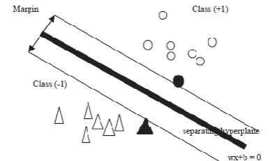
w is the weight vector, b the offset. Since the classes aredefined as +-1 the equation for the line dividing the classes will be:
The distance from the hyper-plan ( w + b = 0 ) to the origin is , where || w || is the norm of w. The distance
from the hyper-plane to the origin is:
Where M is the margin. So, the maximum margin is
obtained by minimizing .
Fig-6: SVM classification (the separating margin between the two classes)
Classification is the process where a given test sample is assigned a class by the classifier during training. We have used the SVM classifier [9].
1. Performance measures
Classification, the sensitivity, specificity and accuracy were calculated using below formulas:
• True Positive (TP): Abnormal brain correctly identified as abnormal.
• True Negative (TN): Normal brain correctly identified as normal.
• False Positive (FP): Normal brain incorrectly identified as abnormal.
• False Negative (FN): Abnormal brain incorrectly identified as normal.
1) Sensitivity = TP/ (TP+FN) *100% 2) Specificity = TN/ (TN+FP) * 100%
3) Accuracy = (TP+ TN)/ (TP+ TN+FP+FN) * 100%
All these three parameters are used to check the classifiers performance.
4. RESULTS
In this paper, SVM technique with fuzzy c-means is used for segmentation and classification of brain MRI images. Real data set of 124 MRI brain images has been used to detect 'tumor' and 'non-tumor' MRI images. The soft tissues in brain MRI images are segmented with Double
Thresholding, Morphological operations and fuzzy c-means algorithm for clustering and gray level run length matrix for feature extraction. The SVM classifier is trained using 100 brain MRI images, after that the remaining 24 brain MRI images was used for testing the trained SVM. The result for classification provides accurate for large data sets.
Table 1: SVM Classification results
Sr. No. Kernel
function Specificity Sensitivity Accuracy
1 Linear 100% 88.45% 91.77%
2 RBF 100% 87.36% 90.01%
5. CONCLUSIONS
In this proposed system brain MRI methods verified to be a significant way to find the brain tumor. The hybrid methodology of gathering support vector machine and fuzzy c-means clustering for classification gives precise result for identifying the brain tumor. For future work, to get better accuracy rate and less error rate a hybrid SVM algorithm is to be proposed. In future work, different data mining techniques can be used to train using different kernel functions in order to improve the performance of the classifiers and the data sets can also be increased.
REFERENCES
[1] Prakash Mahindrakar and Dr. M.Hanumanthappa, “Data Mining In Healthcare: A Survey of Techniques and Algorithms with Its Limitationsand Challenges”, Int. Journal of Engineering Research and Applications,ISSN : 2248-9622, Vol. 3, Issue 6, Nov-Dec 2013, pp.937-941
[2] Kailash Sinha, G.R.Sinha, “Efficient Segmentation Methods for Tumor Detection in MRI Images”, 2014 IEEE Student’s Conference on Electrical, Electronics and Computer Science, 978-1-4799-2526- 1/14/$31.00 ©2014 IEEE
[3] R. S. Raj Kumar and G. Niranjana, “Image Segmentation and Classification of MRI Brain Tumor Based on Cellular Automata and Neural Networks”, IJREAT International Journal of Research in Engineering & Advanced Technology, Volume 1, Issue 1, March, 2013
ISSN: 2320 – 8791.
[image:4.595.64.258.270.385.2]© 2017, IRJET | Impact Factor value: 5.181 | ISO 9001:2008 Certified Journal
| Page 796
[5] A. Padma and R. Sukanesh, “SVM based classification of soft tissues in brain CT images using wavelet based dominant gray level run length texture features”, middle-east journal of scientific research, 2013, 13(7): 883-888.
[6] S.H.S.A. Ubaidillah, R. Sallehuddinand N.A. Ali, “Cancer detection using artificial neural network and support vector machine: A Comparative study”, jurnal teknologi (science & engineering), 2013, 65:1.
[7] O.P. Verma, M. Hammandlu, S. Susan, M. Kulkami and P.K. Jain, "A simple single seeded region growing algorithm for color image segmentation using adaptive thresholding," 2011 International Conference on Communication Systems and Network Technologies, ©2011 IEEE.
[8] G.V. Kumar and Dr G.V. Raju, “Biological early brain cancer detection using artificial neural network”, International Journal on Computer Science and Engineering Vol. 02, No. 08, 2010, 2721-2725