Empathy is the ability to identify with or vicariously experi-ence another person’s feelings or thoughts.1 Previous studies have reported that various brain regions are involved in em-pathy, such as the anterior cingulate cortex (ACC), anterior in-sula, inferior parietal lobe, premotor cortex, posterior superior temporal sulcus, medial prefrontal cortex (mPFC), posterior cingulate cortex, precuneus, temporal pole, and temporopari-etal junction.2-5 However, the intrinsic interactions of these
em-pathy-related regions has not been reported, as studies have typically been conducted using participants performing spe-cific empathy-related tasks.6 To overcome this limitation, rest-ing state functional neuroimagrest-ing studies of empathy should be performed.
The default mode network (DMN) consists of the ACC, mPFC, posterior cingulate cortex, inferior parietal lobule, and precu-neus7 and, accordingly, overlaps with several empathy-related regions, as shown in previous task-based magnetic resonance imaging (MRI) studies.5,8 The DMN is thought to play a role in internally focused thought processes, including the construc-tion of mental simulaconstruc-tions based on previous personal experi-ences.9,10 Given that empathy is achieved by simulating the mental processes that are likely to be operating in others11 and/ or by using personal memories to understand situations of oth-er,12 the DMN is a clear candidate network underlying empa-thy. On this premise, we hypothesized that low resting-state functional connectivity within the DMN might exist in
low-em-Altered Functional Connectivity of the Default Mode
Network in Low-Empathy Subjects
Seung Jun Kim
1,2,3, Sung-Eun Kim
1,4, Hyo Eun Kim
5, Kiwan Han
6, Bumseok Jeong
7,
Jae-Jin Kim
3, Kee Namkoong
3, and Ji-Woong Kim
1,21Department of Psychiatry, Konyang University College of Medicine, Daejeon; 2Konyang University Myunggok Medical Research Institute, Daejeon; 3Department of Psychiatry, Yonsei University College of Medicine, Seoul; 4Yuseung Psychiatry Clinic, Daejeon;
5Department of Psychiatry, Daejeon Sun Hospital, Daejeon;
6Department of Rehabilitative & Assistive Technology, National Rehabilitation Center Research Institute, Seoul;
7Graduate School of Medical Science and Engineering, Korea Advanced Institute of Science and Technology, Daejeon, Korea.
Empathy is the ability to identify with or make a vicariously experience of another person’s feelings or thoughts based on memory and/or self-referential mental simulation. The default mode network in particular is related to self-referential empathy. In order to elucidate the possible neural mechanisms underlying empathy, we investigated the functional connectivity of the default mode network in subjects from a general population. Resting state functional magnetic resonance imaging data were acquired from 19 low-empathy subjects and 18 medium-empathy subjects. An independent component analysis was used to identify the default mode network, and differences in functional connectivity strength were compared between the two groups. The low-empathy group showed lower functional connectivity of the medial prefrontal cortex and anterior cingulate cortex (Brodmann areas 9 and 32) within the default mode network, compared to the medium-empathy group. The results of the present study suggest that empathy is related to functional connectivity of the medial prefrontal cortex/anterior cingulate cortex within the default mode network. Func-tional decreases in connectivity among low-empathy subjects may reflect an impairment of self-referential mental simulation.
Key Words: Empathy, magnetic resonance imaging, functional neuroimaging, medial prefrontal cortex, anterior cingulate cortex
pISSN: 0513-5796 · eISSN: 1976-2437
Received: February 15, 2017 Revised: June 26, 2017
Accepted: June 27, 2017
Corresponding author: Dr. Ji-Woong Kim, Department of Psychiatry, Konyang University College of Medicine, 158 Gwanjeodong-ro, Seo-gu, Daejeon 35365, Korea. Tel: 82-42-600-9160, Fax: 82-42-600-9251, E-mail: [email protected] •The authors have no financial conflicts of interest.
© Copyright: Yonsei University College of Medicine 2017
This is an Open Access article distributed under the terms of the Creative Com-mons Attribution Non-Commercial License (http://creativecomCom-mons.org/licenses/ by-nc/4.0) which permits unrestricted non-commercial use, distribution, and repro-duction in any medium, provided the original work is properly cited.
pathy participants. To address this question, we used resting-state functional MRI to evaluate DMN functional connectivity in low- versus medium-empathy individuals.
A total of 484 student participants from a local university were screened using the Korean version of the Interpersonal Reactivity Index (IRI).13 Lower IRI scores indicated a weaker degree of empathy. After all individual IRI scores were calculat-ed, the participants with IRI scores in the 15th percentile and the 30th to 70th percentile were selected as low- or medium-empa-thy candidates, respectively. The medium-empamedium-empa-thy group was chosen as a control group, because this group was more likely to represent the general population. Due to a significant sex dif-ference in IRI scores, different cutoff scores were applied ac-cording thereto. Sex was balanced across groups in final anal-ysis. All participants were healthy with normal cognitive function and showed no signs of psychiatric illness (as confirmed by a psychiatric interview). The exclusion criteria included a histo-ry of psychiatric or neurologic disorder, left-handedness, and the presence of an MRI-incompatible implant.
Twenty low-empathy participants and 19 medium-empa-thy participants were included in the study. However, the data of only 19 low-empathy participants and 18 medium-empathy participants were included in the final analysis: two partici-pants (one from each group) were excluded due to excessive head movement during the MRI scan. Verbal IQ was assessed using the verbal scales of the Wechsler Adult Intelligence Scale. As depression and anxiety may be associated with abnormal levels of empathy14,15 and abnormal DMN connectivity,16,17 the Hamilton Depression Rating Scale (HAM-D)18 and Hamilton Anxiety Scale (HAM-A)19 were used to assess current symp-toms of depression and anxiety. All participants provided writ-ten informed consent prior to study participation. The study protocol was approved by the Konyang University Hospital Medical Ethics Committee.
The IRI is a self-reported instrument that measures trait em-pathy.20 It is composed of four subscales: Perspective Taking, Fantasy, Personal Distress, and Empathic Concern. Perspective Taking measures the tendency to voluntarily think from anoth-er individual’s psychological panoth-erspective. Fantasy examines the tendency to feel the emotions of people in fictional situations. Personal Distress measures self-oriented anxiety and interper-sonal discomfort. Empathic Concern evaluates other-oriented feelings, including compassion and sympathy. Each subscale contains eight items with each item measured on a five-point Likert scale ranging from 0 (“Does not describe me well”) to 4 (“Describes me very well”).
During resting state scanning, participants were instructed to lie still with their eyes open. Functional MRI data were ac-quired with a Philips 3T scanner (Philips Intera, Philips Medi-cal System, Best, the Netherlands) equipped with an eight-channel SENSE head coil. Resting state functional images were acquired using a gradient echo-planar imaging sequence with the following parameters: 116 volumes (348 s); repetition time
(TR)=3000 ms; echo time (TE)=35 ms; 33 slices; no gap; flip angle=90°; field of view (FOV)=230 mm; voxel size=1.80×1.80×4 mm. Structural images were also acquired with the following parameters: TR=536 ms; TE=10 ms; flip angle=70°; 33 slices; voxel size 0.45×0.45×4 mm; FOV=230 mm.
Preprocessing was performed using Statistical Parametric Mapping software (SPM5; Wellcome Department of Imaging Neuroscience, London, UK). The first five images were discard-ed to allow for the equilibration of longitudinal magnetization. Individual scans were realigned and slice time-corrected, nor-malized to a standard SPM5 template based upon the Mon-treal Neurological Institute (MNI) reference brain, and spa-tially smoothed using a 10-mm isotropic Gaussian kernel with standard SPM methods. Motion parameters for each individ-ual were visindivid-ually inspected, and only data with translation mo-tion less than 2 mm and rotamo-tional movement less than 2° in any direction were included.
Independent component analysis (ICA) is a data-driven meth-od to identify spatially independent components of brain areas with hemodynamic time courses that closely covary. Thus, the regions comprising each component are conceptualized as parts of a specific network with highly synchronous time cours-es.21 For group comparisons, a separate group ICA may not be optimal, because it is biased towards false-positive results.21,22 Therefore, images of all participants were decomposed into sets of independent components using the Group ICA FMRI Toolbox (GIFT) and the Infomax algorithm.21
The group ICA was carried out in three stages: data reduc-tion, application of the ICA algorithm, and backwards recon-struction for each participant. First, data from each participant underwent a principal component analysis to reduce compu-tational complexity. Next, the reduced participant data were concatenated over the time domain. To determine the number of independent components, a dimensionality estimation was performed using the minimum description length criteria.23 The ICA estimation identified 18 components. In the second stage of the analysis, we used the Infomax algorithm to run the ICA and a mask based on all participants. In the final stage of backwards reconstruction, time courses and spatial maps were reconstructed and converted to Z-scores to normalize signals.24
Selection of the DMN component was completed in two stages. First, each of the 18 components identified by the group ICA was manually inspected for the presence of obvious arti-facts. Then, the individual independent components were spatially sorted using a DMN mask provided within the GIFT toolbox. The mask consisted of the posterior cingulate cortex [Brodmann areas (BAs) 23 and 31], posterior parietal cortex (BAs 7, 39, and 40), dorsolateral and superior frontal cortices (BAs 8, 9, and 10), ACC (BAs 11 and 32), and inferior temporal gyrus (BAs 19 and 37). The component that showed the highest correlation with the DMN template was selected as the DMN (r=0.58).
test for significant group differences in demographic and clin-ical variables. Selected best-fit components were entered into a second level random-effects analysis in SPM5. Two sample t-tests examined group differences in the degree of regional functional connectivity. The statistical threshold for these analyses was set at p<0.001, uncorrected, with an extent thresh-old of 20 voxels. Anatomical regions and denominations are reported according to the atlases of Talairach and Tournoux.25 All coordinates are reported in the MNI space.
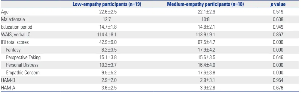
Table 1 shows the demographic and clinical data of the 19 low-empathy participants and 18 medium-empathy partici-pants that were included in our study. There were no signifi-cant differences in age, sex, education, or verbal IQ, HAM-D, and HAM-A scores between the two groups. For the IRI, the overall scores of the medium-empathy group were significantly higher than those of the low-empathy group; moreover, the IRI subscale scores of the medium-empathy group were higher than those of the low-empathy group for all subscales, except for Perspective Taking.
Table 2 shows the anatomical location and Talairach coordi-nates for the peak activation voxel in each brain region (x, y, z), as well as the t scores from our random effects analyses [p<0.05
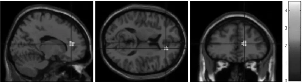
family-wise error (FWE)-corrected, k>40 voxels]. Individuals in the low-empathy group showed a lower functional connec-tivity of the mPFC/ACC within the DMN (BAs 9 and 32, x=15, y=39, z=15, t=4.51, k=23 voxels, uncorrected p<0.001) than that of the medium-empathy group (Fig. 1).
The functional connectivity strength of the mPFC/ACC was positively correlated with the scores of the IRI and all IRI sub-scales (r=0.59, p<0.001 for IRI; r=56, p<0.001 for Empathic Con-cern; r=0.48, p<0.01 for Fantasy; r=0.37, p<0.05 for Personal Distress), with the exception of Perspective Taking.
The aim of the present study was to investigate resting state differences in DMN connectivity between low- and medium-empathy participants. Direct group comparisons revealed low-er functional connectivity of the mPFC/ACC within the DMN of low-empathy participants. Therefore, a decreased function-al connectivity of the mPFC/ACC within the DMN might un-derlie specific deficits in empathy.
[image:3.595.55.558.371.527.2]According to the internal mentation hypothesis, the DMN consists of two distinct interacting subsystems;10 one is the medial temporal lobe subsystem, which is activated during the successful retrieval of old information from memory,26,27 and the other is the mPFC subsystem, which is activated during
Table 1. Demographic and Clinical Characteristics
Low-empathy participants (n=19) Medium-empathy participants (n=18) p value
Age 22.6±2.5 22.1±2.9 0.519
Male:female 12:7 10:8 0.638
Education period 14.7±1.8 14.8±2.1 0.949
WAIS, verbal IQ 114.4±8.1 113.9±9.1 0.867
IRI total scores 42.9±9.0 67.5±4.7 0.000
Fantasy 8.2±3.5 17.9±4.2 0.000
Perspective Taking 15.1±3.8 15.6±3.5 0.646
Personal Distress 10.2±3.7 16.4±4.0 0.000
Empathic Concern 9.5±5.2 17.6±3.8 0.000
HAM-D 2.9±2.0 2.9±3.1 0.954
HAM-A 3.6±2.5 3.9±2.8 0.676
[image:3.595.55.555.574.713.2]WAIS, Wechsler Adult Intelligence Scale; IRI, Interpersonal Reactivity Index; HAM-D, Hamilton Depression Rating Scale; HAM-A, Hamilton Anxiety Scale.
Table 2. The Anatomical Location and Talairach Coordinates for Voxels Whose Resting State Time-Course Best Fit the DMN Component (x, y, z), and t Scores from Random Effects Analyses for Low- or Medium-Empathy Group (p<0.05 FWE-Corrected, k>40 Voxels, for the Purpose of Illustration)
Activated regions
Low-empathy group Medium empathy group
Primary peak location Primary peak location
x y z t k* x y z t k*
mPFC (BA 8, 9, 10)
Left -9 45 33 21.05 487 -9 48 30 31.26 392
Right 9 51 27 16.36 287 6 63 24 18.18 295
PCC (BA 23, 31)
Left -3 -51 24 8.56 47 -3 -57 21 9.49 40
Right 3 -57 21 8.07 40 3 -57 21 11.16 46
MTG (BA 39)
Left -54 -69 21 10.42 67
self-referential mental simulation.28,29 Buckner, et al.10 inter-preted self-referential mental simulation as thinking about the complex interactions among people that are perceived as be-ing socially, interactively, and emotively similar to those of one-self. This interpretation suggests that empathy is, at least in part, based on self-referential mental simulation and requires the ability to relate to the feelings or thoughts of others with-out losing sight of one’s own feelings or thoughts.30 Given our results regarding mPFC connectivity and the abovementioned role of the mPFC in self-referential mental simulation, the pres-ent study suggests that low-empathy individuals may have im-paired or decreased self-referential mental simulation during the resting state. In other words, low-empathy individuals may show decreased functional connectivity among regions of the DMN responsible for self-referential mental simulation, where-as connectivity is better sustained in normal medium-empa-thy individuals.
Previous studies have reported altered DMN connectivity in various psychiatric disorders associated with a lack of empa-thy, including autistic spectrum disorder,31 schizophrenia,32 and antisocial personality disorder.33 Of note, the present study identified low DMN connectivity in low-empathy participants of a general population rather than a clinical population. Con-sistent with this finding, a previous study reported altered func-tional connectivity of the DMN in general population partici-pants with alexithymia.34 Therefore, various features of DMN connectivity may be related to individual trait differences among individuals from the general population, making the DMN a useful target for the investigation of personality traits, specifi-cally empathy.
The limitations of our study should be noted. First, partici-pants could have made an error in measuring the degree of their own empathy, thus the IRI, as a self-report assessment, may not have been valid. Using objective measurements for empathy could overcome this limitation. Second, the current sample size should have been larger for more valid results. Third, although we recorded the psychiatric history of the par-ticipants and psychiatrists administered the D and HAM-A, standardized psychiatric interviews were not performed to
assess the occurrence of psychiatric disorders. Finally, although all participants responded to the technologist at the begning and end of the MRI scan and although none of them in-dicated that they slept during the scan, attentional measure-ments during the scan were not performed.
In conclusion, we found that low-empathy individuals ex-hibit diminished functional connectivity of the mPFC/ACC within the DMN, which may reflect decreased self-referential mental simulation as an underlying cause of empathic deficits. Accordingly, the functional connectivity of the mPFC/ACC with-in the DMN may be a major factor underlywith-ing trait empathy.
ACKNOWLEDGEMENTS
We would like to thank Bum Hee Park for his methodological advice.
This work was supported by a grant of the Korea Health 21 R&D Project, Ministry of Health & Welfare, Republic of Korea (A050495).
REFERENCES
1. Deutsch F, Madle RA. Empathy: historic and current conceptual-izations, measurement, and a cognitive theoretical perspective. Hum Dev 1975;18:267-87.
2. Singer T, Lamm C. The social neuroscience of empathy. Ann N Y Acad Sci 2009;1156:81-96.
3. Lamm C, Decety J, Singer T. Meta-analytic evidence for common and distinct neural networks associated with directly experienced pain and empathy for pain. Neuroimage 2011;54:2492-502. 4. Zaki J, Ochsner KN. The neuroscience of empathy: progress,
pit-falls and promise. Nat Neurosci 2012;15:675-80.
5. Spreng RN, Mar RA, Kim AS. The common neural basis of autobi-ographical memory, prospection, navigation, theory of mind, and the default mode: a quantitative meta-analysis. J Cogn Neurosci 2009;21:489-510.
6. Cole DM, Smith SM, Beckmann CF. Advances and pitfalls in the analysis and interpretation of resting-state FMRI data. Front Syst Neurosci 2010;4:8.
[image:4.595.42.541.72.207.2]7. Raichle ME, MacLeod AM, Snyder AZ, Powers WJ, Gusnard DA, Shulman GL. A default mode of brain function. Proc Natl Acad Sci U S A 2001;98:676-82.
Fig. 1. Coordinates of a voxel exhibiting lower functional connectivity within the default mode network in the low-empathy empathy group, compared to the medium empathy group (x=15, y=39, z=15, t=4.51, k=23 voxels, uncorrected p<0.001).
4
3
2
1
8. Wagner DD, Kelley WM, Heatherton TF. Individual differences in the spontaneous recruitment of brain regions supporting mental state understanding when viewing natural social scenes. Cereb Cortex 2011;21:2788-96.
9. Buckner RL, Carroll DC. Self-projection and the brain. Trends Cogn Sci 2007;11:49-57.
10. Buckner RL, Andrews-Hanna JR, Schacter DL. The brain’s default network: anatomy, function, and relevance to disease. Ann N Y Acad Sci 2008;1124:1-38.
11. Gordon RM. Folk psychology as simulation. Mind Lang 1986;1: 158-71.
12. Gopnik A, Astington JW. Children’s understanding of representa-tional change and its relation to the understanding of false belief and the appearance-reality distinction. Child Dev 1988;59:26-37. 13. Kang I, Kee SW, Kim SE, Jeong BS, Hwang JH, Song JE, et al.
Reli-ability and validity of the Korean-version of Interpersonal Reac-tivity Index. J Korean Neuropsychiatr Assoc 2009;48:352-8. 14. Korgaonkar MS, Fornito A, Williams LM, Grieve SM. Abnormal
structural networks characterize major depressive disorder: a con-nectome analysis. Biol Psychiatry 2014;76:567-74.
15. Schreiter S, Pijnenborg GH, Aan Het Rot M. Empathy in adults with clinical or subclinical depressive symptoms. J Affect Disord 2013; 150:1-16.
16. Guo W, Liu F, Zhang J, Zhang Z, Yu L, Liu J, et al. Abnormal default-mode network homogeneity in first-episode, drug-naive major de-pressive disorder. PLoS One 2014;9:e91102.
17. Andreescu C, Sheu LK, Tudorascu D, Walker S, Aizenstein H. The ages of anxiety--differences across the lifespan in the default mode network functional connectivity in generalized anxiety disorder. Int J Geriatr Psychiatry 2014;29:704-12.
18. Hamilton M. Development of a rating scale for primary depressive illness. Br J Soc Clin Psychol 1967;6:278-96.
19. Hamilton M. The assessment of anxiety states by rating. Br J Med Psychol 1959;32:50-5.
20. Davis MH. Measuring individual differences in empathy: evidence for a multidimensional approach. J Personal Soc Psychol 1983; 44:113-26.
21. Calhoun VD, Adali T, Pearlson GD, Pekar JJ. A method for making group inferences from functional MRI data using independent component analysis. Hum Brain Mapp 2001;14:140-51.
22. Otti A, Guendel H, Läer L, Wohlschlaeger AM, Lane RD, Decety J,
et al. I know the pain you feel-how the human brain’s default mode predicts our resonance to another’s suffering. Neuroscience 2010; 169:143-8.
23. Li YO, Adali T, Calhoun VD. Estimating the number of independent components for functional magnetic resonance imaging data. Hum Brain Mapp 2007;28:1251-66.
24. Beckmann CF, DeLuca M, Devlin JT, Smith SM. Investigations into resting-state connectivity using independent component analysis. Philos Trans R Soc Lond B Biol Sci 2005;360:1001-13. 25. Talairach J, Tournoux P. Co-planar stereotaxic atlas of the human
brain: 3-dimensional proportional system : an approach to cerebral imaging. New York: Thieme; 1988.
26. Greicius MD, Menon V. Default-mode activity during a passive sensory task: uncoupled from deactivation but impacting activa-tion. J Cogn Neurosci 2004;16:1484-92.
27. Vincent JL, Snyder AZ, Fox MD, Shannon BJ, Andrews JR, Raichle ME, et al. Coherent spontaneous activity identifies a hippocam-pal-parietal memory network. J Neurophysiol 2006;96:3517-31. 28. Gusnard DA, Akbudak E, Shulman GL, Raichle ME. Medial
pre-frontal cortex and self-referential mental activity: relation to a de-fault mode of brain function. Proc Natl Acad Sci U S A 2001;98: 4259-64.
29. Mitchell JP, Macrae CN, Banaji MR. Dissociable medial prefrontal contributions to judgments of similar and dissimilar others. Neu-ron 2006;50:655-63.
30. Decety J, Jackson PL. The functional architecture of human em-pathy. Behav Cogn Neurosci Rev 2004;3:71-100.
31. Assaf M, Jagannathan K, Calhoun VD, Miller L, Stevens MC, Sahl R, et al. Abnormal functional connectivity of default mode sub-net-works in autism spectrum disorder patients. Neuroimage 2010; 53:247-56.
32. Garrity AG, Pearlson GD, McKiernan K, Lloyd D, Kiehl KA, Cal-houn VD. Aberrant “default mode” functional connectivity in schizophrenia. Am J Psychiatry 2007;164:450-7.
33. Juárez M, Kiehl KA, Calhoun VD. Intrinsic limbic and paralimbic networks are associated with criminal psychopathy. Hum Brain Mapp 2013;34:1921-30.