ABSTRACT
Ubiquitylated developmental membrane signaling proteins are often internalized for endocytic trafficking, through which endosomal sorting complexes required for transport (ESCRT) act sequentially to deliver internalized cargos to lysosomes. The ESCRT function in endocytic sorting is well established; however, it is not fully understood how the sorting machinery itself is regulated. Here, we show that Ubiquitin isopeptidase Y (Ubpy) plays a conserved role in vivo in the homeostasis of an essential ESCRT-0 complex component Hrs. We find that, in the absence of Drosophila Ubpy, multiple membrane proteins that are essential components of important signaling pathways accumulate in enlarged, aberrant endosomes. We further demonstrate that this phenotype results from endocytic pathway defects. We provide evidence that Ubpy interacts with and deubiquitylates Hrs. In Ubpy-null cells, Hrs becomes ubiquitylated and degraded in lysosomes, thus disrupting the integrity of ESCRT sorting machinery. Lastly, we find that signaling proteins are enriched in enlarged endosomes when Hrsactivity is abolished. Together, our data support a model in which Ubpy plays a dual role in both cargo deubiquitylation and the ESCRT-0 stability during development.
KEY WORDS: Developmental signaling, Endocytic machinery, ESCRT-0, Hrs, Ubpy, Drosophila
INTRODUCTION
Many developmental signaling events are initiated through ligand binding to respective receptors at the plasma membrane. Following uptake into endocytic vesicles referred to as early or sorting endosomes, membrane receptors may recycle back to cell surface via tubular recycling endosomes. However, when directed by a ubiquitin (Ub) signal, receptor complexes are captured and sorted towards lysosomal degradation (Henne et al., 2011). The Ub moieties of protein cargos are recognized by ESCRT complexes, and are sorted into invaginating multivesicular bodies (MVBs). Mature MVBs fuse with lysosomes to deliver protein cargos for degradation (Raiborg and Stenmark, 2009). ESCRT-0 is composed of heterodimers of two subunits: Hrs and Stam. Both subunits bind Ub and clathrin, and Hrs additionally has an FYVE zinc-finger domain that binds phosphatidylinositol 3-phosphate. The ability to bind both lipid and Ub allows ESCRT-0 to initiate endosomal sorting (Clague et al., 2012).
Ubpy (or USP8), a USP family deubiquitinase (DUB), participates in sorting of ubiquitylated receptors through its RESEARCH REPORT
1State Key Laboratory of Biomembrane and Membrane Biotechnology, Ministry of
Education Key Laboratory of Cell Proliferation and Differentiation, Peking-Tsinghua Center for Life Sciences, College of Life Sciences, Peking University, Beijing 100871, China. 2Department of Cellular & Molecular Medicine, Lerner
Research Institute, Cleveland Clinic, Cleveland, OH 44195, USA.
*Author for correspondence ([email protected])
Received 28 May 2013; Accepted 28 January 2014
interaction with ESCRT-0 (Mizuno et al., 2005; Row et al., 2006). Most studies of Ubpy focus on endosomal trafficking of growth factor receptor tyrosine kinases (RTKs) in cultured vertebrate cells. However, conflicting data have been reported. In some cases, reduced Ubpy activity results in accumulation of ubiquitylated cargos (Bowers et al., 2006; Mizuno et al., 2006; Row et al., 2006; Alwan and van Leeuwen, 2007). Other studies suggest that Ubpy promotes RTK stability (Mizuno et al., 2005; Niendorf et al., 2007; Berlin et al., 2010). The role of Ubpy in Drosophiladevelopment is equally controversial. It was suggested that the Hedgehog (Hh) signaling activator Smoothened (Smo) is subject to Ubpy control (Li et al., 2012; Xia et al., 2012). However, Mukai et al. (Mukai et al., 2010) found that Smo abundance is unchanged when Ubpy is depleted.
In view of these discrepancies, we examined the in vivo consequence of loss of Ubpyactivity in Drosophila. Our results show that, in addition to the reported function on deubiquitylating membrane proteins, Ubpy deubiquitylates Hrs, a key component of ESCRT-0. Altered Hrs ubiquitylation leads to Hrs degradation in lysosomes, thereby regulating subcellular localization of multiple signaling molecules important for Drosophiladevelopment.
RESULTS AND DISCUSSION
Ubpy is required for localization of multiple signaling proteins in developing Drosophila wing
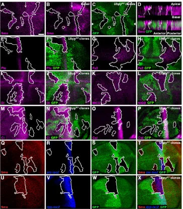
In an in vivoRNAi screen targeting DrosophilaUb-proteasome system (UPS) genes (Du et al., 2011; Zhang et al., 2012), we found that inhibiting Ubpyactivity resulted in larval wing disc deformation and adult fly lethality, consistent with an essential role of Ubpyin the developing wing (Mukai et al., 2010). Correct patterning of adult wing relies on interplay among several signaling systems, including Hh, Notch (N) and Wingless (Wg) signaling. To explore which pathway(s) is regulated by Ubpy, we examined the expression of core components of these pathways in UbpyRNAi-expressing wing discs. Surprisingly, we found that multiple signaling molecules, including Smo and the Hh signaling receptor Patched (Ptc), the N signaling ligand Delta (Dl) and receptor N, and the Wg signaling receptor Frizzled2 (Fz2), were all mislocalized as large puncta in wing epithelial cells (supplementary material Fig. S1A-H). We excluded the possibility that this phenotype was due to RNAi off-target effects as two additional UbpyRNAi targeting distinct regions of Ubpy exhibited the same effect (supplementary material Fig. S1B-F,H). Moreover, we generated UbpyKO (Mukai et al., 2010) somatic clones in wing discs to eliminate Ubpyactivity completely. Consistent with RNAi results, Ptc, Smo, N, Dl and Fz2 accumulated in puncta in Ubpy-null cells (Fig. 1A-L). Similar results were obtained in eye discs, suggesting a general requirement of Ubpy for signaling protein localization (supplementary material Fig. S2A-F).
Our results add another layer of complexity to Ubpy regulation of Smo as other groups observed either no change (Mukai et al., 2010) or reduced Smo expression (Li et al., 2012; Xia et al., 2012) in Ubpy
Ubpy controls the stability of the ESCRT-0 subunit Hrs in
development
Junzheng Zhang1,2, Juan Du1,2, Cong Lei1, Min Liu1,2and Alan Jian Zhu1,2,*
mutant cells. As wing discs are composed of columnar epithelial cells and Smo subcellular localization is biased towards basolateral domains (Denef et al., 2000), we speculate that images acquired from a single focal plane may not faithfully reflect the distribution of actively trafficking protein cargos. Therefore, we re-examined Smo localization in UbpyRNAi and Ubpy-null cells. We found that the reported Smo downregulation (Li et al., 2012; Xia et al., 2012) could only be detected at the basal-most focal plane in the posterior compartment of wing discs (Fig. 1B; supplementary material Fig. S1E). By contrast, in the same cell, Smo aggregates were evident when moving up to apical domains (Fig. 1A; supplementary
[image:2.612.47.411.59.473.2]as well as ptc-lacZ and dpp-lacZ reporters. Surprisingly, the expression of these markers was not obviously affected by Ubpy RNAi in dorsal compartment of wing discs (supplementary material Fig. S1I-L). Note that a slight expansion of ptc-lacZand dpp-lacZ expression domains (<15% penetrance, n>50) was observed in cells in which Ubpy was massively knocked down (supplementary material Fig. S1M,N). Nevertheless, our result is inconsistent with a previous report that UbpyRNAi downregulates Hh signaling when the same condition was applied (Xia et al., 2012). To address this discrepancy, we generated Ubpy-null clones in wing discs. Consistent with our RNAi result, Hh signaling was not altered in
Fig. 1.Ubpyregulates subcellular localization of developmental signaling proteins in wing discs.(A-C) Smo accumulation as puncta in UbpyKOclones
(marked by absence of GFP). Note downregulated Smo at basal-most focal plane in the posterior compartment (B). Anterior-posterior border is indicated by arrows. (D) Optical cross-sections along the anterior-posterior axis (as indicated by arrowheads in C) shows Smo
accumulation in both anteriorally and posteriorally localized clones. (E-L) Similar localization defect for Ptc (E,F), Fz2 (G,H), Dl (I,J) and N (K,L) in wing discs. (M-X) Ci (M,N) and Col (O,P) expression is unaltered in UbpyKOclones. Similarly, no
Developmental signaling proteins are trapped in enlarged endosomal vesicles
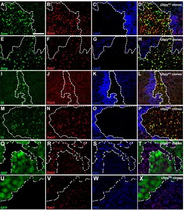
Our observation of mis-localized signaling proteins in UbpyKOcells raises an intriguing possibility that endocytic trafficking itself may be affected. To test this hypothesis, we first examined the distribution of early and late endosomes (marked by Rab5 and Rab7, respectively) in UbpyKOcells. Both Rab5-positive early endosomes and Rab7-postive late endosomes were significantly enlarged in UbpyKOcells compared with those in wild-type cells (Fig. 2A-P). Utilizing an FK2 antibody, we found that ubiquitylated proteins were accumulated in these enlarged vesicles (Fig. 2Q-X). The aberrant appearance of these vesicles indicates that Ubpy is required for a key step in the endocytic pathway.
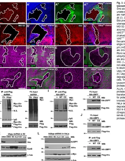
We next used Smo and Dl as examples to examine if accumulated signaling proteins were mis-localized in enlarged vesicles. We found that both Rab5 and Rab7 were present in >80% of Smo- or Dl-containing puncta. This result indicates that sorting of membrane signaling molecules is stalled in enlarged, aberrant vesicles that may have mixed endosomal identities. To explore this possibility further, we co-labeled Smo in UbpyKOclones with another routinely used early endosomal marker, Hrs (Raiborg et al., 2001). To our surprise, Hrs protein was largely undetectable in UbpyKOcells (Fig. 3A-A′′′). This result is in direct contrast to Rab5 accumulation in enlarged
vesicles (Fig. 2), suggesting that Ubpy may directly regulate endocytic sorting at the level of Hrs.
Ubpy protects Hrs from lysosomal degradation
[image:3.612.47.408.328.739.2]To examine if Ubpy regulates Hrs protein stability, we treated wing discs bearing UbpyKOclones with either lysosomal or proteasomal inhibitors. Blocking lysosomal (E64 and chloroquine; Fig. 3C; supplementary material Fig. S4C-E) but not proteasomal (MG132; Fig. 3B; supplementary material Fig. S4B) activity stabilized Hrs, suggesting that Ubpy protects Hrs from lysosomal degradation. To validate our data obtained from pharmacological inhibition, we utilized well-characterized mutant alleles to disrupt proteasome function or specific steps of endocytic lysosomal trafficking. Disrupting the 20S proteasome core component b6 (Pros26) activity by overexpressing dominant-negative DTS5(Belote and Fortier, 2002) failed to rescue UbpyRNAi-induced Hrs degradation (supplementary material Fig. S4H). The DrosophilaVps18 homolog Deep orange (Dor) is a member of the class C Vps/HOPS complex that regulates late endosome-to-lysosome transition (Sevrioukov et al., 1999; Sriram et al., 2003). Consistent with a role of Dor in cargo delivery to lysosomes, Hrs was found accumulated in dor8mutant wing cells (Fig. 3D). Next, we examined Hrs in vps22ZZ13or vps2PP6mutant clones, which disrupt ESCRT-II and -III function, respectively
Fig. 2. Signaling proteins are trapped in aberrant vesicles in UbpyKOcells. (A-H) Accumulated Smo colocalizes with the early- and late-endosomal markers Rab5 (A-D) and Rab7 (E-H). Note that both Rab5- and Rab7-positive vesicles are enlarged in UbpyKOcells (marked by
absence of lacZ). (I-P) Dl is trapped in aberrant vesicles that are positive for both Rab5 (I-L) and Rab7 (M-P). Overall, >80% of Smo- or Dl-bearing vesicles are positive for Rab5 or Rab7 in UbpyKOcells,
compared with <15% random colocalization in which one of the two images is rotated [n(field of view)=5-7]. (Q-X) In UbpyKO
clones (marked by absence of GFP), ubiquitylated cargos are enriched in enlarged vesicles positive for Rab5 (Q-T) or Rab7 (U-X). Somatic clones are circled by dashed lines. Scale bar: 20 μm.
(Vaccari et al., 2009). In both cases, Hrs accumulated in aggregates,
[image:4.612.54.458.55.572.2]although the size of aggregates in vps2PP6cells (Fig. 3F) was much abundance, localization or activity. The observation that endogenousUbpy removes Ub from its substrates to regulate substrate Fig. 3.Ubpyprotects Hrs from lysosomal degradation.(A-C′) Despite Smo accumulation (A), Hrs (A′) is largely undetectable in UbpyKOclones, which
are marked by lack of GFP (A″) or lacZ (B′,C′). Disrupting lysosome function by E64 prevents Hrs degradation (C), whereas inhibiting the UPS function by MG132 (B) has no effect. (D-G′) Hrs accumulation as puncta in dor8(D),
vps22ZZ13(E) or vps2PP6(F) clones
(marked by lack of GFP). UbpyKO,
vps2PP6double mutant clones (G) show
the same phenotype as that of vps2PP6
Hrs was stable (supplementary material Fig. S5B). However, Hrs accumulation was greatly reduced when Ubpywas depleted by RNAi (Fig. 3K). Interestingly, this UbpyRNAi-mediated Hrs degradation is independent of Hrs association with vesicle membrane or ESCRT-I (supplementary material Fig. S5D). Consistent with in vivoresults, Hrs was protected from degradation when lysosomal activity was inhibited in S2 cells (Fig. 3K). Previous studies show that disrupted Ubpy activity leads to moderate reduction of Hrs expression in vertebrate cells (Row et al., 2006; Niendorf et al., 2007; Berlin et al., 2010). However, the physiological relevance and molecular nature of the Ubpy regulation on Hrs is not known. We found that shRNA knockdown of human UBPY resulted in lysosomal degradation of Hrs in HeLa cells (Fig. 3L), highlighting a conserved function of Ubpy on Hrs stability that may be important for the ESCRT-0 homeostasis on endosomal sorting.
Loss of Hrs activity mimics UbpyKOdefects in Drosophila Hrs-mediated ESCRT-0 initiates cargo sorting on endosomes and is required for Drosophiladevelopment (Lloyd et al., 2002; Seto and Bellen, 2006; Chanut-Delalande et al., 2010). Our biochemical data suggest that Hrs serves as a Ubpy substrate. Thus, reduced Ubpy activity in wing discs may result in defective ESCRT-0 activity and membrane protein cargo sorting. If this were true, we would expect that removing Hrsactivity would mimic the endosomal defects observed in UbpyKOcells. Indeed, signaling molecules accumulated
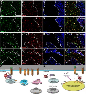
in enlarged vesicles in hrsD28clones induced in wing (supplementary material Fig. S6) and eye discs (supplementary material Fig. S2J-O). Similarly, these vesicles were positive for both Rab5 and Rab7 (Fig. 4A-P). Our observation is consistent with a report that signaling molecules are mis-localized in hrsD28egg chamber cells (Jékely and Rørth, 2003).
[image:5.612.46.409.341.739.2]The canonical role of Ubpy in endocytic sorting is to regulate cargo protein ubiquitylation. Indeed, overexpressed Ubpysuffices to modulate cargo ubiquitylation, leading to cargo stabilization, including Smo and Fz2 (supplementary material Fig. S7) (Mukai et al., 2010; Li et al., 2012; Xia et al., 2012). Our genetic and biochemical analyses led us to propose an additional role of Ubpy in Hrs stabilization in order to maintain the ESCRT-0 homeostasis essential for subsequent cargo sorting towards lysosomes (Fig. 4Q). In the absence of Ubpy, ubiquitylated Hrs (Ub-Hrs) may still function to mediate the formation of internal vesicles as incorporation of both Hrs and cargos into enlarged vesicles was observed (Fig. 2; supplementary material Fig. S8H). But because Hrs is not deubiquitylated, incorporated Ub-Hrs is ultimately degraded. Unlike Ub-Hrs, cargos are trapped in enlarged vesicles (Fig. 4R). These seemingly paradoxical fates of Hrs and cargo proteins could be explained when we consider complex roles of Ubpy and Hrs in endosomal sorting. Ubpy deubiquitylation in early endosomes is required for displacement of ESCRT-0 by ESCRT-III, which facilitates cargo sorting into MVBs (Hasdemir et al., 2009;
Fig. 4. Signaling proteins are trapped in enlarged endosomes in hrsD28cells. (A-P) Accumulated Smo colocalizes with Rab5 (A-D) and Rab7 (E-H), forming enlarged, aberrant endosomal vesicles in hrsD28clones (marked by absence of lacZ).
Similarly, Dl colocalizes with Rab5 (I-L) and Rab7 (M-P). Overall, >80% of Smo- or Dl-bearing vesicles are positive for Rab5 or Rab7 in hrsD28cells [n(field of view)=5-7].
Somatic clones are circled by dashed lines. Scale bar: 20 μm. (Q,R) Shown is a dual role model of Ubpy functioning in endocytic trafficking. Apart from the canonical role of Ubpy on ubiquitylated cargos in early endosomes, Ubpy may play an addition role in protecting Hrs from ESCRT-mediated lysosomal degradation (Q). In the absence of Ubpy, Ub-Hrs may still be capable of mediating endocytic sorting. As the result, incorporated Ub-Hrs is degraded in the lysosome. Upon Hrs depletion, cargo proteins are no longer able to complete endosomeal trafficking for degradation, leading to cargo accumulation in enlarged vesicles with mixed endosomal identity (R).
Ali et al., 2013). In the absence of Ubpy, Ub-Hrs might be inefficient to sort its cargos into MVBs due to its reduced ability to interact with cargos (Polo et al., 2002; Miller et al., 2004). Furthermore, upon Hrs depletion, endosomal sorting may also be stalled. Thus, cargos may fail to complete endosomal sorting for degradation, resulting in cargo accumulation in enlarged vesicles. At the same time, Ub-Hrs may be recognized by as yet unknown factors that facilitate Hrs degradation in the lysosome. Further experiments are needed to explore these two possibilities.
Hrs degradation upon Ubpydepletion seems to have occurred earlier than cargo accumulation in enlarged vesicles (supplementary material Fig. S8A-F), suggesting that these two events might take place sequentially. This interpretation is consistent with aberrant endosomal vesicles associated with reduced Hrs activity (Fig. 4) (Komada and Soriano, 1999; Kanazawa et al., 2003; Hanyaloglu et al., 2005; Lu and Bilder, 2005; Rives et al., 2006; Raiborg et al., 2008; Chanut-Delalande et al., 2010; Li et al., 2012). The Hrsand Ubpymutant phenotypes are intriguing in the context of current understanding of sequential ESCRT actions in cargo sorting. Hrs-mediated ESCRT-0 recruits ESCRT-I through interactions with Tsg101 (Bache et al., 2003; Katzmann et al., 2003; Lu et al., 2003). However, reduced Tsg101 activity results in a different phenotype; MVB formation is inhibited (Doyotte et al., 2005; Razi and Futter, 2006). We believe that Hrs and Ubpy must play additional roles other than simply recruiting ESCRT-I. This notion is supported by a recent report that ESCRT-0 and Ubpy work together with ESCRT-III to facilitate cargo sorting to MVBs (Ali et al., 2013).
MATERIALS AND METHODS Fly genetics
ap-Gal4 (Du et al., 2011), MS1096-Gal4 (Zhang et al., 2012), ptc-lacZ, dpp-lacZ(Su et al., 2011), dor8(gift of Helmut Krämer) (Sevrioukov et al., 1999), hrsD28(gift of Hugo Bellen) (Lloyd et al., 2002), UbpyKO(gift of Satoshi Goto)
(Mukai et al., 2010), vps2PP6and vps22ZZ13(gift of David Bilder) (Vaccari et
al., 2009) alleles were described previously. Transgenic RNAi flies targeting different regions of Ubpy gene were obtained from Dr Satoshi Goto (Mitsubishi-Kagaku Institute of Life Sciences, Machida, Japan) (5798R-1 and 5798R-2) and the Vienna DrosophilaRNAi Center (VDRC #107623).
UAS-DTS5(Bloomington #6786), HrsRNAi (TRiP #28026) and Vps25RNAi (VDRC #38821) transgenic flies were obtained from Bloomington and VDRC, respectively. UAS-FLAG-Ubpy transgenic fly was a gift of Dr Jianhang Jia (Xia et al., 2012).
All fly crosses were maintained at 25°C unless noted otherwise. UbpyKO
and vps2PP6alleles were recombined to chromosome III by homologous
recombination. Loss-of-function somatic clones were induced in the wing and eye discs by Flp/FRT-mediated homologous recombination; second-instar larvae from parental crosses were heat-shocked at 37°C for one hour. Specific fly strains and cross conditions as shown in figures are listed in supplementary material Table S1. All phenotypes in wing and eye discs are fully penetrant (n>20) except for those shown in supplementary material
Rab7 (1:2000; gift of Akira Nakamura, Institute of Molecular Embryology and Genetics, Kumamoto, Japan), mouse anti-Smo (1:20; 20C6; DSHB), chicken anti β-galactosidase (1:200; ICL Lab), rabbit anti β-galactosidase (1:4000; Cappel) and mouse anti-ubiquitylated proteins (1:1000; FK2; Enzo). Alexa Fluor-conjugated secondary antibodies (1:400; Invitrogen) were used. In some experiments, third-instar larvae were dissected and incubated at 25°C for 4 hours in complete Drosophilaclone-8 cell medium [Shields and Sang M3 insect medium (Sigma) supplemented with 2% heat-inactivated fetal bovine serum (FBS; Invitrogen), 5 mg/ml insulin (Sigma) and 2.5% fly extract, 100 U/ml penicillin and 100 mg/ml streptomycin] supplemented with either lysosomal [E64 (50 μM; Sigma); chloroquine (10 mg/ml; MP Biomedicals)] or proteasomal [MG132 (50 μM; Sigma)] inhibitors before fixation. Fluorescence images were acquired with a Zeiss Axio Imager Z1 microscope equipped with an ApoTome or a Leica SP5 confocal microscope. The figures were assembled in Adobe Photoshop CS5. Minor image adjustments (brightness and/or contrast) were performed in AxioVision 4.8.1 or Adobe Photoshop.
Cell culture, transfection and RNAi treatment
DrosophilaSchneider S2 cells were cultured in Schneider’s Drosophila
Medium (Invitrogen) supplemented with 10% FBS, 100 U/ml penicillin and 100 mg/ml streptomycin at 25°C. DNA transfection was carried out using a standard calcium phosphate protocol. FLAG-Hrsplasmid was generated by fusing an FLAG tag at the N-terminus of the full-length fly HrscDNA and then cloned into a pUAST vector. Hrs mutants lacking the FYVE motif and PSAP domain, respectively, were generated by PCR. Wild-type (WT) and catalytically dead (CS) HA-Ubpyplasmids were gifts of Dr Jianhang Jia (Xia et al., 2012). Myc-Ubplasmid was provided by Dr Shunsuke Ishii (Dai et al., 2003).
dsRNA was generated using the MEGAscript High Yield Transcription Kit (Ambion) according to the manufacturer’s instructions. A DNA template targeting Ubpy(encoding amino acids 333-435) was generated by PCR and used for dsRNA synthesis. dsRNA targeting yeast gal80coding sequence was used as a negative control (Su et al., 2011). For RNAi knockdown, S2 cells were cultured in full medium containing 40 nM indicated dsRNA for 4 days. dsRNA-treated cells were then split and incubated with fresh dsRNA for additional 4 days before harvesting for further experiments.
HeLa cells were cultured in DMEM (Invitrogen) supplemented with 10% FBS, 100 U/ml penicillin and 100 mg/ml streptomycin at 37°C. Lipofectamine 2000 (Invitrogen) was used for transfection. RNAi knockdown of human UBPY was achieved by transfecting a shRNA plasmid (TRCN0000007436, Sigma) that has been shown to specifically target UBPYin HeLa cells.
In both S2 and HeLa cells, MG132 (50 μM; Sigma) and ALLN (50 μM; Sigma) were used to inhibit the proteasome activity, and E64 (50 μM; Sigma) and leupeptin (50 μM; Sigma) were used to inhibit lysosome function.
Immunoblotting, immunoprecipitation and ubiquitylation assays
hot-lysed in 100 μl of denaturing buffer (1% SDS, 50 mM Tris, pH 7.5, 0.5 mM EDTA) by boiling for five minutes at 100°C. Lysates were then diluted 1:10 with NP-40 lysis buffer and subjected to immunoprecipitation using anti-Flag M2 affinity gel (Sigma).
Acknowledgements
We thank Hugo Bellen, David Bilder, Marcos González-Gaitán, Satoshi Goto, Shunsuke Ishii, Jianhang Jia, Helmut Krämer, Cheng-Yu Lee, Akira Nakamura, Alain Vincent, and the Bloomington DrosophilaStock Center, the Developmental Studies Hybridoma Bank (DSHB) and the Vienna DrosophilaRNAi Center (VDRC) for fly stocks, antibodies and plasmids.
Competing interests
The authors declare no competing financial interests.
Author contributions
J.Z. and A.J.Z. designed experiments; J.Z., J.D., C.L. and M.L. performed experiments; J.Z. and A.J.Z. analyzed the data and wrote the manuscript.
Funding
This work was supported by a National Institutes of Health grant [R01GM085175 to A.J.Z.]; a Ministry of Science and Technology of China grant [2014CB942802 to A.J.Z.]; a National Natural Science Foundation of China grant [31371410 to A.J.Z.]; an American Heart Association Postdoctoral Fellowship [10POST4110011 to J.Z.]; and a Peking-Tsinghua Center for Life Sciences Postdoctoral Fellowship (to M.L.). Deposited in PMC for release after 12 months.
Supplementary material
Supplementary material available online at
http://dev.biologists.org/lookup/suppl/doi:10.1242/dev.099564/-/DC1
References
Ali, N., Zhang, L., Taylor, S., Mironov, A., Urbé, S. and Woodman, P.(2013). Recruitment of UBPY and ESCRT exchange drive HD-PTP-dependent sorting of EGFR to the MVB. Curr. Biol.23, 453-461.
Alwan, H. A. and van Leeuwen, J. E.(2007). UBPY-mediated epidermal growth factor receptor (EGFR) de-ubiquitination promotes EGFR degradation. J. Biol. Chem.282, 1658-1669.
Bache, K. G., Brech, A., Mehlum, A. and Stenmark, H. (2003). Hrs regulates multivesicular body formation via ESCRT recruitment to endosomes. J. Cell Biol.
162, 435-442.
Belote, J. M. and Fortier, E. (2002). Targeted expression of dominant negative proteasome mutants in Drosophila melanogaster. Genesis34, 80-82.
Berlin, I., Higginbotham, K. M., Dise, R. S., Sierra, M. I. and Nash, P. D.(2010). The deubiquitinating enzyme USP8 promotes trafficking and degradation of the chemokine receptor 4 at the sorting endosome. J. Biol. Chem.285, 37895-37908. Bowers, K., Piper, S. C., Edeling, M. A., Gray, S. R., Owen, D. J., Lehner, P. J. and
Luzio, J. P.(2006). Degradation of endocytosed epidermal growth factor and virally ubiquitinated major histocompatibility complex class I is independent of mammalian ESCRTII. J. Biol. Chem.281, 5094-5105.
Chanut-Delalande, H., Jung, A. C., Baer, M. M., Lin, L., Payre, F. and Affolter, M. (2010). The Hrs/Stam complex acts as a positive and negative regulator of RTK signaling during Drosophila development. PLoS ONE5, e10245.
Clague, M. J., Liu, H. and Urbé, S.(2012). Governance of endocytic trafficking and signaling by reversible ubiquitylation. Dev. Cell23, 457-467.
Dai, P., Akimaru, H. and Ishii, S. (2003). A hedgehog-responsive region in the Drosophila wing disc is defined by debra-mediated ubiquitination and lysosomal degradation of Ci. Dev. Cell4, 917-928.
Denef, N., Neubüser, D., Perez, L. and Cohen, S. M.(2000). Hedgehog induces opposite changes in turnover and subcellular localization of patched and smoothened. Cell102, 521-531.
Doyotte, A., Russell, M. R., Hopkins, C. R. and Woodman, P. G.(2005). Depletion of TSG101 forms a mammalian “Class E” compartment: a multicisternal early endosome with multiple sorting defects. J. Cell Sci.118, 3003-3017.
Du, J., Zhang, J., Su, Y., Liu, M., Ospina, J. K., Yang, S. and Zhu, A. J.(2011). In vivo RNAi screen reveals neddylation genes as novel regulators of Hedgehog signaling. PLoS ONE6, e24168.
Gregory, M. A. and Hann, S. R.(2000). c-Myc proteolysis by the ubiquitin-proteasome pathway: stabilization of c-Myc in Burkitt’s lymphoma cells. Mol. Cell. Biol.20, 2423-2435.
Hanyaloglu, A. C., McCullagh, E. and von Zastrow, M.(2005). Essential role of Hrs in a recycling mechanism mediating functional resensitization of cell signaling.
EMBO J.24, 2265-2283.
Hasdemir, B., Murphy, J. E., Cottrell, G. S. and Bunnett, N. W.(2009). Endosomal deubiquitinating enzymes control ubiquitination and down-regulation of protease-activated receptor 2. J. Biol. Chem.284, 28453-28466.
Henne, W. M., Buchkovich, N. J. and Emr, S. D.(2011). The ESCRT pathway. Dev. Cell21, 77-91.
Jékely, G. and Rørth, P.(2003). Hrs mediates downregulation of multiple signalling receptors in Drosophila. EMBO Rep.4, 1163-1168.
Kanazawa, C., Morita, E., Yamada, M., Ishii, N., Miura, S., Asao, H., Yoshimori, T. and Sugamura, K.(2003). Effects of deficiencies of STAMs and Hrs, mammalian class E Vps proteins, on receptor downregulation. Biochem. Biophys. Res. Commun.309, 848-856.
Katzmann, D. J., Stefan, C. J., Babst, M. and Emr, S. D.(2003). Vps27 recruits ESCRT machinery to endosomes during MVB sorting. J. Cell Biol.162, 413-423. Komada, M. and Soriano, P.(1999). Hrs, a FYVE finger protein localized to early
endosomes, is implicated in vesicular traffic and required for ventral folding morphogenesis. Genes Dev.13, 1475-1485.
Li, S., Chen, Y., Shi, Q., Yue, T., Wang, B. and Jiang, J.(2012). Hedgehog-regulated ubiquitination controls smoothened trafficking and cell surface expression in Drosophila. PLoS Biol.10, e1001239.
Lloyd, T. E., Atkinson, R., Wu, M. N., Zhou, Y., Pennetta, G. and Bellen, H. J. (2002). Hrs regulates endosome membrane invagination and tyrosine kinase receptor signaling in Drosophila. Cell108, 261-269.
Lu, H. and Bilder, D.(2005). Endocytic control of epithelial polarity and proliferation in Drosophila. Nat. Cell Biol.7, 1232-1239.
Lu, Q., Hope, L. W., Brasch, M., Reinhard, C. and Cohen, S. N.(2003). TSG101 interaction with HRS mediates endosomal trafficking and receptor down-regulation.
Proc. Natl. Acad. Sci. USA100, 7626-7631.
Miller, S. L. H., Malotky, E. and O’Bryan, J. P.(2004). Analysis of the role of ubiquitin-interacting motifs in ubiquitin binding and ubiquitylation. J. Biol. Chem. 279, 33528-33537.
Mizuno, E., Iura, T., Mukai, A., Yoshimori, T., Kitamura, N. and Komada, M.(2005). Regulation of epidermal growth factor receptor down-regulation by UBPY-mediated deubiquitination at endosomes. Mol. Biol. Cell16, 5163-5174.
Mizuno, E., Kobayashi, K., Yamamoto, A., Kitamura, N. and Komada, M.(2006). A deubiquitinating enzyme UBPY regulates the level of protein ubiquitination on endosomes. Traffic7, 1017-1031.
Mukai, A., Yamamoto-Hino, M., Awano, W., Watanabe, W., Komada, M. and Goto, S.(2010). Balanced ubiquitylation and deubiquitylation of Frizzled regulate cellular responsiveness to Wg/Wnt. EMBO J.29, 2114-2125.
Niendorf, S., Oksche, A., Kisser, A., Löhler, J., Prinz, M., Schorle, H., Feller, S., Lewitzky, M., Horak, I. and Knobeloch, K. P.(2007). Essential role of ubiquitin-specific protease 8 for receptor tyrosine kinase stability and endocytic trafficking in vivo. Mol. Cell. Biol.27, 5029-5039.
Polo, S., Sigismund, S., Faretta, M., Guidi, M., Capua, M. R., Bossi, G., Chen, H., De Camilli, P. and Di Fiore, P. P.(2002). A single motif responsible for ubiquitin recognition and monoubiquitination in endocytic proteins. Nature416, 451-455. Raiborg, C. and Stenmark, H.(2009). The ESCRT machinery in endosomal sorting of
ubiquitylated membrane proteins. Nature458, 445-452.
Raiborg, C., Bremnes, B., Mehlum, A., Gillooly, D. J., D’Arrigo, A., Stang, E. and Stenmark, H. (2001). FYVE and coiled-coil domains determine the specific localisation of Hrs to early endosomes. J. Cell Sci.114, 2255-2263.
Raiborg, C., Malerød, L., Pedersen, N. M. and Stenmark, H.(2008). Differential functions of Hrs and ESCRT proteins in endocytic membrane trafficking. Exp. Cell Res.314, 801-813.
Razi, M. and Futter, C. E.(2006). Distinct roles for Tsg101 and Hrs in multivesicular body formation and inward vesiculation. Mol. Biol. Cell17, 3469-3483.
Rives, A. F., Rochlin, K. M., Wehrli, M., Schwartz, S. L. and DiNardo, S.(2006). Endocytic trafficking of Wingless and its receptors, Arrow and DFrizzled-2, in the Drosophila wing. Dev. Biol.293, 268-283.
Row, P. E., Prior, I. A., McCullough, J., Clague, M. J. and Urbé, S.(2006). The ubiquitin isopeptidase UBPY regulates endosomal ubiquitin dynamics and is essential for receptor down-regulation. J. Biol. Chem.281, 12618-12624. Seto, E. S. and Bellen, H. J.(2006). Internalization is required for proper Wingless
signaling in Drosophila melanogaster. J. Cell Biol.173, 95-106.
Sevrioukov, E. A., He, J. P., Moghrabi, N., Sunio, A. and Krämer, H.(1999). A role for the deep orange and carnation eye color genes in lysosomal delivery in Drosophila. Mol. Cell4, 479-486.
Sriram, V., Krishnan, K. S. and Mayor, S.(2003). deep-orange and carnation define distinct stages in late endosomal biogenesis in Drosophila melanogaster. J. Cell Biol.
161, 593-607.
Su, Y., Ospina, J. K., Zhang, J., Michelson, A. P., Schoen, A. M. and Zhu, A. J. (2011). Sequential phosphorylation of smoothened transduces graded hedgehog signaling. Sci. Signal.4, ra43.
Tokumoto, T., Yamashita, M., Tokumoto, M., Katsu, Y., Horiguchi, R., Kajiura, H. and Nagahama, Y.(1997). Initiation of cyclin B degradation by the 26S proteasome upon egg activation. J. Cell Biol.138, 1313-1322.
Torroja, C., Gorfinkiel, N. and Guerrero, I.(2004). Patched controls the Hedgehog gradient by endocytosis in a dynamin-dependent manner, but this internalization does not play a major role in signal transduction. Development131, 2395-2408. Vaccari, T., Rusten, T. E., Menut, L., Nezis, I. P., Brech, A., Stenmark, H. and Bilder,
D.(2009). Comparative analysis of ESCRT-I, ESCRT-II and ESCRT-III function in Drosophila by efficient isolation of ESCRT mutants. J. Cell Sci.122, 2413-2423. Xia, R., Jia, H., Fan, J., Liu, Y. and Jia, J.(2012). USP8 promotes smoothened
signaling by preventing its ubiquitination and changing its subcellular localization.
PLoS Biol.10, e1001238.
Zhang, J., Liu, M., Su, Y., Du, J. and Zhu, A. J.(2012). A targeted in vivo RNAi screen reveals deubiquitinases as new regulators of Notch signaling. G3 (Bethesda)
2, 1563-1575.
Zhu, A. J., Zheng, L., Suyama, K. and Scott, M. P.(2003). Altered localization of Drosophila Smoothened protein activates Hedgehog signal transduction. Genes