R E S E A R C H A R T I C L E
Open Access
Hepatoprotective activity of
Sonchus asper
against
carbon tetrachloride-induced injuries in male rats:
a randomized controlled trial
Rahmat A Khan
1,2*†, Muhammad R Khan
2†, Sumaira Sahreen
2,3†and Naseer Ali Shah
2†Abstract: Background:Sonchus asper(SAME) is used as a folk medicine in hepatic disorders. In this study, the hepatoprotective effects of the methanol extract of SAME was evaluated against carbon tetrachloride (CCl4)-induced liver injuries in rats.
Methods:To evaluate the hepatoprotective effects of SAME, 36 male Sprague–Dawley rats were equally divided into 6 groups. Rats of Group I (control) were given free access to approved feed and water. Rats of Group II were injected intraperitoneally with CCl4(3 ml/kg) as a 30% solution in olive oil (v/v) twice a week for 4 weeks. Animals of Groups III (100 mg/kg) and IV (200 mg/kg) received SAME, whereas those of Group V were given silymarinviagavage (100 mg/ kg) after 48 h of CCl4treatment. Group VI received SAME (200 mg/kg) twice a week for 4 weeks without CCl4
treatment. Various parameters, such as the serum enzyme levels, serum biochemical marker levels, antioxidant enzyme activities, and liver histopathology were used to estimate the hepatoprotective efficacy of SAME.
Results:The administration of SAME and silymarin significantly lowered the CCl4-induced serum levels of hepatic marker enzymes (aspartate aminotransferase, alanine aminotransferase, and lactate dehydrogenase), cholesterol, low-density lipoprotein, and triglycerides while elevating high-density lipoprotein levels. The hepatic contents of glutathione and activities of catalase, superoxide dismutase, glutathione peroxidase, glutathione S-transferase, and glutathione reductase were reduced. The levels of thiobarbituric acid-reactive substances that were increased by CCl4were brought back to control levels by the administration of SAME and silymarin. Liver histopathology showed that SAME reduced the incidence of hepatic lesions induced by CCl4in rats.
Conclusion:SAME may protect the liver against CCl4-induced oxidative damage in rats.
Keywords:Carbon tetrachloride,Sonchus asper, Liver histopathology, Antioxidant, Lipid peroxidation, Liver enzymes
Background
Hepatotoxicity is the most widespread pathology world-wide, representing up to 83% of all cases. Hepatitis, viral infections, food additives, alcohol, toxic industrial che-micals, and air and water pollutants are the major risk factors of liver toxicity. There is increasing evidence that free radicals and reactive oxygen species play a crucial role in various steps that initiate and regulate the pro-gression of liver diseases independently of the original agent [1]. Carbon tetrachloride (CCl4) is a potent
environmental hepatotoxin [2] that, in addition to hep-atic problems, causes dysfunction of the kidneys, lungs, testis, brain, and blood by generating free radicals [3,4].
CCl4 requires bioactivation in phase I of the
cyto-chrome P450 system to form the reactive metabolic trichloromethyl radical (•CCl3) and trichloromethyl per-oxy radical (•OOCCl3). These free radicals can bind with polyunsaturated fatty acids to produce alkoxy (R•) and peroxy radicals (ROO•) that, in turn, generate lipid per-oxides that are highly reactive, change enzyme activity, and finally induce injury or necrosis with corresponding health problems [5,6].
CCl4is known to decrease glutathione (GSH) of phase
II enzymes and reduce antioxidant enzymes and sub-strates to induce oxidative stress, which is an important factor in acute and chronic injuries in various tissues [7,8].
* Correspondence:[email protected]
†Equal contributors 1
Department of Biotechnology, Faculty of Biological Sciences, University of Science and Technology Bannu, Khyber Pakhtunkhwa, Pakistan
2
Department of Biochemistry, Faculty of Biological Sciences, Quaid-I-Azam University Islamabad, Islamabad, Pakistan
Full list of author information is available at the end of the article
Free radicals of CCl4reduce the GSH contents and activ-ities of antioxidant enzymes, leading to hepatic injury [9]. The depletions of these antioxidant enzymes occur sec-ondary to the controlling action against peroxy radicals produced by CCl4. Reactive oxygen species cause oxidative DNA damage in the form of DNA adducts, genetic muta-tion, strand breakage, and chromosomal alterations [10]. Some recent investigations revealed that free radicals increased the number of argyrophilic nucleolar organizer regions (AgNORs) and activity of telomerase in kidney tis-sues [11], caused depletion of cytochrome P450 2E1, and increased the 8-hydroxyl-2 deoxyguanosine concentration [12]. DNA fragmentation causes p53 gene expression, blocks the cell cycle, and gives additional time to repair DNA. However, severe DNA damage triggers apoptosis [13].
Sonchus asper(SAME), locally called Mahtari, is a com-mon herb that grows wildly and abundantly in open fields. Chemical studies of SAME indicated a high content of vita-min C (ascorbic acid) [14], carotenoids, and typeω-3 fatty acids [15,16]. Phenolic compounds, which are secondary metabolites in plants, are one of the most widely oc-curring groups of phytochemicals that exhibit a wide range of physiological properties, such as antioxidant, allergenic, microbial, artherogenic, anti-thrombotic, anti-inflammatory, vasodilatory, and cardiopro-tective effects [17,18]. Chemical characterization of SAME has shown the presence of ionone derivatives of glycosides and sesquiterpene lactone glycosides [19]. These bioactive compounds have been shown to possess strong antioxidant and anti-inflammatory properties [20]. SAME has diuretic, refrigerant, sedative, and antiseptic properties that are used in the treatment of cough, bronchitis, and asthma [21] as well as kidney inflammation [22], and its decoction is used in the treatment of impotence (erectile dysfunction) [23]. With increasing recognition of herbal medicine and phy-totherapy as alternative forms of health care, the objectives of this study were to evaluate the antioxidant and hepato-protective properties of SAME against CCl4-induced hep-atic injuries in Sprague–Dawley male rats.
Methods
Chemicals
Reduced glutathione (GSH), oxidized glutathione (GSSG), glutathione reductase, gamma-glutamyl p-nitroanilide, gly-cylglycine, bovine serum albumin (BSA), 1,2-dithio-bis nitro benzoic acid (DTNB), 1-chloro-2,4-dinitrobenzene (CDNB), reduced nicotinamide adenine dinucleotide phosphate (NADPH), CCl4, flavine adenine dinucleotide (FAD), glucose-6-phosphate, Tween-20, 2,6-dichlorophe-nolindophenol, thiobarbituric acid (TBA), picric acid, sodium tungstate, sodium hydroxide, trichloroacetic acid (TCA) and perchloric acid (PCA) were purchased from Sigma Chemicals Co. USA.
Plant collection
Plants ofSonchus asper at maturity were collected from
Wah Cantt, District Rawalpindi (Pakistan) during June, 2010. Plants were identified by Prof. Dr. Mir Ajab Khan (Dean Faculty of Biological Sciences, Quaid-i-Azam Uni-versity Islamabad, Pakistan) and a specimen was submit-ted vide #147 at Herbarium of Pakistan (Quaid-i-Azam University Islamabad, Pakistan). Whole plant (leaves, stem, flowers, roots and seeds) were shades dried at room temperature for two weeks, chopped, ground mechanically of 1 mm mesh size.
Preparation of plant extracts
Five kg powder ofSonchus asperwas socked in 10 liters of methanol with shaking a number of times. After a week of socking the extract was filtered through Whatmann filter paper # 45, and the filtrate was evaporated through rotary vacuum evaporator to get 407 g of methanol crude extract (SAME), stored at 4°C until further use.
Animals
Six week old, 36 Sprague Dawley male rats (190–200 g)
were provided by National Institute of Health Islamabad and were kept in ordinary cages at room temperature of 25 ± 3°C with a 12 h dark/light cycle. They were allowed to standard laboratory feed and water. The study protocol was approved by Ethical Committee of Quaid-i-Azam University Islamabad for laboratory animal feed and care.
Experimental design
To study the antioxidant effects of SAME, rats were equally divided into 6 groups (6 rats). Group 1 received only vehicles olive oil; (1 ml/kg bw) and DMSO (1 ml/kg bw) and have free access to food materials. Animals of group II, III and IV received CCl43 ml/kg (30% in olive oil; v/v) intraperitoneally twice a week for four weeks.
Group II was treated with CCl4 only while group III
(100 mg/kg bw) and IV received (200 mg/kg bw) SAME (intragastric, in DMSO) after 48 h of CCl4. Rats of group V were given silymarin (intragastric, in DMSO) at a dose
of 100 mg/kg bw, after 48 h of CCl4treatment. Animals
of a group VI were given SAME (200 mg/kg bw) intra-gastrically. Experimental period was of four weeks. After 24 h of the last treatment, all the animals were weighted, sacrificed; collected their blood, weighted and perfuse a liver in ice-cold saline solution. Half of liver tissues was treated with liquid nitrogen for further enzymatic and DNA damage analysis while the other portion was pro-cessed for histology.
Assessment of serum markers
(LDH) and biochemical markers; level of total cholesterol (TC), high-density lipoproteins (HDL), low-density lipopro-teins (LDL) and triglycerides (TG) were estimated by using standard AMP diagnostic kits (Stattogger Strasse 31b 8045 Graz, Austria).
Assessment of antioxidant status
70 mg of liver tissue was homogenized in 10 volumes of
100 mmol KH2PO4 buffer containing 1 mmol EDTA
(pH 7.4) and centrifuged at 12,000 × g for 30 min at 4°C. The supernatant was collected and used for determin-ation of antioxidant status as described below using con-centration of protein estimated following the method of Lowry et al. [24]. Antioxidant status, including activity of catalase [25], superoxide dismutase [26], glutathione-S-transferase assay [27], glutathione reductase [28], glutathione peroxidase [29], reduced glutathione assay [30] and lipid peroxidation assay [31] were performed on various hepatic samples.
DNA fragmentation% assay
DNA fragmentation assay was conducted using the pro-cedure of Wu et al. [32] with some modifications. The liver tissue (50 mg) was homogenized in 10 volumes of a
TE solution pH 8.0 (5 mmol Tris–HCl, 20 mmol EDTA)
and 0.2% triton X-100. 1.0 ml aliquot of each sample was centrifuged at 27,000 × g for 20 min to separate the intact chromatin (pellet, B) from the fragmented DNA (supernatant, T). The pellet and supernatant fractions were assayed for DNA content using a freshly prepared DPA (Diphenylamine) solution for reaction. Optical density was read at 620 nm with (SmartSpecTM Plus
Spectrophotometer catalog # 170–2525)
spectrophotom-eter. The results were expressed as an amount of % frag-mented DNA by the following formula;
%Fragmented DNA¼T100=ðTþBÞ
AgNORs count
Silver staining technique was used according to the Trere et al. [33]. The AgNORs technique was performed on dried slides as follows; unstained fixed slides were dewaxed by dipping for 3 minutes in xylene. After complete removal of wax, the slides were hydrated in de-creasing ethanol concentration (90, 70 and 50%) and washed in distilled water for 10 min and dried in an oven. After drying slides were treated with one drop of colloidal solution (2% gelatin and 1% formic acid) and
two drops of 50% AgNO3solution on the slide and
incu-bated at 35°C for about 8–12 min. The progressive stain-ing was followed under a microscope to get golden colored nuclei and brown/black NORs. Then, the slide was washed in distilled water, treated for 1 min with 1% sodium thiosulphate at room temperature to stop the re-action, and washed in tap water. The cells were exam-ined under a light microscope at 100 × magnifications and number of AgNORs was counted per cell.
Histopathological studies
For microscopic evaluation, liver were fixed in a fixative (absolute ethanol 60%, formaldehyde 30%, and glacial acetic acid 10%) and embedded in paraffin, sectioned at
4 μm and subsequently stained with hematoxylin/eosin.
Sections were studied under a light microscope (DIA-LUX 20 EB) at 40 and 100 magnifications. Slides of all the treated groups were studied and photographed.
Statistical analysis
[image:3.595.56.549.597.704.2]Data were expressed as mean and standard error (SE) and ANOVA test were used to analyze the difference among various treatments, with least significance differ-ence (LSD) at 0.05 and 0.01 as a level of significance. SPSS ver. 14.0 (Chicago, IL, USA) and Microsoft Excel 2007 (Roselle, IL, USA) were used for the statistical and graphical evaluations.
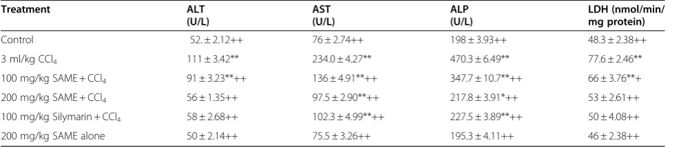
Table 1 Effect of SAME on liver function test in rat
Treatment ALT AST ALP LDH (nmol/min/
(U/L) (U/L) (U/L) mg protein)
Control 52. ± 2.12++ 76 ± 2.74++ 198 ± 3.93++ 48.3 ± 2.38++ 3 ml/kg CCl4 111 ± 3.42** 234.0 ± 4.27** 470.3 ± 6.49** 77.6 ± 2.46**
100 mg/kg SAME + CCl4 91 ± 3.23**++ 136 ± 4.91**++ 347.7 ± 10.7**++ 66 ± 3.76**+
200 mg/kg SAME + CCl4 56 ± 1.35++ 97.5 ± 2.90**++ 217.8 ± 3.91*++ 53 ± 2.61++
100 mg/kg Silymarin + CCl4 58 ± 2.68++ 102.3 ± 4.99**++ 227.5 ± 3.89**++ 50 ± 4.08++
200 mg/kg SAME alone 50 ± 2.14++ 75.5 ± 3.26++ 195.3 ± 4.11++ 46 ± 2.38++
Mean ± SE (n = 6 number).
Results
Effect of SAME on alanine aminotransferase, aspartate aminotransferase, alkaline phosphatase, and lactate dehydrogenase
The levels of certain serum marker enzymes, including ala-nine aminotransferase (ALT), aspartate aminotransferase (AST), alkaline phosphatase (ALP), and lactate dehydrogen-ase (LDH), are highly susceptible to hepatotoxins. They serve as markers of liver damage and oxidative stress, which promote the release of aminotransferases from hepatocytes into the bloodstream. The protective effects of SAME and silymarin on the activity of liver serum marker enzymes are shown in Table 1. Administration of CCl4 to rats signifi-cantly (P<0.01) increased the activity of liver serum mar-ker enzymes compared with controls. The levels of ALT, AST, ALP, and LDH were significantly (P<0.01) and dose-dependently restored by treatment with SAME and
sily-marin compared with the CCl4 group. However,
non-significant variation was observed by SAME treatment alone compared with the control group.
Effect of SAME on triglycerides, total cholesterol, high-density lipoprotein, and low-high-density lipoprotein
The effect of SAME on the cholesterol profile is
shown in Table 2. Administration of CCl4 significantly
(P<0.01) increased the concentration of triglycerides, total cholesterol, and low-density lipoprotein, but decreased the high-density lipoprotein level compared with the control group. The CCl4-induced alteration of the cholesterol profile was significantly (P<0.01) restored with SAME treatment at both doses (100 mg/kg and 200 mg/kg) compared with the control group. Silymarin resulted in a degree of recovery of the cholesterol profile comparative with that achieved with the high dose of SAME.
Effect of SAME on antioxidant and lipid peroxidation enzymes in liver
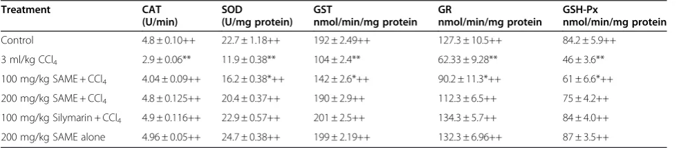
The toxicity of CCl4 significantly decreased the liver
catalase, superoxide dismutase, GSH S-transferase, GSH peroxidase (GSH-Px), and GSH reductase levels of the control group (Table 3). Addition of SAME at doses of 100 and 200 mg/kg and silymarin at 100 mg/kg reversed the decreased hepatic catalase and superoxide dismutase levels. In particular, the high
dose of SAME (200 mg/kg) almost completely
[image:4.595.57.539.100.207.2]restored the catalase and superoxide dismutase activ-ities to the control levels. The hepatic levels of GSH S-transferase, GSH-Px, and GSH reductase were par-tially reversed by SAME at 200 mg/kg.
Table 2 Effect SAME on serum level of biochemical markers in rat
Treatment Triglycerides
(mg/dl)
Total cholesterol (mg/dl)
HDL (mg/dl)
LDL (mg/dl)
Control 7.8 ± 0.45++ 6.1 ± 0.25++ 3.6 ± 0.21++ 2.48 ± 0.32++ 3 ml/kg CCl4 11.3 ± 0.58** 11.2 ± 0.23** 2.8 ± 0.18** 8.4 ± 0.17**
100 mg/kg SAME + CCl4 9.2 ± 0.41**++ 7.7 ± 0.21**++ 3.08 ± 0.09**++ 4.2 ± 0.21**++
200 mg/kg SAME + CCl4 8.3 ± 0.18++ 6.4 ± 0.27++ 3.5 ± 0.20++ 2..53 ± 0.35++
100 mg/kg Silymarin + CCl4 7.2 ± 0.44++ 5.7 ± 0.19++ 3.7 ± 0.21++ 2.21 ± 0.31++
200 mg/kg SAME alone 7.5 ± 0.44++ 5.8 ± 0.44++ 3.75 ± 0.21++ 2.36 ± 0.12++
Mean ± SE (n = 6 number).
** Significance from the control group atP<0.01 probability level. ++ Significance from the CCl4group atP<0.01 probability level.
Table 3 Effect of SAME on antioxidant defense system in rat liver
Treatment CAT SOD GST GR GSH-Px
(U/min) (U/mg protein) nmol/min/mg protein nmol/min/mg protein nmol/min/mg protein
Control 4.8 ± 0.10++ 22.7 ± 1.18++ 192 ± 2.49++ 127.3 ± 10.5++ 84.2 ± 5.9++ 3 ml/kg CCl4 2.9 ± 0.06** 11.9 ± 0.38** 104 ± 2.4** 62.33 ± 9.28** 46 ± 3.6**
100 mg/kg SAME + CCl4 4.04 ± 0.09++ 16.2 ± 0.38*++ 142 ± 2.6*++ 90.2 ± 11.3*++ 61 ± 6.6*++
200 mg/kg SAME + CCl4 4.8 ± 0.125++ 20.4 ± 0.37++ 190 ± 2.9++ 112.3 ± 6.5++ 75 ± 4.2++
100 mg/kg Silymarin + CCl4 4.9 ± 0.116++ 22.9 ± 0.57++ 201 ± 2.5++ 134.3 ± 5.7++ 84 ± 4.0++
200 mg/kg SAME alone 4.96 ± 0.05++ 24.7 ± 0.38++ 199 ± 2.19++ 132.3 ± 6.96++ 87 ± 3.5++
Mean ± SE (n = 6 number).
[image:4.595.56.549.601.708.2]Effect of SAME on GSH and thiobarbituric acid-reactive substances in liver
The hepatic level of GSH was decreased while the thiobarbituric acid-reactive substances (TBARS) were increased in the CCl4-treated animals compared with the controls (Table 4). The CCl4-induced changes in these parameters were dose-dependently diminished by oral addition of various doses of SAME and sily-marin. No significant changes in GSH or TBARS were found with SAME alone compared with control rats.
Effect of SAME on body weight, absolute liver weight, relative liver weight, % DNA fragmentation, and AgNORs in rat liver
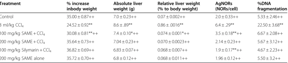
The NORs stained by silver ion and the AgNOR-associated proteins, called AgNOR proteins, play a sig-nificant role in differentiation of tumor cells from nor-mal cells, as shown along with increases in liver weight, relative liver weight, % DNA fragmentation, and % increases in body weight (Table 5). Administra-tion of CCl4 significantly (P<0.01) amplified the num-ber of NORs per cell, increased the liver weight, increased the relative liver weight, increased the % DNA fragmentation, and decreased the % increase in
body weight compared with untreated rats. However, 50 mg/kg of silymarin and various doses of SAME augmented the CCl4-induced intoxication and
signifi-cantly (P<0.01) recovered the NORs per cell, body
weight, liver weight, and relative liver weight. The %
DNA fragmentation was markedly (P<0.01) decreased.
Effect of SAME on liver histology
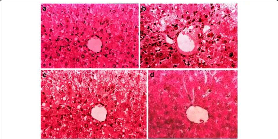
Histopathological investigation results showed significant correlations with the biochemical study results. The graded observations of hematoxylin and eosin-stained sec-tions are shown in Table 6 and Figure 1. Administration of CCl4caused marked increases in fatty changes and cellular hypertrophy; cellular aggregation caused benign tumor formation, and inflammatory cell infiltration, ballooning, and congestion of blood vessels were observed compared with controls. Administration of SAME attenuated the hepatic injuries with little or no fatty changes; dilation of blood vessels and uniform hepatocyte morphology nearly identical to that of the control group was found. Similar observations were recorded after silymarin treatment. These results suggest a positive correlation of SAME and silymarin with the activities of serum aminotransferases and hepatic lipid peroxidation and hepatic antioxidant enzymes.
Discussion
[image:5.595.56.292.114.219.2]A number of drugs, toxic industrial chemicals, and viral infections have been reported to cause severe hepatic in-juries, which are sometimes difficult to manage by medical therapies. It is important to evaluate plant extracts that can be used for improved treatment of hepatic failure caused by severe oxidative stress and necrosis [34]. Estima-tion of serum enzymes is a useful quantitative marker of the extent and type of hepatocellular damage. Increases in serum AST, ALT, ALP, and LDH levels have been attribu-ted to damaged structural integrity of the liver, because these are cytoplasmic in location and are released into the circulation after autolytic breakdown or cellular necrosis
Table 4 Effect SAME on TBARS and GSH contents in rat liver
Treatment GSH (μmol/g tissue) TBARS (nmol /min /mg protein)
Control 1.23 ± 0.02++ 28.33 ± 1.12++ 3 ml/kg CCl4 0.60 ± .012** 52.17 ± 2.61**
100 mg/kg SAME + CCl4 1.09 ± 0.02**++ 43.0 ± 2.76**++
200 mg/kg SAME + CCl4 1.81 ± 0.50++ 30.0 ± 1.77++
100 mg/kg Silymarin + CCl4 1.27 ± 0.013++ 27.17 ± 1.22++
200 mg/kg SAME alone 1.25 ± 0.018++ 27.50 ± 1.82++
Mean ± SE (n = 6 number).
** Significance from the control group atP<0.01 probability level. ++ Significance from the CCl4group atP<0.01 probability level.
Table 5 Effect of SAME on absolute liver weight, relative liver weight, and % DNA fragmentation and AgNORs in rat liver
Treatment % increase
inbody weight
Absolute liver weight (g)
Relative liver weight (% to body weight)
AgNORs (NORs/cell)
%DNA fragmentation
Control 35.00 ± 0.87++ 7.0 ± 0.23++ 0.07 ± 0.002++ 2.0 ± 0.33++ 5.33 ± 2.46++ 3 ml/kg CCl4 24.52 ± 0.92** 8.6 ± .89** 0.86 ± .0016** 6.4 ± .29** 22.50 ± 3.68**
100 mg/kg SAME + CCl4 30.08 ± 0.81**++ 7.4 ± 0.10*++ 0.074 ± 0.001*++ 3.5 ± 0.18**++ 6.67 ± 2.08++
200 mg/kg SAME + CCl4 35.64 ± 0.73++ 7.04 ± 0.23++ 0.070 ± 0.0023++ 2.14 ± 0.23++ 5.67 ± 3.12++
100 mg/kg Silymarin + CCl4 36.82 ± 0.69++ 6.83 ± 0.07++ 0.068 ± 0.007++ 1.9 ± 0.17**++ 4.67 ± 2.23++
200 mg/kg SAME alone 35.72 ± 0.70++ 6.8 ± 0.12++ 0.068 ± 0.011++ 1.96 ± 0.12++ 5.50 ± 3.2++
Mean ± SE (n = 6 number).
[image:5.595.58.540.602.708.2][35]. Therefore, marked release of AST, ALT, ALP, and LDH into the circulation indicates severe damage to hep-atic tissue membranes during CCl4intoxication [36]. The reversal of increased serum enzymes in CCl4-induced liver damage by SAME may occur secondary to prevention of leakage of intracellular enzymes by the membrane stabilization and antioxidant activity of SAME; this was supported by the histological results in the present study. Thus, the anti-lipid peroxidation and/or adaptive nature of the systems brought about by SAME acted against the damaging effects of free radicals produced by CCl4. Similar hepatoprotective effects have been described forS. arvensis [37]. High levels of serum cholesterol, low-density lipopro-tein, and triglycerides and lower concentrations of high-density lipoprotein were induced with CCl4 in rats, con-sistent with results of other studies [36,38,39].
Catalase, superoxide dismutase, and GSH-Px constitute a “mutually supportive team” of antioxidant defenses and play a key role in detoxification of free radicals [40,41]. Ad-ministration of CCl4into rat livers increased lipid peroxida-tion, resulting in accumulation of superoxide radicals and consequently decreased their activities in the liver [42]. Our data revealed that CCl4 treatment significantly decreased the activities of catalase, superoxide dis mutase, GSH-Px, and GSH reductase in liver tissues. Co-administration of
SAME markedly decreased the toxicity of CCl4and
[image:6.595.57.543.99.207.2]enzym-atic activities. Ameliorating effects of different plant metabolites on these enzymes against the toxicity of CCl4 have also been documented [42]. TBARS is a major reactive aldehyde resulting from the peroxidation of polyunsatur-ated fatty acids. It is a useful indicator of tissue damage, in-cluding a series of chain reactions [43]. High hepatic levels
Figure 1(a) Hemotoxylin and Eosin-stained liver section of control rat (b) Liver section of CCl4treated rats showing marked fatty
[image:6.595.57.538.454.695.2]changes, cellular hypertrophy, degeneration of the lobular architecture and blood vessel congestion with disturbed epithelium (c) Liver section of CCl4+ SAME showing less injuries (d) Liver section of CCl4+ Silymarin showing less injuries.
Table 6 Effects of SAME on histology of liver
Treatment Fatty changes Cellular aggregation
hypertrophy
Blood vessel congestion
Inflammatory cells infiltrations
Control - - -
-3 ml/kg CCl4 +++ ++ ++ +++
100 mg/kg SAME + CCl4 - −/+ - −/+
200 mg/kg SAME + CCl4 - - -
-100 mg/kg Silymarin + CCl4 - - -
-200 mg/kg SAME alone - - -
of TBARS and nitrite with CCl4treatment indicate exten-sive oxidative damage to liver tissues. Restoration of nitrite and TBARS with SAME may suggest a role of SAME in controlling various pathological conditions, including oxi-dative stress [20]. Histopathologic results in the present study revealed that CCl4 induces extensive fatty change, blood vessel congestion, cellular hypertrophy, necrotic foci, destruction of the lobular architecture, fibrosis, and nuclear degeneration in some areas, all of which were markedly
diminished by induction of SAME.These data are in good
agreement with the results of the serum aminotransferase activities and hepatic lipid peroxidation levels. Similar pro-tection was evident with respect to necrosis, which is a more severe form of injury, in that it was minimized by treatment with SAME. Other studies on the protective effect of Carissa opacaextract against CCl4-induced hep-atotoxicity in rats have revealed similar findings that are in agreement with the present findings [44].
Conclusions
This study evaluated the pharmacological use of SAME against CCl4induced liver injuries. Low lipid peroxidation and increased activities of various antioxidant enzymes in-dicate that SAME is able to protect various pathological conditions, including oxidative stress. It is suggested, to iso-late the antioxidants from SAME that could be utilized as therapeutics against the oxidative diseases.
Competing interests
The authors declare that they have no competing interests.
Author details
1Department of Biotechnology, Faculty of Biological Sciences, University of
Science and Technology Bannu, Khyber Pakhtunkhwa, Pakistan.2Department of Biochemistry, Faculty of Biological Sciences, Quaid-I-Azam University Islamabad, Islamabad, Pakistan.3Botanical Sciences Division, Pakistan Museum of Natural History, Islamabad, Pakistan.
Authors’contributions
RAK made significant contribution to acquisition of data, analysis, drafting of the manuscript. MRK SS and NAS have made substantial contribution to conception and design, interpretation of data, drafting and revising the manuscript for intellectual content. All authors read and approved the final manuscript.
Received: 13 December 2011 Accepted: 9 July 2012 Published: 9 July 2012
References
1. Jemal A, Siegel R, Ward E:Cancer statistics.Cancer J Clin2007,57:43–66. 2. Guven A, Guven U, Gulmez M:The effect of kefir on the activities of GSH,
GSHPX, GST, CAT, GSH and LPO levels in carbon tetrachloride-induced mice tissue.J Vet Med2003,50:412–416.
3. Ozturk F, Ucar M, Ozturk IC, Vardi N, Batcioglu K:Carbon tetrachloride induced nephrotoxicity and protective effect of betaine in Sprague Dawley rats.Urology2003,62:353–356.
4. Khan MR, Rizvi W, Khan GN, Khan RA, Shaheen S:Carbon tetrachloride induced nephrotoxicity in rat: Protective role ofDigera muricata.
J Ethnopharmacol2009,122:91–99.
5. Weber LW, Boll M, Stampfl A:Hepatotoxicity and mechanism of action of haloalkanes: carbon tetrachloride as a toxicological model.Cr Rev Toxicol
2003,33:105–136.
6. Ogeturk M, Kus I, Colakoglu N, Zararsiz I, Ilhan N, Sarsilmaz M:Caffeic acid phenyl ester protects kidney against carbon tetrachloride toxicity in rats.
J Ethnopharmacol2005,97:273–280.
7. Szymonik-lesiuk S, Czechowska G, Stryjecka-Zimmer M, Słomka M, Madro A, Celinski K, Wielosz M:Catalase, superoxide dismutase, and glutathione peroxidase activities in various rat tissues after carbon tetrachloride intoxication.J Hepato-Biliary-Pancreatic Surg2003,10:309–315. 8. Preethi KC, Kuttan R:Hepato and reno protective action ofCalendula
officinalisL. flower extract.Indian J Exp Biol2009,47:163–168.
9. Ichi I, Kamikawa C, Nakagawa T, Kobayashi K, Kataoka R, Nagata E, Kitamura Y, Nakazaki C, Matsura T, Kojo S:Neutral sphingomyelinase-induced ceramide accumulation by oxidative stress during carbon tetrachloride intoxication.
Toxicology2009,261:33–40.
10. Jia X, Han C, Chen J:Effect of tea on preneoplastic lesions and cell cycle regulators in rat liver.Cancer Epidemiol Biomark Prevent2002,
11:1663–1667.
11. Khanna AK, Ansari MA, Kumar M, Khanna A:Correlation between AgNOR quantity in needle biopsy specimens of prostatic adenocarcinomas: correlation with proliferation state, Gleason score, clinical stage, and DNA content.Clin Mol Pathol2003,49:209–213.
12. Fahmy SR, Hamdi SAH, Abdel-Salam HA:Curative effect of dietary freshwater and marine crustacean extracts on carbon tetrachloride-induced nephrotoxicity.Aust J Basic Appl Sci2009,3:2118–2129. 13. Van Gijssel HE, Maassen CB, Mulder GJ, Meerman JH:P53Protein
expression by hepatocarcinogens in the rat liver and its potential role in mitoinhibition of normal hepatocytes as a mechanism of hepatic tumor promotion.Carcinogenesis1997,18:1027–1032.
14. Afolayan AJ, Gimo FO:Nutritional quality of some wild leafy vegetables in South Africa.Int J Food Sci Nutr2009,60:424–431.
15. Guil-Guerrero JS, Gimenez-Gimenez A, Rodrguez-Garc I, Torija-Isasa ME: Nutritional composition ofSonchusspecies (S. asperL.,S. oleraceusL., andS. tenerrimusL.).J Sci Food Agric1998,76:628–632.
16. El-Zalabani SM, Mahmoud II, Ahmed FI, Shehab NG:Protein, carbohydrate, mineral and vitamin contents ofSonchus oleraceusL. growing in Egypt, Al-Azhar.J Pharm Sci1999,23:46–54.
17. Middleton E, Kandaswami C, Theoharides TC:The effects of plant flavonoids on mammalian cells: Implications for inflammation, heart disease and cancer.Pharmacol Rev2000,52:673–751.
18. Manach C, Mazur A, Scalbert A:Polyphenols and prevention of cardiovascular diseases.Curr Opinion Lipid2005,16:77–84. 19. Helal AM, Nakamura N, El-Askary H, Hattori M:Sesquiterpene lactone
glucosides fromSonchus asper.Phytochemistry2000,53:473–477. 20. Alpinar K, Ozyurek M, Kolak U, Guclu K, Aras C, Altun M, Celik SE, Berker KI,
Bektasoglu B, Apak R:Antioxidant capacities of some food plants wildly grown in Ayvalik of Turkey.Food Sci Tech Res2009,15:59–64. 21. Koche DK, Shirsat RP, Imran S, Nafees M, Zingare AK, Donode KA:
Ethnomedicinal Survey of Nageria Wild Life Sanctuary, District Gondia (M.S.). India- Part II.Ethnobot Leaflets2008,12:532–537.
22. Zabihullah Q, Rashid A, Akhter N:Ethanobotnical survey in Kot Manzary Baba valley malakand agency, Pakistan.Pakistan J Plant Sci2006, 12:115–121.
23. Kareru PG, Kenji GM, Gachanja AN, Keriko JM, Mungai G:Traditional medicine among the Embu and Mbeere peoples of Kenya.Afr J Trad Compl Alter Med2007,4:75–86.
24. Lowry OH, Rosenberg NJ, Farr AL, Randall RJ:Protein measurement with Folin Phenol reagent.J Biol Chem1974,193:265–275.
25. Chance B, Maehly AC:Assay of catalase and peroxidases.Methods Enzymol
1955,11:764–775.
26. Kakkar P, Das B, Viswanathan PN:A modified spectrophotometric assay of superoxide dimutase.Indian J Biochem Biophys1984,21:130–132. 27. Habig WH, Pabst MJ, Jakoby WB:Glutathione-S-transferases: the first
enzymatic step in mercapturic acid formation.J Biol Chem1974, 249:7130–7139.
28. Carlberg I, Mannervik EB:Glutathione level in rat brain.J Biol Chem1975, 250:4475–4480.
29. Mohandas J, Marshal JJ, Duggin GG, Horvath JS, Tiller DJ:Differential distribution of glutathione and glutathione-related enzymes in rabbit kidney. Possible implications in analgesic nephropathy.Biochem Pharmacol1984,33:1801–1807.
3,4-bromobenzene oxide as a hepatotoxic metabolite.Pharmacology1974, 11:151–169.
31. Iqbal M, Sharma SD, Zadeh HR, Hasan N, Abdulla M, Athar M:Glutathione metabolizing enzymes and oxidative stress in ferric nitrilotriacetate (Fe-NTA) mediated hepatic injury.Redox Rep1996,2:385–391. 32. Wu B, Ootani A, Iwakiri R, Sakata Y, Fujise T, Amemori S, Yokoyama F,
Tsunada S, Fujimoto KL:T cell deficiency leads to liver carcinogenesis in Azoxymethane-treated rats.Exp Biol Med2005,231:91–98.
33. Trere D, Zilbering A, Dittus D, Kim P, Ginsberg PC, Daskal I:AgNOR quantity in needle biopsy specimens of prostatic adenocarcinomas: correlation with proliferation state, Gleason score, clinical stage, and DNA content.
Clin Mol Pathol1996,49:209–213.
34. Toklu HZ, Tunali AT, Velioglu-Ogunc A, Ercan F, Gedik N, Keyer-Uysal M, Sener G:Silymarin, the antioxidant component ofSilybum marianum, prevents sepsis-induced acute lung and brain injury.J Surg Res2008, 145:214–222.
35. Recknagel RO, Glende EA, Dolak JA, Waller RZ:Mechanisms of carbon tetrachloride toxicity.Pharmacol Therap1989,43:139–154.
36. Lin HM, Tseng HC, Wang CJ, Lin JJ, Lo CW, Chou FP:Hepatoprotective effects ofSolanum nigrumLinn. Extract against CCl4-induced oxidative
damage in rats.Chem Biol Inter2008,171:283–293.
37. Guo Y, Zhou X, Fan W, Qu G:Protective action ofSonchus arvensisL. against hepatic injury.Shenyang Yaoxueyuan Xebio1994,11:278–281. 38. Khan RA, Khan MR, Sahreen S:Prevention of CCl4-induced nephrotoxicity
withSonchus asperin rat.Food Chem Toxicol2010,122:91–99.
39. Kamalakkannan N, Rukkumani R, Aruna K, Varma PS, Viswanathan P, Menon VP: Protective effect of N-acetyl cysteine in carbon tetrachloride-induced hepatotoxicity in rats.Iranian J Pharmacol Therap2005,4:118–123. 40. Feng J, Funk WD, Wang SS, Weinrich SL, Avilion AA, Chiu CP:The RNA
component of human telomerase.Science1995,269:1236–1241. 41. Singh N, Kamath V, Narasimhamurthy K, Rajini PS:Protective effects of
potato peel extract against carbon tetrachloride-induced liver injury in rats.J Environ Toxicol Pharmacol2008,6:242–246.
42. Khan RA, Khan MR, Sahreen S:Protective effect ofSonchus asperextracts against experimentally-induced lung injuries in rats: a novel study.
Exp Toxicol Pathol, . http://dx.doi.org/10.1016/j.bbr.2011.03.031.
43. Ohkawa H, Ohishi N, Yogi K:Assay for lipid peroxidation in animal tissues by thiobarbituric acid reaction.Ann Biochem1989,95:351–358. 44. Sahreen S, Khan MR, Khan RA:Hepatoprotective effects of methanol
extract ofCarissa opacaleaves on CCl4-induced damage in rat.BMC Comp Alter Med2011,11:48.
doi:10.1186/1472-6882-12-90
Cite this article as:Khanet al.:Hepatoprotective activity ofSonchus asperagainst carbon tetrachloride-induced injuries in male rats: a randomized controlled trial.BMC Complementary and Alternative Medicine
201212:90.
Submit your next manuscript to BioMed Central and take full advantage of:
• Convenient online submission
• Thorough peer review
• No space constraints or color figure charges
• Immediate publication on acceptance
• Inclusion in PubMed, CAS, Scopus and Google Scholar
• Research which is freely available for redistribution