Chronic morphine use does not induce
peripheral tolerance in a rat model of
inflammatory pain
Christian Zöllner, … , Christoph Stein, Michael Schäfer
J Clin Invest. 2008;
118(3)
:1065-1073.
https://doi.org/10.1172/JCI25911
.
Although opioids are highly effective analgesics, they are also known to induce cellular
adaptations resulting in tolerance. Experimental studies are often performed in the absence
of painful tissue injury, which precludes extrapolation to the clinical situation. Here we show
that rats with chronic morphine treatment do not develop signs of tolerance at peripheral
µ-opioid receptors (µ-receptors) in the presence of painful CFA-induced paw inflammation. In
sensory neurons of these animals, internalization of µ-receptors was significantly increased
and G protein coupling of µ-receptors as well as inhibition of cAMP accumulation were
preserved. Opioid receptor trafficking and signaling were reduced, and tolerance was
restored when endogenous opioid peptides in inflamed tissue were removed by antibodies
or by depleting opioid-producing granulocytes, monocytes, and lymphocytes with
cyclophosphamide (CTX). Our data indicate that the continuous availability of endogenous
opioids in inflamed tissue increases recycling and preserves signaling of µ-receptors in
sensory neurons, thereby counteracting the development of peripheral opioid tolerance.
These findings infer that the use of peripherally acting opioids for the prolonged treatment of
inflammatory pain associated with diseases such as chronic arthritis, inflammatory
neuropathy, or cancer, is not necessarily accompanied by opioid tolerance.
Research Article
Neuroscience
Find the latest version:
Chronic morphine use does not induce
peripheral tolerance in a rat model
of inflammatory pain
Christian Zöllner,1 Shaaban A. Mousa,1 Oliver Fischer,1 Heike L. Rittner,1 Mohammed Shaqura,1
Alexander Brack,1 Mehdi Shakibaei,2 Waltraud Binder,1 Florian Urban,1
Christoph Stein,1 and Michael Schäfer1
1Klinik für Anaesthesiologie und operative Intensivmedizin, Charité — Universitätsmedizin Berlin, Campus Benjamin Franklin, Berlin, Germany. 2Institut für Anatomie, Ludwig-Maximilians Universität München, Munich, Germany.
Although opioids are highly effective analgesics, they are also known to induce cellular adaptations resulting
in tolerance. Experimental studies are often performed in the absence of painful tissue injury, which precludes
extrapolation to the clinical situation. Here we show that rats with chronic morphine treatment do not develop
signs of tolerance at peripheral
μ
-opioid receptors (
μ
-receptors) in the presence of painful CFA-induced paw
inflammation. In sensory neurons of these animals, internalization of
μ
-receptors was significantly increased
and G protein coupling of
μ
-receptors as well as inhibition of cAMP accumulation were preserved. Opioid
receptor trafficking and signaling were reduced, and tolerance was restored when endogenous opioid peptides
in inflamed tissue were removed by antibodies or by depleting opioid-producing granulocytes, monocytes,
and lymphocytes with cyclophosphamide (CTX). Our data indicate that the continuous availability of
endog-enous opioids in inflamed tissue increases recycling and preserves signaling of
μ
-receptors in sensory neurons,
thereby counteracting the development of peripheral opioid tolerance. These findings infer that the use of
peripherally acting opioids for the prolonged treatment of inflammatory pain associated with diseases such as
chronic arthritis, inflammatory neuropathy, or cancer, is not necessarily accompanied by opioid tolerance.
Introduction
Opioids are the most widely used drugs in acute and chronic pain. Long-term application of opioids can result in pharmacological tolerance in animals, i.e., a decreased effect with prolonged admin-istration of a constant dose (1, 2). However, surprisingly little data document opioid tolerance in humans (3, 4). Some clinical publi-cations claim that opioid tolerance does not develop frequently in patients with chronic pain resulting from cancer (5, 6) or nonma-lignant tissue injury (7, 8), both of which are usually accompanied by inflammation. In inflammatory pain a substantial component of opioid analgesia is mediated via opioid receptors on peripheral sensory neurons (9, 10). Consequently, we chose to examine the development of tolerance at peripheral μ-opioid receptors (μ -recep-tors) in animals with and without chronic inflammatory pain.
Regulation of intracellular receptor trafficking is of fundamental importance for the function of opioid receptors. Receptor internal-ization and recycling to the membrane following agonist exposure is a well-documented response for a wide variety of G protein coupled receptors (11) and has been proposed to underlie the rapid recov-ery of opioid responsiveness after acute agonist application (12). The enhancement of opioid receptor recycling provides receptor recuperation and counteracts the development of opioid tolerance (13). However, there are differences between ligands and between in vitro and in vivo conditions. For example, morphine-activated
opioid receptors in heterologous cells (14) and neurons (15) are relatively resistant to this regulatory process. Potential mecha-nisms include a lower degree of μ-receptor phosphorylation (16) or slower phosphorylation kinetics (17) than receptors activated by the endogenous opioid peptides endorphin or enkephalin.
In inflammatory pain endogenous ligands of peripheral opi-oid receptors have been identified in resident immune cells of the injured tissue (9, 10). These cells exhibit precursor mRNA, process-ing enzymes, vesicular localization, and calcium-regulated release of opioid peptides (9, 18). In the present study, we therefore investigated the effect of ongoing inflammatory pain on the development of μ - receptor tolerance, endocytosis, and signaling in dorsal root gan-glion (DRG) neurons. Chronic s.c. morphine treatment, a standard regimen to induce opioid tolerance, was used in animals with and without paw inflammation. Paw withdrawal thresholds to noxious pressure were measured after acute intraplantar (i.pl.) injections of small, systemically ineffective doses of fentanyl, an agonist capable of activating peripheral μ-receptors both in inflamed and noninflamed tissue (19). We then examined opioid receptor trafficking, signaling, and cellular adaptations using immunohistochemistry, opioid recep-tor binding, G protein coupling, and cAMP formation. To study the role of endogenous opioid peptides in the inflamed tissue, they were eliminated by antibodies or by depleting opioid-producing immune cells with cyclophosphamide (CTX). Our results indicate that the continuous presence of endogenous opioids in inflamed tissue increases recycling and preserves signaling of μ-receptors in sensory neurons and thereby counteracts the development of tolerance.
Results
Morphine pretreatment induces tolerance at peripheral opioid receptors in animals without inflammatory pain but not in animals with
inflamma-Nonstandard abbreviations used: Bmax G protein, total number of G protein binding;
Bmax μ-receptor, total number of μ-receptor binding; CTX, cyclophosphamide; DAMGO,
[d-Ala2,N-Me-Phe4,Gly5-ol]enkephalin; DRG, dorsal root ganglion(s); END, β
-endor-phin; FSK, forskolin; [35S]GTPγS, [35S]guanosine-5′-O-(γ-thio)-triphosphate; i.pl.,
research article
tory pain. First we pretreated animals with morphine (10 mg/kg s.c. twice daily) for 4 days. Subsequently, higher doses (ED50) of acutely
applied i.pl. fentanyl were necessary to achieve the same peripheral analgesic effects (i.e., elevations of paw pressure thresholds [PPTs]) as in animals without s.c. morphine treatment, indicating the devel-opment of peripheral opioid tolerance (Figure 1A and Table 1). We then examined animals with paw inflammation induced by i.pl. CFA, which leads to reduced baseline PPT (hyperalgesia) and to increased peripheral opioid analgesia (reviewed in ref. 9). In CFA animals, chronic pretreatment with s.c. morphine did not significantly change the ED50 of i.pl. fentanyl antinociception,
indicating a lack of peripheral opioid tolerance (Figure 1B and Table 1). We obtained similar results after repeated pretreatment with i.pl. fentanyl. Fentanyl (2 μg i.pl. twice daily) was used because it is a lipophilic opioid that readily penetrates the perineural bar-rier to access opioid receptors on sensory neurons in noninflamed subcutaneous tissue (19). Chronic i.pl. fentanyl pretreatment for 4 days shifted the dose-response curve significantly to the right for animals without CFA inflammation, indicating the development of peripheral opioid tolerance (Figure 1C and Table 1). This shift was not detected in animals with CFA inflammation, indicating a lack of peripheral opioid tolerance
(Figure 1D and Table 1).
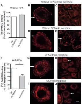
Morphine pretreatment promotes internalization of μ-receptors in DRG neurons of animals with inflammatory pain . To examine whether the avail-ability of μ-receptors on the surface of DRG neurons was changed by chronic morphine pretreatment or paw inflammation, we determined
μ-receptor binding in membrane preparations of these neurons. This
assay quantifies surface receptors but not endocytosed recep-tors. Saturation binding of the μ-receptor ligand [3H][d-Ala2,N
-Me-Phe4,Gly5-ol]enkephalin ([3H]DAMGO) showed similar K d in
all DRG regardless of CFA or morphine pretreatment (data not shown). In rats without CFA, the total number of μ -receptor bind-ing (Bmax μ-receptor) on DRG did not differ between animals with and
without chronic morphine pretreatment (t test, P > 0.05) (Table 2 and Figure 2A). However, in rats with CFA inflammation, the
Bmax μ-receptor
on DRG was significantly decreased by morphine pre-treatment (t test, P < 0.001; Table 2 and Figure 2F). We obtained similar results after repeated pretreatment with i.pl. fentanyl. In animals without CFA inflammation, the Bmax μ-receptor did not differ
(i.pl. saline versus fentanyl pretreatment, 25 ± 1.7 versus 29 ± 1.8 fmol/mg protein; t test, P > 0.05), whereas in rats with CFA inflam-mation the Bmax μ-receptor was significantly lowered by i.pl. fentanyl
[image:3.585.43.380.81.356.2]pretreatment (i.pl. saline versus fentanyl pretreatment, 47 ± 4.8 versus 36 ± 2.0 fmol/mg protein; t test, P < 0.05). To examine whether the surface availability of μ-receptors was correlated to receptor trafficking, we assessed their internalization in cultured and native DRG neurons. In DRG from animals without inflam-mation, μ-receptors were predominantly located at the cell surface,
Table 1
Antinociceptive potency (ED50) of i.pl. fentanyl after pretreatment with s.c. morphine or i.pl. fentanyl
Parameter No inflammation CFA inflammation CFA inflammation without endogenous opioids Saline Morphine Saline Morphine Saline/CTX Morphine/CTX ED50 (μg) 1.0 ± 0.1 1.6 ± 0.1A 0.5 ± 0.03 0.6 ± 0.02 0.8 ± 0.03 1.9 ± 0.1A
Saline Fentanyl Saline Fentanyl Saline/Ab Morphine/Ab ED50 (μg) 1.3 ± 0.1 2.2 ± 0.3A 0.6 ± 0.2 0.7 ± 0.1 0.4 ± 0.1 1.8 ± 0.3A
AStatistically significant difference from animals without morphine. ANOVA, P < 0.05, Tukey test.
Figure 1
Dose-response curves of acute i.pl. fentanyl antinociception in animals without and with hindpaw CFA inflammation. (A) Without paw inflammation the ED50 for elevation of PPT
was significantly lower in s.c. saline–pretreat-ed than in s.c. morphine–pretreatsaline–pretreat-ed animals (ANOVA, P < 0.001). (B) In the presence of paw inflammation, no significant difference in ED50 was detectable between chronic
s.c. morphine and s.c. saline pretreatment (ANOVA, P > 0.05). SEM was occasionally smaller than symbol size. (C) Repeated pre-treatment with i.pl. fentanyl (2 μg twice daily) for 4 days shifted the dose-response curve after acute i.pl. fentanyl application signifi-cantly to the right, confirming the develop-ment of peripheral opioid tolerance (ANOVA, P < 0.05). (D) In CFA animals pretreated with chronic i.pl. fentanyl, the ED50 of acute i.pl.
[image:3.585.197.534.659.731.2]both without (Figure 2, B and C) and with morphine pretreatment (Figure 2, D and E). However, in animals with CFA inflammation,
μ-receptors were, for the most part, removed from the neuronal cell surface. This endocytosis appeared more pronounced in the presence (Figure 2, I and J) than in the absence (Figure 2, G and H) of chronic morphine pretreatment.
Morphine pretreatment does not change μ-agonist–induced G protein coupling and cAMP reduction in DRG neurons of animals with inflammatory pain. To examine whether morphine pretreat-ment influenced opioid receptor signaling, we assessed
G protein coupling and cAMP production. Saturation analysis of DAMGO-stimulated [35S]guanosine-5′-O
-(γ-thio)-triphosphate ([35S]GTPγS) binding was used to
determine the equilibrium dissociation constant (Kd G protein) of [35S]GTPγS for the activated G protein and the
total number of G protein binding (Bmax G protein) to the μ
-receptor (see also Supplemental Methods; supplemental material available online with this article; doi:10.1172/ JCI25911DS1). Similar Kd for G proteins at μ
-recep-tors were found in all DRG regardless of pretreatment
(data not shown). The total number of DAMGO-activated G proteins (apparent
Bmax G protein) was significantly lower after
chronic morphine than after saline pre- treatment, regardless of the absence (Fig-ure 3A) or presence (Figure 3B) of paw inflammation (t test, P < 0.05; Table 2 and Figure 3, A and B). However, since after chronic morphine pretreatment the num-ber of surface μ-receptors was reduced in animals with (but not without) CFA inflammation, the relative number of activated G proteins per surface μ -recep-tor (amplification factor) did not decrease in the CFA-treated group, whereas it did decrease in the group without CFA (Table 2 and Supplemental Methods).
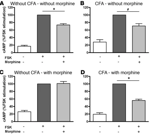
Regardless of pretreatment, the cAMP content in DRG increased significantly after forskolin (FSK) stimulation (Figure 4, A–D). This cAMP increase was significantly reduced by the acute appli-cation of morphine both in DRG of animals without (Figure 4A) and with (Figure 4B) CFA-induced inflammation. In DRG of animals chronically pretreated with s.c. morphine, acute opioid application no longer decreased the cAMP accumulation in the absence of CFA inflammation (ANOVA, P > 0.05; Figure 4C) but
Table 2
Bmax μ-receptor and Bmax G protein in animals with or without CFA inflammation,
pretreated with s.c. saline or morphine
Parameter Without CFA Without CFA/ With CFA With CFA/ with morphine with morphine Bmaxμ-receptor (fmol/mg) 26 ± 1.7 28 ± 2.0 50 ± 4.1 31 ± 5.2 BmaxG protein (fmol/mg) 335 ± 33 230 ± 21A 488 ± 62 290 ± 29B
Amplification factor 13 8 10 9
Bmax μ-receptor, determined by saturation binding of [3H]DAMGO, and Bmax G protein, determined by [35S]GTPγS saturation binding. The amplification factor was calculated by dividing the Bmax of [35S]GTPγS by that of [3H]DAMGO (i.e., Bmax G protein / Bmax
[image:4.585.261.545.377.739.2]μ-receptor) (see also Supplemental Methods). AStatistically significant difference from animals without morphine; t test, P < 0.05. BStatistically sig-nificant difference from CFA animals without morphine; t test, P < 0.05.
Figure 2
μ-Receptors on DRG neurons. [3H]DAMGO saturation
research article
did so in the presence of CFA inflammation (ANOVA, P < 0.01; Figure 4D). Thus, during chronic morphine pretreatment both G protein coupling to μ-receptors and μ-agonist–induced inhibition of cAMP production are preserved in sensory neurons of animals with but not without painful paw inflammation.
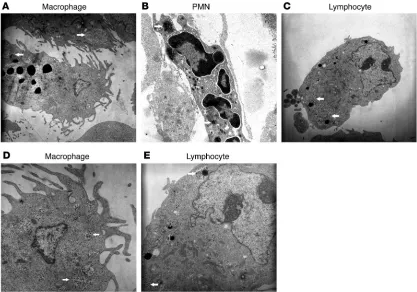
Role of endogenous opioid peptides in the development of opioid toler-ance. CTX treatment depletes opioid-containing immune cells and counteracts morphine-induced internalization of μ -recep- tors in DRG neurons of animals with inflammatory pain. Consis-tent with our previous studies (18), immunoelectron microscopy revealed β-endorphin–containing (END-containing) macrophages (Figure 5, A and D), polymorphonuclear cells (PMNs) (Figure 5B), and lymphocytes (Figure 5, C and E) in s.c. tissue of inflamed paws. END was contained in secretory granules packed in membranous structures. Preabsorption of antibody against END with 5
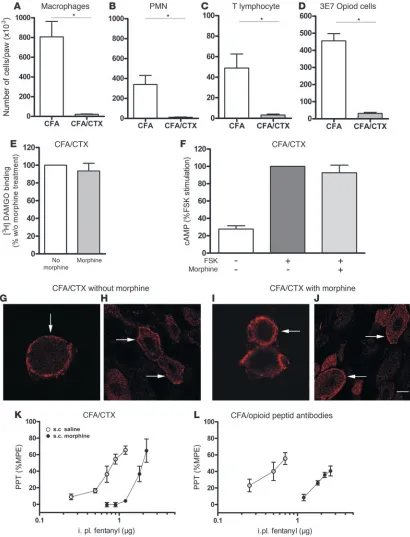
μg/ml purified END completely abolished immunostaining (data not shown). To assess the involvement of these opioid peptide-containing immune cells in tolerance, we first treated animals with CFA inflammation by systemic i.p. application of CTX. As quantified by flow cytometry, CTX treatment completely depleted macrophages, PMNs, T lymphocytes, and opioid-containing cells in inflamed paws (all groups,
t test, P < 0.01) (Figure 6, A–D). To examine whether the lack of endogenous opioid peptides can interfere with the avail-ability of μ-receptors on the surface of DRG neurons after
[image:5.585.100.481.82.244.2]chronic s.c. morphine treatment, we determined μ -receptor bind-ing on membrane preparations of these cells. In rats with CFA inflammation and CTX treatment, the number of μ-receptors on DRG cells did not differ between animals with (46.4 ± 5.3 fmol/ mg protein) and without (43.6 ± 4.0 fmol/mg protein) chronic morphine pretreatment (t test, P > 0.05; Figure 6E). Consistently, immunohistochemistry in cultured DRG neurons from animals with CFA inflammation and CTX revealed that μ-receptors were largely located near the plasma membrane both in the absence (Figure 6, G and H) and presence (Figure 6, I and J) of chronic s.c. morphine pretreatment. This is in opposition to our findings in CFA animals without CTX (Figure 2, G–J). Thus, immunosuppres- sion with CTX abolished morphine-induced opioid receptor inter-nalization in DRG neurons of animals with inflammatory pain.
Figure 3
Analysis of DAMGO-stimulated (10 μM) [35S]GTPγS saturation binding in DRG membranes from animals without and with CFA-induced
inflamma-tion pretreated with daily injecinflamma-tions of s.c. saline or morphine for 4 days. (A) Without CFA-induced inflammation. (B) With CFA-induced inflammation. DAMGO-stimulated specific [35S]GTPγS binding was significantly lower in morphine-treated than in saline-treated animals in both cases (t test,
[image:5.585.282.544.508.742.2]P < 0.05). Insets: Data were plotted according to the traditional Scatchard analysis (see Supplemental Methods), in which the x axis shows specific binding and the y axis shows specific binding divided by free radioligand concentration. From the Scatchard line, Bmax G protein is the x intercept.
Figure 4
Content of cAMP in DRG cells from animals without and with hindpaw CFA inflammation pretreated with s.c. morphine. (A–D) Basal cAMP was significantly lower in the absence of FSK in comparison with FSK treatment. Acute opioid application signifi-cantly decreased FSK-stimulated cAMP production in compari-son with FSK treatment alone in DRG cells of animals with CFA (B) and CFA/morphine pretreatment (D) (ANOVA, *P < 0.01), but not in morphine–pretreated animals without CFA inflammation (C) (ANOVA, #P > 0.05). Values are expressed as percentages
Removal of immune cell–derived opioid peptides inhibits μ-receptor func-tion and restores tolerance. First we performed cAMP experiments to study the functional relevance of the observed lack of μ-receptor internalization in animals with CFA inflammation and CTX treat-ment. Following chronic s.c. morphine treatment, acute application of morphine to DRG neurons of these animals no longer decreased FSK-stimulated cAMP, indicating that μ-receptors were not fully active (ANOVA, P > 0.05) (Figure 6F). In contrast to our initial find-ings (Figure 1B), in CTX-treated CFA animals chronic s.c. morphine shifted the dose-response curve of acute i.pl. fentanyl-induced anti-nociception significantly to the right (Figure 6K and Table 1). To corroborate this finding, rats were locally pretreated with i.pl. anti-bodies against endogenous opioid peptides. CFA animals treated daily with s.c. morphine and a combination of i.pl. anti-END and anti–met-enkephalin developed tolerance. This was evident from a significant rightward shift of the dose-response curve of acute i.pl. fentanyl antinociception in antibody-treated versus vehicle-treated animals (Figure 6L and Table 1). Thus, immune cell–derived opioid peptides apparently counteract the development of tolerance.
Discussion
Our results demonstrate that during inflammatory pain, chronic morphine treatment does not result in antinociceptive tolerance at
peripheral opioid receptors. Rather, we showed increased μ -recep-tor endocytosis as well as intact G protein coupling and cAMP inhibition, indicating fully preserved opioid receptor function in sensory neurons. However, when endogenous ligands of these receptors were removed by treatment with CTX or antibodies, decreased μ-receptor endocytosis/function and tolerance ensue.
[image:6.585.85.502.83.375.2]A spectrum of cellular adaptations resulting from chronic expo-sure to opioids is responsible for the development of tolerance (20) in experimental animals or in human addicts (21). However, many of those previous findings were obtained in the absence of pain-ful tissue injury. This is a shortcoming because patients usually do not consume opioids when they are not in pain, which may explain some of the discrepancies between experimental (20) and clinical (6, 22) studies. For this reason, we studied the development of opi-oid tolerance in animals with painful hindpaw inflammation, a model that resembles postoperative pain, arthritis, and other types of inflammatory pain (9). The analgesic effects of systemically applied opioid agonists can have both a peripheral and a central component of action. In injury-induced pain, a substantial compo-nent of opioid analgesia is mediated via peripheral opioid receptors (9, 10, 23). Therefore, even in the presence of central tolerance, the peripheral component of opioid action might still produce clini-cally sufficient analgesia. To specifically investigate tolerance at
Figure 5
research article
Figure 6
Removal of immune cell–derived opioid peptides by CTX. (A–D) Quantification of immune cells in inflamed paw tissue by flow cytometry. CTX treat-ment depleted macrophages (A), PMNs (B), T lymphocytes (C), and opioid peptide–containing cells (D). *P < 0.01. (E)[3H]DAMGO saturation binding
in DRG cells from animals with paw inflammation and CTX treatment. The number of μ-receptors did not differ significantly between untreated (set at 100%) and s.c. morphine–pretreated animals (t test, P > 0.05). (F) Content of cAMP in DRG cells from CFA animals with CTX treatment. Acute opioid application (+/+) did not decrease FSK-stimulated cAMP production (+/–) after chronic s.c. morphine pretreatment (ANOVA, P > 0.05). Values are expressed as percentages of FSK-stimulated (100%) cAMP levels. (G, H, I, and J) Confocal laser scanning microscopy of cultured DRG cells (G and I) and native DRG sections (H and J) obtained from CFA animals immunosuppressed with CTX. Immunohistochemically labeled μ-receptors are primarily localized to the plasma membrane in animals treated with vehicle (G and H) and with chronic s.c. morphine (I and J) Scale bar: 5 μM (G and I); 10 μM (H and J). (K and L) Dose-response curves of acute i.pl. fentanyl antinociception in CFA animals immunosuppressed with CTX (K) or injected with i.pl. opioid peptide antibodies (L) with and without chronic s.c. morphine pretreatment. In both conditions the ED50 for elevation of PPT
peripheral opioid receptors, we used acute local (i.pl.) injections of small, systemically ineffective doses of fentanyl. This precludes that the behavioral effects were affected by central neural circuits. Previous studies (24, 25) and our current behavioral experiments have shown that in animals without tissue injury chronic opioid treatment can induce profound tolerance at peripheral μ-receptors within several days. However, in the presence of painful inflamma-tion, we did not find such tolerance in vivo. This is in contrast to a recent study by Fernandez-Duenas et al. (26), in which the antino-ciceptive potency of acutely administered s.c. morphine decreased more pronouncedly in mice with CFA inflammation than in mice without inflammation. However, apart from the species difference, in the latter study s.c. morphine pellets were implanted 4 days after i.pl. CFA inoculation. Thus, the tissue injury commenced 4 days before the animals began to receive morphine and it is conceivable that the conditions for intraneuronal opioid receptor trafficking and signaling (e.g., cAMP upregulation) were different after these 4 days of ongoing inflammation without morphine treatment. We aimed to mimic the clinical situation by initiating morphine treat-ment simultaneously with the onset of tissue injury. In addition, changes in the antinocieptive potency of s.c. morphine might have been due to events at supraspinal, spinal, and peripheral levels, since naloxone-methiodide (a peripherally restricted opioid receptor antagonist) did not completely abolish the acute morphine effects in Fernandez-Duenas’s experiments. In contrast, we used a specific dose range of i.pl. fentanyl that only acted peripherally. Thus, our study examines mechanisms at the level of the peripheral sensory neuron exclusively. In line with our findings, a previous study in rats receiving formalin injections into the paw (27) and clinical studies in patients with non-cancer pain (reviewed in ref. 28) have suggested that the treatment of inflammatory pain with opioids is not necessarily associated with the development of pharmacologi-cal tolerance. However, the underlying molecular mechanisms of these unexpected effects have not been elucidated so far.
A fundamental mechanism of opioid receptor regulation involves rapid endocytosis of receptors via clathrin-coated pits and functional resensitization (20). Our studies of μ -receptor redistri- bution in DRG cells support previous observations that morphine-activated receptors are relatively resistant to rapid endocytosis in cultured cells and native neurons (14, 15), although morphine is highly efficient in inducing tolerance in healthy animals. It was suggested that this failure of μ-receptor endocytosis contributes to the development of tolerance and dependence (29). These find-ings led to the current hypothesis that μ-receptor endocytosis and recycling even prevent the development of tolerance (13, 30). Using a well-characterized cell culture model, it was shown that opioid receptor mutations that facilitate receptor endocytosis can attenu-ate, whereas blocking endocytosis can exacerbate, the development of morphine tolerance (2). In our animals with CFA inflammation, chronic morphine pretreatment was associated with enhanced
μ-receptor endocytosis and redistribution toward the intracellular compartment, consistent with the notion that a reduction of sur-face receptors is not necessarily connected to tolerance and that endocytosis serves a protective role and reduces the development of tolerance in an inflammatory painful condition.
Tolerance to morphine may also occur as a result of opioid recep-tor desensitization or receptor downregulation (i.e., a reduction of total receptor number). We examined the basis for the appar-ent protective effect of inflammation against opioid tolerance by investigating downstream signaling mechanisms. Previous studies
have shown that a decrease in G protein coupling to μ-receptors can substantially contribute to the development of opioid toler-ance (31). Indeed, we found a significant decrease in the number of G proteins activated per μ-receptor (amplification factor) in the DRG of opioid-tolerant animals without inflammation. In contrast, both μ-receptor–stimulated G protein levels and surface
μ-receptor numbers decreased to the same degree (unchanged amplification factor) in nontolerant animals with CFA inflamma-tion. This suggests that, in noninjured animals, chronic morphine pretreatment promotes receptor–G protein uncoupling but does not change surface receptor number (i.e., there is no endocytosis). In CFA-treated animals morphine pretreatment decreases sur-face receptor number (because of endocytosis) but the receptors remaining on the surface stay coupled.
Inhibition of adenylyl cyclase–mediated cAMP formation is a hallmark of the cellular actions of opioids (32). Acute activa-tion of Gαi-coupled opioid receptors typically inhibits cAMP
accumulation, but after withdrawal of ongoing opioid treat- ment adenylyl cyclase activity can be enhanced (33). This com-pensatory superactivation in the adenylyl cyclase pathway may contribute to tolerance (32). Our experiments indicate that the acute inhibitory effect of morphine on cAMP formation in DRG cells did not differ between animals with and without CFA inflammation. However, in line with previous studies (34), the absolute content of cAMP after FSK was significantly higher in animals treated with CFA (data not shown). Thus it is reason-able to assume that, consistent with the increased number of
μ-receptors, the acute inhibitory effect of morphine in fact increases but, due to a parallel increase in cAMP during inflam- mation, the relative effect is similar to that in noninjured ani-mals. After chronic morphine pretreatment, the ability of acutely applied opioids to inhibit adenylyl cyclase was abolished in DRG of rats without CFA inflammation, but was preserved in animals with inflammation. Together with our quantitative opioid recep-tor binding assay, this indicates that despite chronic morphine treatment, more functionally active receptors are available on DRG neurons in animals with compared with animals without inflammation. Hence it appears that during paw inflammation enhanced opioid receptor internalization restores the functional-ity of μ -receptors on DRG neurons and thus decreases the devel-opment of behavioral tolerance.
Several studies have shown that opioid receptors are particularly rapidly internalized after activation by their native peptide ligands (14, 35). All physiologically expressed opioids tested so far produce
successfully depleted PMNs, macrophages, T lymphocytes, and opi-research article
oid peptide–containing cells in the inflamed tissue. In these ani-mals, chronic morphine-induced desensitization of μ-receptors and behavioral signs of tolerance were restored. This was corroborated by an additional experiment, in which animals receiving repeated local injections of opioid peptide antibodies developed behavioral signs of tolerance. In line with studies showing that inflammatory pain (42) or the release of endogenous opioid peptides in the spinal cord (36) can increase opioid receptor internalization in vivo, our results thus indicate that the lack of tolerance and enhanced recy-cling/resensitization of opioid receptors in sensory neurons is due to the tonic release of endogenous opioid peptides from resident PMNs, macrophages, and lymphocytes within inflamed paws.
In summary, experimental studies on opioid tolerance are often performed in the absence of painful tissue injury, which precludes extrapolation to the clinical situation. In the current investigation, we found that persistent painful inflammation prevents the devel-opment of tolerance at peripheral opioid receptors by enhancing
μ -receptor endocytosis, recycling, and recovery of opioid respon-siveness after prolonged morphine treatment. Our data indicate that the release of endogenous opioid peptides from inflammatory cells can facilitate the ability of exogenous opioids to stimulate
μ-receptor endocytosis in sensory neurons and thereby reduce the development of morphine tolerance. This is consistent with the notion that opioids promoting receptor endocytosis (e.g., endog- enous opioid peptides) prevent tolerance induction by non-inter-nalizing opioid agonists (e.g., morphine) and that cross-tolerance occurs more likely when different non-internalizing opioid recep-tor ligands are used. Our findings infer that the use of peripherally acting opioid agonists for the prolonged treatment of inflamma- tory pain, such as pain associated with chronic arthritis, inflam-matory neuropathy, or cancer, is not necessarily accompanied by opioid tolerance. In addition, our data help to elucidate the hitherto enigmatic discrepancies between experimental and clini-cal observations on opioid tolerance. Because peripherally acting opioid analgesics have attracted much interest and have become increasingly relevant in daily clinical practice, uncovering mecha-nisms determining tolerance at peripheral opioid receptors will hopefully open new avenues for pain research and therapy.
Methods
Subjects. Experiments were performed using individually housed male Wistar rats (180–200 g). The animal protocol was approved by the state animal care and use committee (Landesamt für Arbeitsschutz, Gesundheit und Tech- nische Sicherheit Berlin), and the guidelines on ethical standards for inves-tigations of experimental pain in animals were followed. All i.pl. injections were performed under brief isoflurane (Willy Rüsch GmbH) anesthesia.
Induction of paw inflammation and chronic opioid pretreatment. All chemicals and drugs were purchased from Sigma-Aldrich unless otherwise indicat-ed. Control animals were treated with s.c. saline injections (twice/day) for 4 days. Chronic opioid pretreatment was performed by injection of mor-phine (Merck; 10 mg/kg body weight) s.c. twice/day (8 am and 6 pm) in the back of the animals from day 1 through day 4. On day 5 animals were injected once with morphine (s.c.), and behavioral experiments were per-formed 3 hours later. On day 1 inflammation was induced using 0.15 ml of CFA (Calbiochem) administered i.pl. into the right hind paw. In sepa-rate experiments (n = 6–8 rats/group), fentanyl (2 μg) was injected into the plantar surface of the hindpaw (i.pl.) twice/day (8 am and 6 pm) from day 1 through day 4 to induce tolerance. On day 5 animals were injected once with i.pl. fentanyl, and behavioral experiments were performed 3 hours later as described.
CTX treatment. CTX (Endoxan) (Baxter Oncology) was dissolved in sterile distilled water to a concentration of 25 mg/ml and injected i.p. 72 hours (100 mg/kg body weight) and 24 hours (50 mg/kg body weight) prior to CFA treatment. An additional injection was performed 24 hours (25 mg/kg body weight) after CFA injection.
Antibody treatment against opioid peptides. Antibodies (Bachem) were injected i.pl. into the inflamed paw once daily for 4 days. Based on our previous study (43), the most effective i.pl. doses of anti-END (2 μ g) and anti–met-enkephalin (0.25 μg) were combined in a total volume of 0.1 ml. Control animals received rabbit IgG (8 μg) i.pl. in a volume of 0.1 ml.
Measurement of PPT. Mechanical nociceptive thresholds were assessed in rats (n = 6–8 per group) 3 hours after saline/morphine injection and before (base-line) and after i.pl. administration of the μ -receptor agonist fentanyl (Janssen-Cilag) (in 100 μ l) using the paw pressure algesiometer (modified Randall-Selit-to test; Ugo Basile). Control animals received i.pl. saline in the same volume. The pressure required to elicit paw withdrawal, the PPT (cutoff at 250 g), was determined immediately after fentanyl injection by averaging 3 consecutive trials separated by 10 s. The sequence of left and right paws was alternated between animals to avoid bias. The experimenter was blind to the treatment.
Membrane preparations. Membranes were prepared as described (44). Briefly, rats were killed and lumbar (L3–L5) DRG were removed. The tissue was placed immediately on ice in cold assay buffer (50 mM Tris-HCl, 1 mM EGTA, pH 7.4) Tissue was homogenized and centrifuged at 42,000 g and 4°C for 20 min. The pellet was resuspended in assay buffer followed by a 10-min incubation at 37°C to degrade endogenous ligands. The homogenate was centrifuged again at 42,000 g and resuspended in assay buffer.
Cultures of DRG neurons. DRG (L3–L5) were removed and placed in sterile modified MEM (Biochrom AG) at 4°C. DRG were digested with collagenase type 2 (37°C for 50 min) and trypsin (37°C for 10 min). After digestion, DRG were centrifuged at 500 g for 5 min and at 300 g for another 5 min. The cells were maintained for 1 h in MEM growth media supplemented with 10% horse serum, 50 μg/ml penicillin and streptomycin.
Saturation μ-receptor binding. Plasma membranes from DRG neurons were prepared as described (44). Saturation binding was performed using the
μ-receptor ligand [3H]DAMGO (0.02–2 nM) (65 Ci/mmol; Amersham
Pharmacia Biotech) to determine Kd and Bmax μ-receptor. See Supplemental
Methods online for more details.
[35S]GTPγS saturation binding at μ-receptors . Saturation analysis of DAMGO-stimulated [35S]GTPγ
S (1250 Ci/mmol; New England Nuclear Corp.) bind-ing was used as previously described (45) to determine the apparent Kd of
[35S]GTPγS for the activated Gα-subunit (apparent Kd G protein
) and the appar-ent Bmax G protein. The relative amplification factor (Bmax G protein / Bmax μ-receptor)
was calculated according to methods described in ref. 45 and represents the number of G proteins activated per μ-receptor. See Supplemental Methods online for more details.
cAMP accumulation. cAMP accumulation was measured immediately after dissociation of DRG cells. Cells were incubated for 20 min in the pres- ence of 50 mM Tris (1 ml) containing the phosphodiesterase inhibitor iso-buthylmethylxanthine (2 mM) and FSK (1 μM). Acute opioid effects were determined by incubating cells with morphine (10 μ M) for 15 min. Follow-ing this incubation, cells were homogenized and boiled for 3 min. The cell suspension was centrifuged at 4,000 g for 4 min. The levels of cAMP in the supernatant were determined by a [3H]cAMP assay kit (5 μCi; Amersham
Pharmacia Biotech) as previously described (46).
Immunofluorescence. Immunofluorescence imaging of DRG neurons was performed in cultures and sections as described previously (47). See Sup-plemental Methodsonline for more details.
Flow cytometry. Cells suspensions from inflamed hindpaws were prepared and stained as described previously (48). See Supplemental Methods online.
Statistics. Data are expressed as means ± SEM. Unpaired 2-tailed Student’s t test and 1-way ANOVA were performed for statistical comparisons. For all tests, a P value of less than 0.05 was considered to be significant. See Supplemental Methods online for more details.
Acknowledgments
This work was supported by DFG grant KFO 100. We thank P. Heppenstall for critical reading and comments on the manuscript, N. Siegemund for technical assistance, and S. Schulz and V. Höllt for μ-receptor antibodies.
Received for publication June 9, 2005, and accepted in revised form November 28, 2007. Address correspondence to: Christian Zöllner, Klinik für Anaes- thesiologie und operative Intensivmedizin, Charité–Universitäts-medizin Berlin, Campus Benjamin Franklin, Hindenburgdamm 30, 12200 Berlin, Germany. Phone: 49-30-8445-3678; Fax: 49-30-8445-4469; E-mail: [email protected]. Waltraud Binder’s present address is: School of Physiology and Pharmacology, University of New South Wales, Sydney, New South Wales, Australia. 1. Bohn, L.M., Gainetdinov, R.R., Lin, F.T., Lefkowitz, R.J., and Caron, M.G. 2000. Mu-opioid receptor desen- sitization by beta-arrestin-2 determines morphine tol-erance but not dependence. Nature. 408:720–723. 2. Finn, A.K., and Whistler, J.L. 2001. Endocytosis of the
mu opioid receptor reduces tolerance and a cellular hallmark of opiate withdrawal. Neuron. 32:829–839. 3. Carroll, I.R., Angst, M.S., and Clark, J.D. 2004.
Management of perioperative pain in patients chronically consuming opioids. Reg. Anesth. Pain Med. 29:576–591.
4. Furlan, A.D., Sandoval, J.A., Mailis-Gagnon, A., and Tunks, E. 2006. Opioids for chronic noncan-cer pain: a meta-analysis of effectiveness and side effects. CMAJ. 174:1589–1594.
5. Portenoy, R.K., Moulin, D.E., Rogers, A., Inturrisi, C.E., and Foley, K.M. 1986. I.v. infusion of opioids for cancer pain: clinical review and guidelines for use. Cancer Treat. Rep. 70:575–581.
6. Zech, D.F., Grond, S., Lynch, J., Hertel, D., and Lehmann, K.A. 1995. Validation of World Health Organization Guidelines for cancer pain relief: a 10-year prospective study. Pain. 63:65–76. 7. Adriaensen, H., Vissers, K., Noorduin, H., and
Meert, T. 2003. Opioid tolerance and dependence: an inevitable consequence of chronic treatment? Acta. Anaesthesiol. Belg. 54:37–47.
8. Portenoy, R.K. 2004. Appropriate use of opioids for persistent non-cancer pain. Lancet. 364:739–740. 9. Stein, C., Schäfer, M., and Machelska, H. 2003.
Attacking pain at its source: new perspectives on opioids. Nat. Med. 9:1003–1008.
10. Tegeder, I., et al. 2003. Peripheral opioid analge-sia in experimental human pain models. Brain. 126:1092–1102.
11. Drake, M.T., Shenoy, S.K., and Lefkowitz, R.J. 2006. Trafficking of G protein-coupled receptors. Circ. Res. 99:570–582.
12. Qiu, Y., Law, P.Y., and Loh, H.H. 2003. Mu-opioid receptor desensitization: role of receptor phos-phorylation, internalization, and representation. J. Biol. Chem. 278:36733–36739.
13. Koch, T., et al. 2005. Receptor endocytosis coun-teracts the development of opioid tolerance. Mol. Pharmacol. 67:280–287.
14. Keith, D.E., et al. 1996. Morphine activates opioid receptors without causing their rapid internaliza-tion. J. Biol. Chem. 271:19021–19024.
15. Sternini, C., et al. 1996. Agonist-selective endocyto-sis of mu opioid receptor by neurons in vivo. Proc. Natl. Acad. Sci. U. S. A. 93:9241–9246.
16. Yu, Y., et al. 1997. Mu opioid receptor phosphory-lation, desensitization, and ligand efficacy. J. Biol. Chem. 272:28869–28874.
17. Schulz, S., et al. 2004. Morphine induces terminal mu- opioid receptor desensitization by sustained phos-phorylation of serine-375. EMBO J. 23:3282–3289. 18. Mousa, S.A., Shakibaei, M., Sitte, N., Schafer, M.,
and Stein, C. 2004. Subcellular pathways of beta-endorphin synthesis, processing, and release from immunocytes in inflammatory pain. Endocrinology.
145:1331–1341.
19. Antonijevic, I., Mousa, S.A., Schäfer, M., and Stein, C. 1995. Perineurial defect and peripheral opioid analgesia in inflammation. J. Neurosci. 15:165–172. 20. Williams, J.T., Christie, M.J., and Manzoni, O. 2001. Cellular and synaptic adaptations mediating opi-oid dependence. Physiol. Rev. 81:299–343. 21. Kissin, I. 2005. Tolerance to opioid analgesia: why do
we differ from rats? Anesth. Analg. 101:1727–1729. 22. Gutstein, H.B. 1996. The effects of pain on opioid
tolerance: how do we resolve the controversy? Phar-macol. Rev. 48:403–407.
23. Kayser, V., Chen, Y.L., and Guilbaud, G. 1991. Behavioural evidence for a peripheral component in the enhanced antinociceptive effect of a low dose of systemic morphine in carrageenin-induced hyperalgesic rats. Brain Res. 560:237–244.
24. Aley, K.O., Green, P.G., and Levine, J.D. 1995. Opi-oid and adenosine peripheral antinociception are subject to tolerance and withdrawal. J. Neurosci. 15:8031–8038.
25. Kolesnikov, Y., and Pasternak, G.W. 1999. Topical opioids in mice: analgesia and reversal of tolerance by a topical N-methyl-D-aspartate antagonist. J. Pharmacol. Exp. Ther. 290:247–252.
26. Fernandez-Duenas, V., et al. 2007. Tolerance to the antinociceptive and antiexudative effects of mor- phine in a murine model of peripheral inflamma-tion. J. Pharmacol. Exp. Ther. 322:360–368.
27. Vaccarino, A.L., et al. 1993. Morphine fails to pro-duce tolerance when administered in the presence of formalin pain in rats. Brain Res. 627:287–290. 28. Kalso, E., Edwards, J.E., Moore, R.A., and McQuay,
H.J. 2004. Opioids in chronic non-cancer pain: systematic review of efficacy and safety. Pain. 112:372–380.
29. Whistler, J.L., Chuang, H.H., Chu, P., Jan, L.Y., and von Zastrow, M. 1999. Functional dissociation of mu opioid receptor signaling and endocytosis: implications for the biology of opiate tolerance and addiction. Neuron. 23:737–746.
30. Ferguson, S.S. 2001. Evolving concepts in G pro- tein-coupled receptor endocytosis: the role in recep-tor desensitization and signaling. Pharmacol. Rev. 53:1–24.
31. Sim-Selley, L.J., Selley, D.E., Vogt, L.J., Childers, S.R., and Martin, T.J. 2000. Chronic heroin self- administration desensitizes mu opioid receptor-activated G-proteins in specific regions of rat brain. J. Neurosci. 20:4555–4562.
32. Kim, K.S., et al. 2006. Adenylyl cyclase type 5 (AC5) is an essential mediator of morphine action. Proc. Natl. Acad. Sci. U. S. A. 103:3908–3913.
33. Nestler, E.J., and Aghajanian, G.K. 1997. Molecular and cellular basis of addiction. Science. 278:58–63. 34. Bolyard, L.A., Van Looy, J.W., and Vasko, M.R. 2000.
Sensitization of rat sensory neurons by chronic exposure to forskolin or ‘inflammatory cocktail’ does not downregulate and requires continuous exposure. Pain. 88:277–285.
35. Keith, D.E., et al. 1998. mu-Opioid receptor inter-nalization: opiate drugs have differential effects on a conserved endocytic mechanism in vitro and in the mammalian brain. Mol. Pharmacol. 53:377–384. 36. Song, B., and Marvizon, J.C. 2003. Peptidases pre-vent mu-opioid receptor internalization in dorsal horn neurons by endogenously released opioids. J. Neurosci. 23:1847–1858.
37. He, L., and Whistler, J.L. 2005. An opiate cocktail that reduces morphine tolerance and dependence. Curr. Biol. 15:1028–1033.
38. Hashimoto, T., et al. 2006. Enhancement of morphine analgesic effect with induction of mu-opioid receptor endocytosis in rats. Anesthesiology. 105:574–580.
39. Bailey, C.P., et al. 2003. Mu-opioid receptor desen- sitization in mature rat neurons: lack of interac-tion between DAMGO and morphine. J. Neurosci. 23:10515–10520.
40. He, L., Fong, J., von Zastrow, M., and Whistler, J.L. 2002. Regulation of opioid receptor trafficking and morphine tolerance by receptor oligomerization. Cell. 108:271–282.
41. Muranski, P., et al. 2006. Increased intensity lym-phodepletion and adoptive immunotherapy — how far can we go? Nat. Clin. Pract. Oncol. 3:668–681. 42. Gendron, L., et al. 2006. Morphine and pain-related
stimuli enhance cell surface availability of somatic delta-opioid receptors in rat dorsal root ganglia. J. Neurosci. 26:953–962.
43. Machelska, H., et al. 2003. Different mechanisms of intrinsic pain inhibition in early and late inflam-mation. J. Neuroimmunol. 141:30–39.
44. Zöllner, C., et al. 2003. Painful inflammation-induced increase in mu-opioid receptor binding and G-protein coupling in primary afferent neu-rons. Mol. Pharmacol. 64:202–210.
45. Shaqura, M.A., Zöllner, C., Mousa, S.A., Stein, C., and Schäfer, M. 2004. Characterization of mu opi-oid receptor binding and G protein coupling in rat hypothalamus, spinal cord, and primary afferent neurons during inflammatory pain. J. Pharmacol. Exp. Ther. 308:712–718.
46. Zaki, P.A., Keith, D.E., Jr., Brine, G.A., Carroll, F.I., and Evans, C.J. 2000. Ligand-induced changes in surface mu-opioid receptor number: relationship to G protein activation? J. Pharmacol. Exp. Ther. 292:1127–1134.
47. Mousa, S.A., Zhang, Q., Sitte, N., Ji, R., and Stein, C. 2001. beta-Endorphin-containing memory-cells and mu-opioid receptors undergo transport to periph-eral inflamed tissue. J. Neuroimmunol. 115:71–78. 48. Rittner, H.L., et al. 2006. Pain control by CXCR2
ligands through Ca2+-regulated release of opioid peptides from polymorphonuclear cells. FASEB J. 20:2627–2629.
49. Abe, K., Honma, S., and Ito, T. 1981. Peritoneal free cells in mice, examined by transmission electron microscopy in thick sections. J. Electron Microsc. (Tokyo) 30:141–147.