IL-12–induced IFN-
gg
is dependent on caspase-1
processing of the IL-18 precursor
Giamila Fantuzzi, … , David A. Reed, Charles A. Dinarello
J Clin Invest.
1999;104(6):761-767. https://doi.org/10.1172/JCI7501.
IL-12 and IL-18 are IFN-
g
–inducing cytokines. In the present study, the role of endogenous
IL-18 in the induction of IFN-
g
by IL-12 was investigated in mice. In the presence of a
specific inhibitor of caspase-1 (also known as IL-1
b
–converting enzyme, or ICE) IL-12–
induced IFN-
g
from splenocytes was reduced by 85%. Using splenocytes from ICE-deficient
mice, IL-12–induced IFN-
g
was reduced by 80%. However, the role of ICE was not through
processing and release of IL-1
b
. Neutralizing anti–IL-18 IgG reduced IL-12–induced IFN-
g
in
splenocytes by 85%. Splenocytes cultured in vitro spontaneously released IL-18 into the
extracellular compartment over time. Extracellular levels of IL-18 significantly correlated with
IL-12–induced IFN-
g
and were reduced in cells obtained from ICE-deficient mice. In vivo,
IL-12 administration increased circulating levels of IL-18 in wild-type mice but not in
ICE-deficient mice. Both neutralization of IL-18 and ICE deficiency significantly reduced
induction of circulating IFN-
g
in mice receiving IL-12. The IL-18 precursor was constitutively
expressed in the livers and spleens of untreated mice. Furthermore, administration of IL-12
significantly increased liver-associated IL-18 levels. These data demonstrate that
endogenous, ICE-cleaved IL-18 significantly contributes to induction of IFN-
g
by IL-12.
Article
Introduction
Two cytokines, IL-12 and IL-18, are currently regarded as the primary inducers of IFN-γ production in an inflammatory reaction (1, 2). However, the relationship between these 2 cytokines is still not fully understood. IL-12 is a heterodimeric cytokine produced mainly by monocytes/macrophages. In addition to inducing IFN-γ, IL-12 stimulates the production of other cytokines, activates natural killer (NK) and T cells, and promotes the development of T-helper type 1 (Th1) responses (1). Administration of IL-12 to mice results in marked splenomegaly, thymic atrophy, macrophage infiltration in tissues, and induction of IFN-γ and TNF-α synthesis (3–5). Most of the toxic effects of IL-12 are due to its abil-ity to induce high levels of IFN-γ(3). Systemic toxicity has also been observed in cancer patients treated with multi-ple doses of IL-12 (6).
Like IL-12, IL-18 is a monocyte/macrophage–derived cytokine that participates in the induction of IFN-γ and other cytokines (2). IL-18 is structurally related to IL-1β (7). Similar to the IL-1βprecursor, the IL-18 pre-cursor (pro–IL-18) also lacks a signal peptide and requires caspase-1 (also known as IL-1β–converting enzyme, or ICE) for cleavage and release of the mature molecule from the intracellular compartment (7–9). Only mature IL-18 is bioactive, whereas pro–IL-18 is biologically inactive (10). Therefore, mice deficient in ICE (ICE KO) have a defect in the production and release of mature, bioactive IL-18, whereas the precur-sor form is normally synthesized (8). The importance of the presence of both IL-12 and IL-18 for optimal
induction of IFN-γhas been demonstrated in IL-18 and ICE KO mice (11–13).
In the absence of a costimulus, IL-18 is a weak inducer of IFN-γ. However, a synergy for IFN-γproduction is observed when cells are cultured with IL-18 in the pres-ence of costimuli (14–16). Different mechanisms may account for the synergy between IL-12 and IL-18. In par-ticular, IL-12 upregulates the expression of the IL-18 receptor, therefore rendering cells more sensitive to IL-18 (17, 18). In addition, IL-12 and IL-18 regulate the tran-scriptional activity of the IFN-γpromoter at different lev-els (19), thus providing 2 distinct signals to the
IFN-γ–producing cell. IL-12 and IL-18 also regulate each other’s production. (20, 21).
In the present report, we investigated the role of IL-18 in the induction of IFN-γby IL-12. IL-12–induced IFN-γ production was evaluated both in vitro and in vivo in the presence of neutralizing anti–IL-18 antibodies. In addi-tion, a specific ICE inhibitor and ICE KO mice were used to evaluate the role of this enzyme in the production and release of mature, bioactive IL-18 in response to IL-12. Finally, we measured levels of IL-18 both in vitro and in vivo in the presence or absence of IL-12 stimulation.
Methods
Reagents and mice. Murine recombinant IL-12 was a kind gift of Genetics Institute Inc. (Andover, Massachusetts, USA). The specific activity of IL-12 was 2.7 ×10⁶U/mg. Human recombinant IL-1 receptor antagonist (IL-1Ra) was a kind gift of Daniel Tracey (Upjohn Co., Kalamazoo, Michigan, USA). The reversible ICE inhibitor
Ac-Tyr-Val-IL-12–induced IFN-
γ
is dependent on caspase-1 processing
of the IL-18 precursor
Giamila Fantuzzi, David A. Reed, and Charles A. Dinarello
Division of Infectious Diseases, University of Colorado Health Sciences Center, Denver, Colorado 80262, USA
Address correspondence to: Giamila Fantuzzi, Division of Infectious Diseases, University of Colorado Health Sciences Center, 4200 East Ninth Avenue B168, Denver, Colorado 80262, USA. Phone: (303) 315-3558; Fax: (303) 315-8054;
E-mail: [email protected].
Received for publication June 3, 1999, and accepted in revised form August 2, 1999.
IL-12 and IL-18 are IFN-γ–inducing cytokines. In the present study, the role of endogenous IL-18 in the induction of IFN-γby IL-12 was investigated in mice. In the presence of a specific inhibitor of cas-pase-1 (also known as IL-1β–converting enzyme, or ICE) IL-12–induced IFN-γfrom splenocytes was reduced by 85%. Using splenocytes from ICE-deficient mice, IL-12–induced IFN-γwas reduced by 80%. However, the role of ICE was not through processing and release of IL-1β. Neutralizing anti–IL-18 IgG reduced IL-12–induced IFN-γin splenocytes by 85%. Splenocytes cultured in vitro spontaneous-ly released IL-18 into the extracellular compartment over time. Extracellular levels of IL-18 signifi-cantly correlated with IL-12–induced IFN-γand were reduced in cells obtained from ICE-deficient mice. In vivo, IL-12 administration increased circulating levels of IL-18 in wild-type mice but not in ICE-deficient mice. Both neutralization of IL-18 and ICE deficiency significantly reduced induction of circulating IFN-γin mice receiving IL-12. The IL-18 precursor was constitutively expressed in the livers and spleens of untreated mice. Furthermore, administration of IL-12 significantly increased liver-associated IL-18 levels. These data demonstrate that endogenous, ICE-cleaved IL-18 significantly contributes to induction of IFN-γby IL-12.
Asp-2,6-dimethylbenzoyloxymethylketone was pur-chased from Alexis Corp. (San Diego, California, USA). RPMI and FBS were from Life Technologies Inc. (Grand Island, New York, USA). The anti-murine CSF antibody was from Endogen Inc. (Woburn, Massachusetts, USA). The generation and genetic background of ICE KO mice have been described previously (22). Six- to 8-week old female mice were used. The wild-type (WT) mice used were of the same genetic background, sex, and age as the ICE KO mice, although they were not littermates.
In vivo experimental studies. Studies were approved by the Animal Use and Care Committee at the University of Col-orado Health Sciences Center. Mice were injected intraperitoneally with either 100 or 400 ng of murine recombinant IL-12 daily for 4 days, between 0900 and 1100 hours. Control groups received 4 daily injections of vehicle (PBS [pH 7.4] containing 0.1% BSA). In some experiments, mice were injected with 200 µL of a neu-tralizing rabbit anti-murine IL-18 antiserum (13) 1 hour before the first and the third injection of IL-12. A group of mice receiving normal rabbit serum (NRS) was includ-ed as the control for mice receiving the anti–IL-18 anti-serum. Two hours after the fourth injection of IL-12, blood was collected from the retro-orbital plexus under methoxyflurane anesthesia. Mice were sacrificed by cer-vical dislocation, and the spleens and livers were removed. Spleens and livers were weighed, homogenized in 4 vol-umes of ice-cold PBS containing 0.2% Tween-20, and cen-trifuged for 5 minutes at 3,000 gin a microfuge. Then the supernatants were collected for cytokine measurements.
Isolation and culture of spleen cells. Spleens were asepti-cally removed, and cell suspensions were prepared according to standard procedures (23). Cells were washed twice in RPMI, resuspended in RPMI contain-ing 10% FBS, and cultured at 5 ×10⁶/mL in 24-well plates. Cultures were incubated for various amounts of time at 37°C in a humidified atmosphere with 5% CO2. At the end of the incubation period, supernatants were collected. The cells were resuspended in the original vol-ume of RPMI and lysed by addition of 0.5% Triton X-100. Both supernatants and cell lysates were frozen at –70°C until they were ready for cytokine measurement.
Cytokine measurements. Both IL-18 and TNF-αlevels were measured using an electrochemiluminescence
(ECL) method (24). The TNF-αassay has been previ-ously described (25). For IL-18 measurement, a similar procedure was employed. An affinity-purified rabbit anti-murine IL-18 polyclonal antibody (R&D Systems Inc., Minneapolis, Minnesota, USA) was labeled with biotin (IGEN Inc., Gaithersburg, Maryland, USA), and a monoclonal anti-murine IL-18 antibody (R&D Sys-tems Inc.) was labeled with ruthenium (II) trisbipyridae chelate (IGEN Inc.). The biotinylated antibody was diluted to 0.5 µg/mL in PBS (pH 7.4) with 0.25% BSA, 0.5% Tween-20, and 0.01% sodium azide (ECL buffer). Twenty-five microliters of this antibody was combined in 6-mL polypropylene tubes with 25 µL of a 1 mg/mL suspension of streptavidin-coated paramagnetic beads (Dynal Inc., Lake Success, New York, USA) diluted in ECL buffer. Tubes were shaken for 15 minutes at room temperature, after which 25 µL of samples or standard concentrations of recombinant murine IL-18 (Pepro-Tech Inc., Rocky Hill, New Jersey, USA) was added. Twenty-five microliters of ruthenylated antibody (1
µg/mL in ECL buffer) was then added, and the tubes were shaken overnight at room temperature. The reac-tion was quenched by addireac-tion of 200 µL/tube of PBS and was quantitated using the Origen 1.5 Analyzer (IGEN Inc.). The ECL IL-18 assay detects both recom-binant murine pro–IL-18 and mature IL-18. The range of detection is 20 pg/mL to 100 ng/mL. A 100 ng/mL concentration of pro–IL-18 is detected in the assay as 20 ng/mL of mature IL-18. The presence of mouse serum or FBS does not interfere with the detection of IL-18. IFN-γand IL-1βwere measured using specific ELISAs (Endogen Inc.).
Western blot analysis. Spleen and livers were weighed, homogenized in 4 volumes of ice-cold PBS containing 0.2% Tween-20, and microfuged for 5 minutes at 3,000
[image:3.612.306.531.549.731.2]g. Then the supernatants were collected for Western blot analysis. SDS-PAGE was performed on 15% gels (Bio-Rad Laboratories Inc., Hercules, California, USA); 5 µL of samples was loaded per lane. Proteins were transferred onto nitrocellulose membranes (Amersham Interna-tional, Amersham, United Kingdom) using a semidry technique. The transfer buffer contained 25 mM Tris-HCl, 192 mM glycine, 15% methanol, and 0.1% SDS. After transfer, nonspecific sites on the membranes were
Figure 1
blocked overnight at 4°C with PBS containing 5% non-fat milk, 1% BSA, and 0.1% Tween-20. Blots were probed with a rabbit anti-murine IL-18 antiserum (26), diluted at 1:400, for 1 hour at room temperature, followed by 3 washes in PBS containing 0.1% Tween-20. A secondary horseradish peroxidase–conjugated antibody (1:6,000 dilution; The Jackson Laboratory, Bar Harbor, Maine, USA) was added for 45 minutes at room temperature. The blot was washed 3 times in PBS/Tween-20, and the antibody was detected by ECL (NEN Life Science Prod-ucts, Boston, Massachusetts, USA).
Results
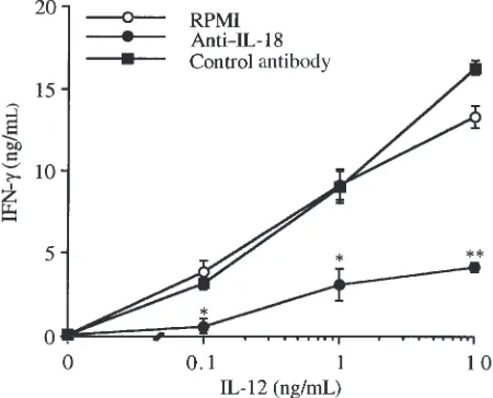
Neutralization of IL-18 inhibits induction of IFN-γby IL-12 in murine splenocytes. Splenocytes were incubated for 48 hours with increasing concentrations of IL-12 in the presence or absence of neutralizing anti–IL-18 IgG (50
µg/mL) (13). As shown in Figure 1, a concentration-dependent increase of IFN-γproduction was observed in cells incubated with IL-12 alone. Neutralization of IL-18 significantly reduced IFN-γlevels at each of the concen-trations of IL-12 tested. On the other hand, when cells were incubated with IL-12 in the presence of an
unrelat-ed IgG (anti-CSF), no decrease in IFN-γ levels was observed. The inhibitory effect of anti–IL-18 IgG was specific for IFN-γ. In fact, in the same samples, neutral-ization of IL-18 did not affect IL-12–induced TNF-α lev-els (data not shown).
Inhibition of ICE activity or ICE deficiency reduces IL-12–induced IFN-γproduction. Splenocytes were incu-bated for 48 hours with increasing concentrations of IL-12 in the presence or absence of a specific ICE inhibitor (27). As shown in Figure 2, inhibition of ICE activity significantly reduced production of IFN-γat each of the concentrations of IL-12. As already observed with anti–IL-18 IgG, the ICE inhibitor did not reduce IL-12–induced TNF-αlevels (data not shown).
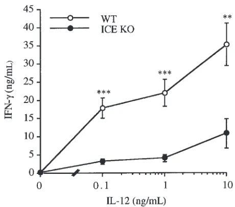
[image:4.612.321.534.50.225.2]To test the effect of ICE deficiency on IL-12–induced IFN-γproduction, splenocytes obtained from WT and ICE KO mice were cultured for 48 hours with increasing concentrations of IL-12. As shown in Figure 3, signifi-cantly lower levels of IFN-γ were observed in cells obtained from ICE KO mice compared with cells derived from WT mice. No differences in IL-12–induced TNF-α levels were observed between WT and ICE KO spleno-cytes (data not shown).
Figure 2
Effect of ICE inhibition on IFN-γinduced by IL-12 in vitro. Splenocytes were cultured for 48 hours with increasing concentrations of IL-12 (0.1–10 ng/mL) in the presence or absence of ICE inhibitor (20 µM).
IFN-γlevels were measured in the supernatants. Data are mean ± SEM of 3 mice per group and are representative of 1 experiment out of 3 per-formed. *P < 0.05, **P < 0.01 by paired Student’s ttest.
Figure 3
[image:4.612.306.536.528.731.2]Blockade of IL-1 receptor does not affect IL-12–induced IFN-γ production. Inhibition of ICE activity leads to reduced lev-els of both mature IL-18 and mature IL-1β(8, 28). How-ever, blocking IL-1 receptors by using saturating concen-trations of IL-1Ra did not affect induction of IFN-γby IL-12 in cultures of murine splenocytes (after stimulation with 10 ng/mL of IL-12, IFN-γlevels were 12.3 ± 0.8 vs. 10.9 ± 1.2 ng/mL in the absence or presence of IL-1Ra, respectively; n = 3). Furthermore, IL-1βwas not detectable (<10 pg/mL) in control or IL-12–stimulated splenocytes obtained from WT mice (data not shown). We can there-fore conclude that, in this system, IL-1βdoes not play a significant role in the induction of IFN-γby IL-12.
IL-18 production by cultured splenocytes. To evaluate whether IL-12 induces IL-18 production in vitro, spleno-cytes were cultured for increasing amounts of time in the presence or absence of IL-12. IL-18 levels were measured separately in the supernatants and in the cell lysates.
As shown in Figure 4, high levels of IL-18 were present in the cell lysates of unstimulated splenocytes cultured for 1 hour (782.00 ± 97.30 pg/mL; n = 9). These intracel-lular levels of IL-18 were not significantly different from those observed in lysates obtained immediately after iso-lation of splenocytes, i.e., without culturing the cells (732.34 ± 54.36 pg/mL; n = 9). As shown in Figure 4, a decrease in the cell-associated concentration of IL-18 was observed as the time of incubation increased.
Low levels of IL-18 were present in the supernatants of cells cultured for 1 hour. However, a greater release of IL-18 into the extracellular compartment occurred over time. TNF-αwas not detectable in cell lysates or supernatants of unstimulated splenocytes at any of the time points tested.
In cells stimulated with IL-12, the amount of intracel-lular and extracelintracel-lular IL-18 did not differ from that of unstimulated splenocytes (Figure 4). On the other hand, in the same IL-12–stimulated samples, high levels of
IFN-γand moderately increased levels of TNF-αwere observed after 24 or 48 hours in culture (data not shown).
A positive correlation was observed between super-natant levels of IL-18 and IFN-γproduction (r2= 0.786,
P = 0.014; n = 9). On the other hand, there was no sig-nificant correlation between IL-18 and TNF-αlevels (data not shown).
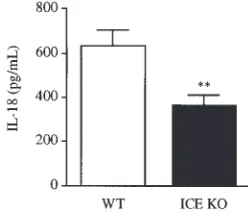
Reduced release of IL-18 in ICE KO mice. To verify whether splenocytes from ICE KO mice have a defective IL-18 release, spleen cells from WT and ICE KO mice were cul-tured for 24 hours in the absence of any exogenous stim-ulation. As shown in Figure 5, significantly lower levels of IL-18 were present in the supernatants of ICE KO splenocytes compared with WT cells.
Reduced serum IFN-γlevels in ICE KO mice after in vivo administration of IL-12. As shown in Figure 6, after daily administration of IL-12 (100 ng/mouse for 4 days), sig-nificantly lower levels of IFN-γwere present in the serum of ICE KO mice compared with WT mice. A similar observation was made when IL-12 was administered at 400 ng/mouse per day. However, with this higher dose of IL-12, the difference between ICE KO and WT mice was no longer statistically significant.
The effect of ICE deficiency is specific for IFN-γ induc-tion, because IL-12–induced serum TNF-αlevels did not differ between WT and ICE KO mice (162.35 ± 4.97 vs. 171.51 ± 14.76 pg/mL in WT and ICE KO mice, respec-tively, after 4 daily injections of IL-12 at 100 ng/mouse per day; n = 5).
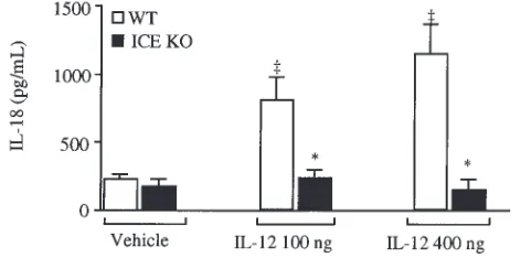
IL-12 administration increases serum and liver-associated IL-18 levels. Consistent with the low IFN-γ levels observed in ICE KO mice, administration of IL-12 at 100 or 400 ng/mouse per day induced a significant increase in circulating IL-18 levels in WT mice but not in ICE KO mice (Figure 7). Measurement of IL-18 in serum versus EDTA or heparin plasma leads to compa-rable results (data not shown).
[image:5.612.60.290.49.203.2]High levels of IL-18 were present in the freshly pre-pared liver homogenates of untreated, healthy mice (29.05 ± 5.6 and 28.55 ± 6.14 ng/g in WT and ICE KO
Figure 4
IL-18 production in unstimulated and IL-12–stimulated splenocytes. Splenocytes were cultured in RPMI-FBS for increasing amounts of time without added exogenous stimulation, or with IL-12 at 10 ng/mL. Cell-associated and released IL-18 levels were measured. Data are mean ± SEM of 9 mice per group.
Figure 5
[image:5.612.389.513.626.734.2]mice, respectively; n = 10). The presence of pro–IL-18 in the livers of untreated mice was confirmed by West-ern blot analysis (Figure 8). Administration of IL-12 at 400 ng/mouse per day significantly increased liver-associated IL-18 in both WT and ICE KO mice (112.79 ± 16.42 and 105.88 ± 38.13 ng/g in WT and ICE KO mice, respectively; n = 10). Contrary to what was observed in the serum, there were no significant dif-ferences between ICE KO mice and WT mice for liver-associated IL-18 production.
The IL-18 assay also detected high levels of IL-18 in the spleen homogenates of untreated mice. The presence of pro–IL-18 in the spleens of untreated mice was con-firmed by Western blot analysis (Figure 8). IL-12 admin-istration did not significantly alter spleen-associated IL-18 levels when expressed as nanograms per gram of tissue (data not shown). However, because IL-12 admin-istration induced a marked splenomegaly (3), the amount of IL-18 produced per spleen was significantly higher in animals treated with 400 ng/d of IL-12 (30.53 ng IL-18/spleen) compared with the control group (6.35 ng/spleen; n = 5; P < 0.001 by unpaired Student’s ttest). No significant differences in spleen IL-18 content were observed between WT and ICE KO mice.
TNF-αwas below detectable limits in both the liver and the spleen homogenates of untreated mice, there-fore demonstrating that the high levels of IL-18 observed in these organs were unlikely to be due to the presence of underlying disease in the mice. In addition, as reported previously (5), IL-12 administration increased spleen-associated TNF-αlevels (0.20 ± 0.05 and 0.22 ng/g in WT and ICE KO mice, respectively, after 4 daily injections of IL-12 at 400 ng/mouse per day; n = 5).
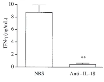
Neutralization of IL-18 reduces IFN-γproduction in vivo. The results suggest that IL-18 significantly contributes to
induction of IFN-γafter in vivo administration of IL-12. To further confirm these results, a neutralizing rabbit anti–IL-18 antiserum was administered before the first and third injection of IL-12 (100 ng/mouse per day). IFN-γ lev-els were measured in the serum 2 hours after the fourth injection. The control group received the same amount of NRS. As shown in Figure 9, neutralization of IL-18 reduced IL-12–induced IFN-γproduction significantly.
Discussion
In the present report, we have shown that endogenous IL-18 participates in the induction of IFN-γby IL-12 in mice, both in vitro and in vivo. The role of endogenous IL-18 in the regulation of IFN-γby microbial stimuli such as endotoxin, zymosan, or live bacteria has been demonstrated previously (13, 29). The novelty of the present report is the demonstration that neutralization of endogenous IL-18 significantly reduces production of IFN-γ when induced directly by IL-12, not by a microbial stimulus.
Our results imply that endogenous IL-18 plays an important and significant role in the induction of
IFN-γby IL-12. Similarly, IL-12–induced NK-cell activity requires endogenous IL-18, because lower cytotoxicity is observed in cells obtained from IL-18 KO mice (11). However, induction of both IFN-γand NK-cell activity by IL-12 are not completely IL-18–dependent, especial-ly when high doses of IL-12 are employed (ref. 11 and this paper).
[image:6.612.315.532.53.175.2]Inhibition of ICE activity or ICE deficiency reduced induction of IFN-γby IL-12, both in vitro and in vivo. The role of ICE in regulating IFN-γ production was through IL-18, not IL-1βprocessing. In fact, IFN-γ pro-duction in response to IL-12 was not affected by block-ade of IL-1 receptors. Moreover, IL-12 did not induce
Figure 6
Effect of in vivo administration of IL-12 on IFN-γlevels in ICE KO and WT mice. WT and ICE KO mice received 4 daily intraperitoneal injections of IL-12 (100 or 400 ng/mouse) or vehicle. Two hours after the fourth injec-tion, blood was collected and serum was prepared for measurement of IFN-γ. Data are mean ± SEM of 10 mice per group. *P < 0.05 vs. WT mice by factorial ANOVA.
Figure 7
Increase in serum IL-18 levels after IL-12 administration. WT and ICE KO mice received 4 daily intraperitoneal injections of IL-12 (100 or 400 ng/mouse) or vehicle. Two hours after the fourth injection, blood was collected and serum was prepared. Data are mean ± SEM of 10 mice per group. *P < 0.05 vs. WT mice. ‡P < 0.01 vs. corresponding vehicle by
[image:6.612.304.540.612.729.2]IL-1βproduction.
A newly developed assay for detection of IL-18 was used in the present study to evaluate the production of this cytokine in response to IL-12. Compared with other proinflammatory cytokines, IL-18 is regulated in a unique way. In fact, constitutive levels of IL-18 mRNA and protein are present in unstimulated human and murine cells and in the tissues of untreat-ed, healthy mice (refs. 20, 26, 30–33, and this paper). These data are consistent with the finding that one of the two IL-18 promoters is constitutively active (30). In unstimulated cells or tissues, IL-18 is present pri-marily in the precursor form, but requires conversion into the bioactive, mature molecule (26, 32). We observed constitutive intracellular levels of precursor IL-18 in freshly isolated spleen cells. Incubation of the cells at 37°C for 8–48 hours resulted in the release of IL-18 into the extracellular compartment in the absence of any exogenous stimulation. Although the assay used in this study for IL-18 determination detects both the precursor and mature forms, IL-18 found in the extracellular compartment is mostly in the mature form. Two sets of data support this con-cept: (a) reduced levels of extracellular IL-18 were observed in cultures of ICE KO splenocytes; and (b) a significant correlation was observed between extracel-lular IL-18 levels and IL-12–induced IFN-γproduction. In vitro, IL-18 is constitutively present and is available to act with a second stimulus, in this case IL-12. IL-18 alone is a weak inducer of IFN-γproduction, possibly owing to a low constitutive expression of IL-18 recep-tors on T cells (17). Therefore, it is not surprising that, even in the presence of mature, bioactive IL-18, no IFN-γis detectable in unstimulated spleen cell cul-tures; a second stimulus, such as IL-12, is necessary for induction of IFN-γ.
Extracellular levels of IL-18 were significantly reduced,
but not absent, in cultures of ICE KO spleen cells. Two explanations are possible for this phenomenon: (a) ICE is not the only enzyme responsible for the cleavage and release of IL-18 from the intracellular compartment; and (b) when ICE is not present or its activity is inhibited, IL-18 can be released from the cell in the precursor form. Both mechanisms have been demonstrated for cleavage and release of IL-1β(34–37).
An increase in circulating IL-18 levels was observed in vivo in IL-12–injected mice. This observation is in agreement with results obtained by Lauw et al. in IL-12–injected chimpanzees (21). Compared with WT mice, significantly lower levels of serum IFN-γwere observed in ICE KO mice after administration of IL-12 at 100 ng/mouse per day. Consistently, circulating IL-18 levels were low in ICE KO mice compared with WT mice, again demonstrating the important role of ICE in the cleavage and release of IL-18. Circulating IL-18 levels remained low in ICE KO mice even when high doses of IL-12 were administered. In this case, IL-12–induced IFN-γproduction was relatively inde-pendent of IL-18, because ICE KO mice had low IL-18 but high IFN-γ levels in their circulation. Similar results were obtained previously by evaluating IL-12–induced NK-cell cytotoxicity in IL-18 KO mice. Also, in that case, the effect of IL-12 was IL-18–depend-ent only when lower doses of IL-12 were used (11).
[image:7.612.322.523.51.144.2]Administration of IL-12, particularly at the higher dose, also significantly increased liver-associated IL-18 levels. The Kupffer cell is the most likely source for the hepatic production of IL-18 (14). Consistent with observations that tissue-associated IL-18 is mostly present in the precursor form (26), no differences in the liver IL-18 content were observed between WT and ICE KO mice. As reported previously (3), administration of IL-12 induced hepatomegaly and splenomegaly. No dif-ferences in these parameters were observed between WT
Figure 8
Constitutive expression of pro–IL-18 in the spleens and livers on untreat-ed mice. Spleen and liver homogenates from 3 untreatuntreat-ed mice were sub-jected to Western blot analysis with a specific rabbit anti-murine IL-18 antiserum. Recombinant murine pro–IL-18 and mature IL-18 were used as standards.
Figure 9
[image:7.612.322.491.606.739.2]and ICE KO mice (data not shown). As a consequence of the increase in organ weight, the total content of IL-18 in the liver, and particularly the spleen, was high-ly increased after administration of IL-12.
In conclusion, the present report demonstrates that ICE-cleaved endogenous IL-18 significantly contributes to induction of IFN-γby IL-12, both in vitro in cultures of murine splenocytes and in vivo in mice chronically inject-ed with IL-12. Although IL-12 administration does increase IL-18 levels, this is not strictly necessary, because IL-18 is constitutively produced in liver and spleen cells. It is possible that the constitutively expressed IL-18 binding protein (38) may serve as a natural mechanism for bind-ing constitutive IL-18 and controllbind-ing its bioactivity.
Acknowledgments
This work was supported by National Institutes of Health grants AI-15614 and CA-46934.
We wish to thank Richard A. Flavell for providing the ICE KO mice.
1. Trinchieri, G., and Gerosa, F. 1996. Immunoregulation by interleukin-12. J. Leukoc. Biol.59:505–511.
2. Dinarello, C.A., et al. 1998. Overview of interleukin-18: more than an interferon-γ inducing factor. J. Leukoc. Biol.63:658–664.
3. Car, B.D., et al. 1995. Role of interferon-γin interleukin 12-induced pathology in mice. Am. J. Pathol.147:1693–1707.
4. Orange, J.S., et al. 1995. Mechanism of interleukin 12-mediated toxici-ties during experimental viral infections: role of tumor necrosis factor and glucocorticoids. J. Exp. Med.181:901–904.
5. Sacco, S., et al. 1997. Protective effect of a single interleukin-12 (IL-12) predose against the toxicity of subsequent chronic IL-12 in mice: role of cytokines and glucocorticoids. Blood.90:4473–4479.
6. Cohen, J. 1995. IL-12 deaths: explanation and a puzzle. Science.270:908. 7. Bazan, J.F., Timans, J.C., and Kastelein, R.A. 1996. A newly defined
inter-leukin-1? Nature.379:591.
8. Gu, Y., et al. 1997. Activation of interferon-γinducing factor mediated by interleukin-1βconverting enzyme. Science.275:206–209.
9. Ghayur, T., et al. 1997. Caspase-1 processes IFN-γ inducing factor and regulates LPS-induced IFN-γproduction. Nature.386:619–623. 10. Puren, A.J., Razeghi, P., Fantuzzi, G., and Dinarello, C.A. 1998.
Inter-leukin-18 enhances lipopolysaccharide-induced interferon-gamma pro-duction in human whole blood cultures. J. Infect. Dis.178:1830–1834. 11. Takeda, K., et al. 1998. Defective NK cell activity and Th1 response in
IL-18-deficient mice. Immunity.8:383–390.
12. Hyodo, Y., et al. 1999. IL-18 up-regulates perforin-mediated NK activity without increasing perforin messenger RNA expression by binding to constitutively expressed IL-18 receptor. J. Immunol.162:1662–1668. 13. Fantuzzi, G., Puren, A.J., Harding, M.W., Livingston, D.J., and Dinarello,
C.A. 1998. Interleukin-18 regulation of interferon γproduction and cell proliferation as shown in interleukin-1β-converting enzyme (caspase-1)-deficient mice. Blood.91:2118–2125.
14. Okamura, H., et al. 1995. Cloning of a new cytokine that induces IFN-γ production by T cells. Nature.378:88–91.
15. Ushio, S., et al. 1996. Cloning of the cDNA for human IFN-γ inducing factor, expression in Escherichia coli, and studies on the biologic activities of the protein. J. Immunol.156:4274–4279.
16. Robinson, D., et al. 1997. IGIF does not drive Th1 development but syn-ergizes with IL-12 for interferon-γproduction and activates IRAK and NFκB. Immunity.7:571–581.
17. Yoshimoto, T., et al. 1998. IL-12 up-regulates IL-18 receptor expression on T cells, Th1 cells, and B cells: synergism with IL-18 for IFNγ produc-tion. J. Immunol.161:3400–3407.
18. Ahn, H.-J., et al. 1997. A mechanism underlying synergy between IL-12 and IFN-γ inducing factor in enhanced production of IFN-γ. J. Immunol.
159:2125–2131.
19. Barbulescu, K., et al. 1998. IL-12 and IL-18 differentially regulate the transcriptional activity of the human IFN-γ promoter in primary CD4+ T lymphocytes. J. Immunol.160:3642–3647.
20. Bohn, E., et al. 1998. IL-18 (IFN-γ inducing factor) regulates early cytokine production in, and promotes resolution of, bacterial infection in mice. J. Immunol.160:299–307.
21. Lauw, F.N., et al. 1999. Interleukin-12 induces sustained activation of multiple host inflammatory mediator systems in chimpanzees. J. Infect. Dis.179:646–652.
22. Kuida, K., et al. 1995. Altered cytokine export and apoptosis in mice defi-cient in interleukin-1βconverting enzyme. Science.267:2000–2003. 23. Coligan, J.E., Kruisbeek, A.M., Margulies, D.H., Shevach, E.M., and
Strober, W. 1992. Current protocols in immunology. Volume 1. John Wiley & Sons. New York, NY. 3.1.3–3.1.5.
24. Deaver, D.R. 1995. A new non-isotopic detection system for immunoas-says. Nature.377:758–760.
25. Fantuzzi, G., Sacco, S., Ghezzi, P., and Dinarello, C.A. 1997. Physiologi-cal and cytokine responses in interleukin-1β-deficient mice after zymosan-induced inflammation. Am. J. Physiol.273:R400–R406. 26. Puren, A.J., Fantuzzi, G., and Dinarello, C.A. 1999. Gene expression,
syn-thesis, and secretion of interleukin 18 and interleukin 1βare differen-tially regulated in human blood mononuclear cells and mouse spleen cells. Proc. Natl. Acad. Sci. USA.96:2256–2261.
27. Thornberry, N.A., et al. 1994. Inactivation of interleukin-1 beta convert-ing enzyme by peptide (acyloxy)methyl ketones. Biochemistry.
33:3934–3940.
28. Black, R.A., et al. 1988. Generation of biologically active interleukin-1 beta by proteolytic cleavage of the inactive precursor. J. Biol. Chem.
263:9437–9442.
29. Mastroeni, P., et al. 1999. Interleukin 18 contributes to host resistance and gamma interferon production in mice infected with virulent Salmo-nella typhimurium. Infect. Immun.67:478–483.
30. Tone, M., Thompson, S.A.J., Tone, Y., Fairchild, P.J., and Waldmann, H. 1997. Regulation of IL-18 (IFN-γ inducing factor) gene expression. J. Immunol.159:6156–6163.
31. Marshall, J.D., et al. 1999. Regulation of human IL-18 mRNA expression.
Clin. Immunol.90:15–21.
32. Olee, T., Hashimoto, S., Quach, J., and Lotz, M. 1999. IL-18 is produced by articular chondrocytes and induces proinflammatory and catabolic responses. J. Immunol.162:1096–1100.
33. Xu, B., et al. 1998. Expression of interleukin-18 in murine contact hyper-sensitivity. J. Interferon Cytokine Res.18:653–659.
34. Fantuzzi, G., et al. 1997. Response to local inflammation of IL-1β con-verting enzyme-deficient mice. J. Immunol.158:1818–1824.
35. Nylander-Lundqvist, E., and Egelrud, T. 1997. Formation of active IL-1 beta from pro-IL-1 beta catalyzed by stratum corneum chymotryptic enzyme in vitro. Acta Derm. Venereol.77:203–206.
36. Miller, B.E., et al. 1995. Inhibition of mature IL-1βproduction in murine macrophages and a murine model of inflammation by WIN 6794, an inhibitor of IL-1βconverting enzyme. J. Immunol.154:1331–1338. 37. Beuscher, H.U., Gunther, C., and Rollinghoff, M. 1990. IL-1βis secreted
by activated murine macrophages as biologically inactive precursor. J. Immunol.144:2179–2183.