systemic lupus erythematosus: an evolving
paradigm
Gary M. Kammer
J Clin Invest.
2005;115(4):836-840. https://doi.org/10.1172/JCI24791.
In systemic lupus erythematosus (SLE), IL-2 production by T lymphocytes in vitro is
impaired. Deficient IL-2 production may be an outcome of a primary SLE T cell disorder that
is due to impaired signal transduction. In this issue of the
JCI
, evidence is presented that an
anti-TCR/CD3 complex autoantibody present in SLE sera can bind to T cells and activate
the Ca
2+-calmodulin kinase IV (CaMKIV) signaling cascade, resulting in downregulation of
IL-2
transcription and IL-2 production. Because IL-2 may contribute to the maintenance of T
cell tolerance, deficient IL-2 production could promote a breach of T cell tolerance that
results in autoantibody production in SLE.
Commentary
Find the latest version:
Acknowledgments
J.A. Helms is supported in part by the Oak Foundation.
Address correspondence to: Jill A. Helms, Department of Surgery, Stanford Uni-versity, 257 Campus Drive, GK 207, Stan-ford, California 94305, USA. Phone: (650) 736-0919; Fax: (650) 736-4374; E-mail: [email protected].
1. Massa, N. 1536. Liber introductorius anatomiae, sive dissectionis corporis humani. Venetiis. Cap.
21:37–38.
2. Riddle, R.D., Johnson, R.L., Laufer, E., and Tabin, C. 1993. Sonic hedgehog mediates the polarizing activity of the ZPA. Cell.75:1401–1416.
3. Litingtung, Y., Dahn, R.D., Li, Y., Fallon, J.F., and Chiang, C. 2002. Shh and Gli3 are dispensable for limb skeleton formation but regulate digit number and identity. Nature.418:979–983.
4. Dahn, R.D., and Fallon, J.F. 2000. Interdigital regulation of digit identity and homeotic trans-formation by modulated BMP signaling. Science.
289:438–441.
5. Colnot, C., et al. 2005. Indian hedgehog synchro-nizes skeletal angiogenesis and perichondrial mat-uration with cartilage development. Development.
132:1057–1067.
6. St-Jacques, B., Hammerschmidt, M., and McMa-hon, A.P. 1999. Indian hedgehog signaling regu-lates proliferation and differentiation of chondro-cytes and is essential for bone formation. Genes Dev.
13:2072–2086.
7. Chung, U.I., Schipani, E., McMahon, A.P., and Kronenberg, H.M. 2001. Indian hedgehog couples chondrogenesis to osteogenesis in endochondral bone development. J. Clin. Invest.107:295–304. 8. Archer, C.W., Dowthwaite, G.P., and Francis-West,
P. 2003. Development of synovial joints. Birth Defects Res. C. Embryo Today.69:144–155. 9. Hartmann, C., and Tabin, C.J. 2001. Wnt-14 plays
a pivotal role in inducing synovial joint forma-tion in the developing appendicular skeleton. Cell.
104:341–351.
10. Niedermaier, M. 2005. An inversion involving the mouse Shh locus results in brachydactyly through dysregulation of Shh expression. J. Clin. Invest.
115:900–909. doi:10.1172/JCI200523675. 11. Vortkamp, A., et al. 1996. Regulation of rate of
cartilage differentiation by Indian hedgehog and PTH-related protein. Science.273:613–622. 12. Hu, D., Marcucio, R.S., and Helms, J.A. 2003. A zone
of frontonasal ectoderm regulates patterning and growth in the face. Development.130:1749–1758. 13. Hu, D., and Helms, J.A. 1999. The role of sonic
hedgehog in normal and abnormal craniofacial morphogenesis. Development.126:4873–4884. 14. Jeong, J., Mao, J., Tenzen, T., Kottmann, A.H., and
McMahon, A.P. 2004. Hedgehog signaling in the neural crest cells regulates the patterning and growth of facial primordia. Genes Dev.18:937–951. 15. Gorlin, R.J., Cohen, M.M., and Levin, L.S. 1990. Syn-dromes of the head and neck. Oxford University Press. New York, New York, USA. 977 pp.
16. Garciadiego-Cazares, D., Rosales, C., Katoh, M., and Chimal-Monroy, J. 2004. Coordination of chondrocyte differentiation and joint formation by alpha5beta1 integrin in the developing appen-dicular skeleton. Development.131:4735–4742. 17. Dahmann, C., and Basler, K. 2000. Opposing
tran-scriptional outputs of Hedgehog signaling and engrailed control compartmental cell sorting at the Drosophila A/P boundary. Cell.100:411–422. 18. Sarkar, L., et al. 2000. Wnt/Shh interactions regulate
ectodermal boundary formation during mamma-lian tooth development. Proc. Natl. Acad. Sci. U. S. A.
97:4520–4524.
19. Larsen, C.W., Hirst, E., Alexandre, C., and Vincent, J.P. 2003. Segment boundary formation in Dro-sophila embryos. Development.130:5625–5635.
Altered regulation of IL-2 production in systemic
lupus erythematosus: an evolving paradigm
Gary M. Kammer
Arthritis Associates Inc., Willoughby, Ohio, USA.
In systemic lupus erythematosus (SLE), IL-2 production by T lymphocytes
in vitro is impaired. Deficient IL-2 production may be an outcome of a
primary SLE T cell disorder that is due to impaired signal transduction.
In this issue of the
JCI
, evidence is presented that an anti-TCR/CD3
com-plex autoantibody present in SLE sera can bind to T cells and activate
the Ca
2+-calmodulin kinase IV (CaMKIV) signaling cascade, resulting in
downregulation of
IL-2
transcription and IL-2 production (see the related
article beginning on page 996). Because IL-2 may contribute to the
mainte-nance of T cell tolerance, deficient IL-2 production could promote a breach
of T cell tolerance that results in autoantibody production in SLE.
Recently, it has been recognized that diverse autoantibodies directed against intra- and extracellular autoantigens exist in patients with systemic lupus erythematosus (SLE) for years before the clinical diagnosis is made (1); this suggests that physiologic mechanisms
that maintain tolerance to self antigens have been breached. Tolerance to self antigens is established and preserved by a subpopula-tion of T lymphocytes known as Tregs (2), and the loss of tolerance is a pathologic pro-cess giving rise to autoimmunity. This cir-cumstance raises the possibility of the exis-tence of abnormal T cell clones that mediate defective helper and suppressor effector functions, which result in autoantibody generation by forbidden B cell clones. In SLE, defective signaling cascades are believed to give rise to a primary T cell disorder that is characterized by impaired effector func-tions (3). These effector dysfuncfunc-tions are, at least in part, a result of skewed expression of
various effector molecules, including CD40 ligand (e.g., CD154) and multiple cytokines, and may reflect an imbalance of gene expres-sion. An extracellular factor(s) in the micro-environment that interacts with T cells and exacerbates these dysfunctions has not been previously identified.
Tregs, skewed cytokine production, and loss of tolerance
Impaired effector T cell functions due to skewed cytokine production may create a microenvironment that promotes a strong Th2 immune response relative to Th1 and Treg activity. Relative overproduction of IL-4, IL-6, and IL-10 by Th2 cells and underpro-duction of IL-2, IL-12, TGF-β, and IFN-γ by Th1 cells and Tregs can result in imbalanced autocrine and paracrine effects on T and B cells in the microenvironment. Because of the reduced numbers of CD4+CD25+ Tregs
(4) as well as the diminished generation of IL-2 and TGF-β, there may be insufficient suppressor activity in SLE to counterbalance the enhanced Th2 effect on B cell antibody production. Taken together, these conditions create a microenvironment that promotes a
Nonstandard abbreviations used: CaMKIV, Ca2+
-calmodulin kinase IV; CRE, cAMP response element; CREB, CRE-binding protein; CREM, CRE modulator; IL-2R, IL-2 receptor; pCREB, phosphorylated CREB; pCREM, phosphorylated CREM; PKA-IIβ, type IIβ PKA; pRIIβ, phosphorylated RIIβ; RIIβ, β type II regulatory subunit; SLE, systemic lupus erythematosus.
Conflict of interest: The author has declared that no conflict of interest exists.
dysregulated immune response driving both physiologic and forbidden B cell clones to overproduce antibodies and autoantibodies, which results in hypergammaglobulinemia. Moreover, these events occur despite the existence of other primary counterregulato-ry mechanisms, including expression of the cell surface molecule cytotoxic T lymphocyte antigen 4 (CTLA-4)(5).
IL-2 is a key cytokine that has been held to function predominantly as a growth factor. This cytokine is largely produced by activated CD4+ and CD8+ T cells and
binds to high-affinity cell surface IL-2 receptors (IL-2Rs) expressed by T cells, B cells, NK cells, and APCs. However, current evidence from analyses of IL-2–/–
and IL-2R–/– knockout mice supports the
notion that IL-2 may operate, not as a principal growth factor in vivo, but as a third signal that stimulates clonal expan-sion of effector cells to promote tolero-genic responses and to regulate develop-ment and function of CD4+CD25+ Tregs
and, possibly, CD8+ Tregs to maintain
tolerance (6, 7). Although much less is known about the mechanisms of IL-2 function in humans, it seems reasonable to suppose that IL-2 may serve a parallel role in immune homeostasis.
Mechanisms of deficient IL-2 production by SLE T cells
Deficient IL-2 production may predispose individuals to impaired
immunoregula-tion, loss of tolerance, and the development of SLE owing to the abrogation of suppres-sor mechanisms that maintain tolerance to self antigens. Two lines of evidence support this concept. First, it has been demonstrat-ed that T cells from animal models of lupus as well as patients with SLE produce low amounts of IL-2 in vitro (8, 9). Second, vac-cination of MRL/lpr lupus mice with live vaccinia recombinant viruses expressing the human IL-2 gene ameliorated disease activity (10). However, at the time that the IL-2 deficiency was discovered, the mecha-nisms leading to deficient IL-2 production by SLE T cells were unknown.
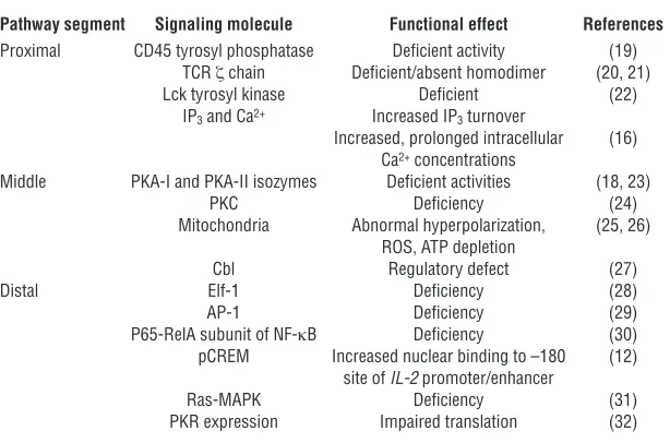
In SLE, a primary T cell disorder has been proposed to exist based on the identifica-tion of multiple discrete signaling abnor-malities at the level of the TCR/CD3 com-plex, the cytosol, and the nucleus (3, 11) (summarized in Table 1). Tsokos, Kammer, and their colleagues first proposed that a primary failure of T cells due to defective signaling could hinder IL-2 gene transcrip-tion and IL-2 productranscrip-tion and contribute to impaired T cell effector functions in SLE (12). To date, the data support this notion (13). Figure 1 presents a schematic of the mechanisms identified to date in SLE T cells that contribute to the downregula-tion of IL-2 producdownregula-tion. However, extra-cellular factors in the microenviron ment that impinge on the SLE T cell to further modify its immunoregulatory functions could also exist.
IgG anti-TCR/CD3 autoantibodies and reduced IL-2 generation
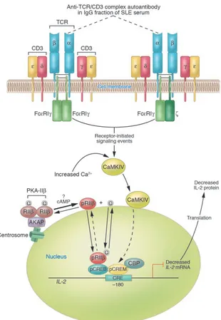
In this issue of the JCI, Tsokos and col-leagues present an incisive series of experi-ments that demonstrate that there is a factor in the microenvironment that can induce inhibition of IL-2 production by SLE T cells (14). Here, these authors show for what is believed to be the first time that the IgG fraction of SLE serum possesses an anti-TCR/CD3 complex autoantibody(s) that stimulates translocation of Ca2+-calmodulin
kinase IV (CaMKIV) from the cytosol to the nucleus. This event induces upregulation of the CREM (cAMP response element [CRE] modulator) transcript and CREM protein, proposed phosphorylation of CREM (to pCREM), binding of pCREM homodimers as well as pCREM–phosphorylated CRE-binding protein (pCREB) heterodimers to the –180region of the IL-2 promoter/ enhancer, and leads to reduced IL-2 tran-scription and IL-2 production (Figure 1).
It has not been previously shown that an IgG autoantibody in SLE serum can activate a signaling cascade in T cells, although it is well known that serum fac-tors can activate signaling pathways in diverse cell types. Indeed, we have previ-ously found that SLE serum does not induce the defects of PKA signaling in normal T cells that have been identified inSLE T cells (15). To determine whether such an activating factor exists in SLE serum, Tsokos and his associates first investigated whether SLE sera could cause increased expression of nuclear pCREM relative to pCREB in normal T cells, just as had been observed in SLE T cells. Indeed, in normal T cells cultured in the presence of SLE sera, but not sera from patients with rheumatoid arthritis or from healthy controls, the authors observed increased levels of the CREM transcript, CREM pro-tein, and pCREM binding to the –180 CRE region of the IL-2 promoter/enhancer. These findings revealed that SLE sera con-tain a factor capable of inducing nuclear CREM expression (14).
In subsequent experiments, the authors identified the protein kinase that medi-ates the overexpression of CREM. Based on their previous recognition that signals from the TCR/CD3 complex significantly stimulate the production of increased con-centrations of intracellular Ca2+ in SLE T
cells (16), they searched for a Ca2+
[image:3.585.54.361.111.314.2]-depen-dent kinase and found that SLE T cells exhibited increased amounts of nuclear CaMKIV compared to nuclear CaMKII in
Table 1
Abnormal signaling molecules in SLE T cells
Pathway segment Signaling molecule Functional effect References
Proximal CD45 tyrosyl phosphatase Deficient activity (19)
TCR ζ chain Deficient/absent homodimer (20, 21)
Lck tyrosyl kinase Deficient (22)
IP3 and Ca2+ Increased IP3 turnover
Increased, prolonged intracellular (16) Ca2+ concentrations
Middle PKA-I and PKA-II isozymes Deficient activities (18, 23)
PKC Deficiency (24)
Mitochondria Abnormal hyperpolarization, (25, 26) ROS, ATP depletion
Cbl Regulatory defect (27)
Distal Elf-1 Deficiency (28)
AP-1 Deficiency (29)
P65-RelA subunit of NF-κB Deficiency (30)
pCREM Increased nuclear binding to –180 (12) site of IL-2 promoter/enhancer
Ras-MAPK Deficiency (31)
PKR expression Impaired translation (32)
AP-1, activator protein–1; IP3, inositol triphosphate; PKR, dsRNA-regulated protein kinase. Modified
normal T cells. When normal T cells were then incubated with SLE or normal con-trol sera, transiently transfected with a CaMKIV expression plasmid, and activated with a phorbol ester and ionomycin, T cells exposed to SLE sera exhibited significantly greater pCREM binding to the –180 region of the IL-2 promoter/enhancer (14). When these T cells were also transiently trans-fected with an IL-2 promoter–reporter
luciferase construct, the cells incubated with SLE sera expressed significantly lower IL-2 promoter luciferase activity and IL-2 production compared with cells incubated in normal sera.Taken together, these results provide firm evidence that an extracellular factor in SLE sera can activate
CaMKIV and induce its nuclear transloca-tion from the cytosol, where it upregulates CREM expression, pCREM binding to the
IL-2 promoter/enhancer, and inhibition of
IL-2 transcription and IL-2 production. To identify the SLE serum factor, the authors postulated that the TCR/CD3 complex is the target for a circulating IgG autoantibody(s) in SLE serum. They per-formed a subtraction experiment using wild-type and mutant Jurkat T cells that do not express the surface TCR/CD3 complex-es. Absorption of the IgG autoantibody(s) by wild-type Jurkat T cells but not TCR/ CD3– mutant Jurkat T cells eliminated
the ligand(s) for the TCR/CD3 complex, reducing CaMKIV nuclear translocation
and pCREM binding to the IL-2 promoter/ enhancer (14). Although the authors did not perform an experiment to identify the sub-unit of the TCR/CD3 complex against which the autoantibody(s) is directed, the results support the notion that SLE sera possess an anti-TCR/CD3 complex autoantibody(s) that can bind to a subunit(s) of the TCR/ CD3 complex and activate the CaMKIV sig-naling cascade in T cells (Figure 1).
[image:4.585.52.362.78.527.2]Movement of signaling molecules between intracellular compartments is a well-known concept in cell biology. Under homeostatic conditions, signal-ing molecules are localized to specific intracellular regions; in response to a stimulus, these molecules can translocate
Figure 1
Diagram of established and proposed mech-anisms contributing to reduced IL-2 produc-tion by SLE T cells. A primary T cell disorder characterized by multiple abnormal signaling molecules has been identified in human SLE. A proportion of TCR ζ chains are replaced with FcεRIγ chains through which signaling can take place. In the microenvironment, anti-TCR/CD3 autoantibody(s) can bind to SLE T cells via the TCR/CD3 complex; however, the subunit(s) of the TCR/CD3 complex against which the autoantibody(s) is directed has not yet been identified. For the purpose of this illustration, the autoantibody is proposed to be against the α subunit of the TCR (dotted lines). Autoantibody binding to the TCR/CD3 complex results in heightened intracellular Ca2+ concentrations and activation of
the CaMKIV signaling cascade. CaMKIV translocates to the nucleus, is proposed to phosphorylate CREM, and induces increased pCREM binding to the CRE located at the –180 region of the IL-2 promoter/enhancer. Because there is overexpression of the RIIβ
to a target to perform a specific function, such as phosphorylation. cAMP-depen-dent activation of type IIβ PKA (PKA-IIβ) releases the β type II regulatory subunit (RIIβ) from its binding site on A-kinase anchor proteins (AKAPs) and its interac-tion with the catalytic subunit, permit-ting its autophosphorylation and trans-location from the cytosol to the nucleus in T cells (17) (Figure 1). Once in the nucleus, phosphorylated RIIβ (pRIIβ) binds to pCREB, inhibits activator pro-tein–1 formation, and strongly inhibits
IL-2 transcription and IL-2 production. In SLE, the accumulation of RIIβ in the nuclei of T cells from some patients seems likely to contribute to reduced IL-2 production (18) (Figure 1). The identi-fication of CaMKIV in the nuclei of SLE T cells is the second example of cytosol-to-nuclear translocation of a protein kinase. Despite our incomplete under-standing of the repressor mechanisms involved at the level of the basal tran-scription machinery, the recognition that both nuclear CaMKIV and pRIIβ have the effect of downregulating IL-2 production suggests complementary functions with a powerful outcome (14, 17).
The weight of evidence to date has led investigators to propose that the signaling abnormalities previously identified in SLE T cells reflect a disorder primary to the T cell (3, 11). Here, Tsokos and his colleagues present the first credible evidence that an extracellular factor(s) in the microenviron-ment can trigger a signaling cascade that ultimately modulates IL-2 transcription in T cells (14). It may be that extracellular fac-tors further hinder an already impaired T cell by activating functional pathways via cell surface receptors.
Reduced IL-2 production, effector and Treg functions, and tolerance in SLE
Two fundamental concepts seem to be emerging from the IL-2–/– and IL-2R–/–
knockout murine models that may become applicable to the study of SLE in humans. The first is that IL-2 is nec-essary for effective Treg maintenance of homeostasis via mechanisms involving anergy and apoptosis. Loss of IL-2 in the murine system is associated with reduced numbers of CD4+CD25+ T cells and
auto-immunity; restoration of IL-2 prevents autoimmunity and generates homeosta-sis. The second is that IL-2 may operate as a third signal for the expansion,
develop-ment, and function of antigen-dependent T cell effectors (6, 7).
In human SLE, reduced IL-2 genera-tion may be a key factor underlying the impaired T cell effector functions and breach of tolerance that result in auto-antibody production. In the murine sys-tem, development of CD4+CD25+ Tregs in
the thymus and their subsequent export to and expansion in the periphery are dependent on IL-2 (7). Whether there is a similar requirement for IL-2 in the devel-opment and expansion of CD4+CD25+
Tregs in humans remains uncertain (6, 7). Although there is still limited informa-tion about CD4+CD25+ Treg function in
human SLE, one might hypothesize that the reduced numbers of these Tregs in the periphery in SLE (4) reflect a defect in their development, export, and/or prolif-eration in the periphery.
Future directions
Clearly there is a paucity of information about IL-2 function in human immunity and tolerance, the molecular mechanisms regulating its expression in T cells, and its contribution to the immunopathogenesis of SLE. This gap in knowledge presents a compelling opportunity to investigate these issues. Among possible future direc-tions, establishing the mechanisms of IL-2 regulation of homeostasis in humans should generally be emphasized. Because there is consensus that a breach of T cell tolerance contributes to the persistence of autoreactive T cells and autoantibody production, mechanisms that may result in this failure should be intensively investi-gated. In this vein, the contribution by Tso-kos and his colleagues in this issue of the
JCI (14) provides a new appreciation and insight into how the microenvironment in SLE can further impinge on a defective T cell to inhibit IL-2 production. From such studies will come the inspiration and novel approaches necessary to develop therapeu-tic tools to abate disease and improve the quality of life of our patients.
Address correspondence to: Gary M. Kam-mer, Arthritis Associates Inc., 36100 Euclid Avenue, Suite 260, Willoughby, Ohio 44094, USA. Phone: (440) 8700; Fax: (440) 953-8796; E-mail: [email protected].
1. Arbuckle, M.R., et al. 2003. Development of autoan-tibodies before the clinical onset of systemic lupus erythematosus. N. Engl. J. Med.349:1526–1533. 2. Thompson, C., and Powrie, F. 2004. Regulatory T
cells. Curr. Opin. Pharmacol.4:408–414.
3. Kammer, G.M., Perl, A., Richardson, B.C., and Tsokos, G.C. 2002. Abnormal T cell signal transduction in systemic lupus erythematosus. Arthritis Rheum.46:1139–1154.
4. Liu, M.F., Wang, C.-R., Fung, L.-L., and Wu, C.-R. 2004. Decreased CD4+CD25+ T cells in peripheral blood of patients with systemic lupus erythemato-sus. Scand. J. Immunol.59:198–202.
5. Jiang, H., and Chess, L. 2004. An integrated view of suppressor T cell subsets in immunoregula-tion. J. Clin. Invest.114:1198–1208. doi:10.1172/ JCI200423411.
6. Nelson, B.H. 2004. IL-2, regulatory T cells, and tol-erance. J. Immunol.172:3983–3988.
7. Malek, T.R., and Bayer, A.L. 2004. Tolerance, not immunity, crucially depends on IL-2. Nat. Rev. Immunol.4:665–674.
8. Wofsy, D., et al. 1981. Deficient interleukin 2 activ-ity in MRL/Mp and C57BL/6J mice bearing the lpr gene. J. Exp. Med.154:1671–1680.
9. Alcocer-Varela, J., and Alarcon-Segovia, D. 1982. Decreased production of and response to inter-leukin-2 by cultured lymphocytes from patients with systemic lupus erythematosus. J. Clin. Invest.
69:1388–1392.
10. Gutierrez-Ramos, J.C., Andreu, J.L., Revilla, Y., Vinuela, E., and Martinez, C. 1990. Recovery from autoimmunity of MRL/lpr mice after infection with an interleukin-2/vaccinia recombinant virus. Nature.346:271–274.
11. Dayal, A.K., and Kammer, G.M. 1996. The T cell enigma in lupus. Arthritis Rheum.39:23–33. 12. Solomou, E.E., Juang, Y.-T., Gourley, M.F., Kammer,
G.M., and Tsokos, G.C. 2001. Molecular basis of deficient IL-2 production in T cells from patients with systemic lupus erythematosus. J. Immunol.
166:4216–4222.
13. Kyttaris, V.C., and Tsokos, G.C. 2004. T lympho-cytes in systemic lupus erythematosus: an update. Curr. Opin. Rheumatol.16:548–552.
14. Juang, Y.-T., et al. 2005. Systemic lupus erythemato-sus serum IgG increases CREM binding to the IL-2 promoter and suppresses IL-2 production through CaMKIV. J. Clin. Invest.115:996–1005. doi:10.1172/ JCI200522854.
15. Kammer, G.M., Haqqi, T.M., Hasler, P., and Mal-emud, C.J. 1993. The effect of circulating serum factors from patients with systemic lupus erythe-matosus on protein kinase A (PKA) activity and PKA-dependent protein phosphorylation in T lym-phocytes. Clin. Immunol. Immunopathol.67:8–16. 16. Vassilopoulos, D., Kovacs, B., and Tsokos, G.C.
1995. TCR/CD3 complex-mediated signal transduction pathway in T cells and T cell lines from patients with systemic lupus erythematosus. J. Immunol.155:2269–2281.
17. Elliott, M.R., et al. 2004. Down-regulation of IL-2 production in T lymphocytes by phosphorylated protein kinase A-RIIb. J. Immunol.172:7804–7812. 18. Mishra, N., Khan, I.U., Tsokos, G.C., and Kammer,
G.M. 2000. Association of deficient type II protein kinase A activity with aberrant nuclear translocation of the RIIb-subunit in systemic lupus erythemato-sus T lymphocytes. J. Immunol.165:2830–2840. 19. Takeuchi, T., Pang, M., Amano, K., Koide, J., and
Abe, T. 1997. Reduced protein tyrosine phosphatase (PTPase) activity of CD45 on peripheral blood lym-phocytes in patients with systemic lupus erythema-tosus. Clin. Exp. Immunol.109:20–26.
20. Liossis, S.N.C., Ding, X.Z., Dennis, G.J., and Tsokos, G.C. 1998. Altered pattern of TCR/CD3-mediated protein-tyrosyl phosphorylation in T cells from patients with systemic lupus erythematosus. Defi-cient expression of the T cell receptor zeta chain. J. Clin. Invest.101:1448–1457.
21. Takeuchi, T., et al. 1998. TCRz chain lacking exon 7 in two patients with systemic lupus erythematosus. Int. Immunol.10:911–921.
expres-sion in peripheral blood lymphocytes from patients with systemic lupus erythematosus. Autoimmunity.
29:111–120.
23. Kammer, G.M., Khan, I.U., and Malemud, C.J. 1994. Deficient type I protein kinase A isozyme activity in systemic lupus erythematosus T lymphocytes. J. Clin. Invest.94:422–430.
24. Tada, Y., Nagasawa, K., Yamauchi, Y., Tsukamoto, H., and Niho, Y. 1991. A defect in the protein kinase C system in T cells from patients with systemic lupus erythematosus. Clin. Immunol. Immunopathol.
60:220–231.
25. Gergely, P., Jr., et al. 2002. Persistent mitochondrial hyperpolarization, increased reactive oxygen inter-mediate production, and cytoplasmic alkaliniza-tion characterize altered IL-10 signaling in patients with systemic lupus erythematosus. J. Immunol.
169:1092–1101.
26. Gergely, P., Jr., et al. 2002. Mitochondrial hyper-polarization and ATP depletion in patients with systemic lupus erythematosus. Arthritis Rheum.
46:175–190.
27. Yi, Y., McNerney, M., and Datta, S.K. 2000. Regula-tory defects in Cbl and mitogen-activated protein kinase (extracellular signal-related kinase) pathways cause persistent hyperexpression of CD40 ligand in human lupus T cells. J. Immunol.165:6627–6634. 28. Juang, Y.-T., Tenbrock, K., Nambiar, M.P., Gourley,
M.F., and Tsokos, G.C. 2002. Defective produc-tion of funcproduc-tional 98-kDa form of Elf-1 is respon-sible for the decreased expression of TCR z-chain in patients with systemic lupus erythematosus. J. Immunol.169:6048–6055.
29. Kyttaris, V.C., Juang, Y.-T., Tenbrock, K., Wein-stein, A., and Tsokos, G.C. 2004. Cyclic adenosine 5′-monophosphate response element
modula-tor is responsible for the decreased expression of c-fos and activator protein-1 binding in T cells from patients with systemic lupus erythematosus. J. Immunol.173:3557–3563.
30. Wong, H.K., Kammer, G.M., Dennis, G., and Tso-kos, G.C. 1999. Abnormal NF-kB activity in T lym-phocytes from patients with systemic lupus ery-thematosus is associated with decreased p65-RelA protein expression. J. Immunol.163:1682–1689. 31. Deng, C., et al. 2001. Decreased
ras-mitogen-acti-vated protein kinase signaling may cause DNA hypomethylation in T lymphocytes from lupus patients. Arthritis Rheum.44:397–407.
32. Grolleau, A., Kaplan, M.J., Hanash, S.M., Beretta, L., and Richardson, B.C. 2000. Impaired translational response and increased protein kinase PKR expres-sion in T cells from lupus patients. J. Clin. Invest.
106:1561–1568.
In hypertension, the kidney is not always
the heart of the matter
Michael E. Mendelsohn
Molecular Cardiology Research Institute, Tufts–New England Medical Center, Boston, Massachusetts, USA.
Blood pressure abnormalities are thought to originate from intrinsic
changes in the kidney, a concept that has been largely unchallenged for
more than 4 decades. However, recent molecular, cellular, and transgenic
mouse studies support an alternative hypothesis: primary abnormalities in
vascular cell function can also directly cause abnormalities of blood
pres-sure. In this issue of the
JCI
, Crowley and coworkers describe the application
of an elegant cross-renal transplant model to type 1A angiotensin (AT
1A)
receptor–deficient mice and their wild-type littermates to explore the
rela-tive contributions of renal and extrarenal tissues to the low blood pressure
seen in the AT
1Areceptor–deficient animals (see the related article beginning
on page 1092). Their studies further support the emerging paradigm that
primary abnormalities of the vasculature can make unique, nonredundant
contributions to blood pressure regulation; the findings have potentially
important implications for the ways we diagnose and treat blood pressure
diseases in humans.
Nonstandard abbreviations used: AT1A, type 1A
angiotensin; BKCa2+ large conductance Ca2+-activated K+
channel; CO, cardiac output; D+R+, donor+ recipient+;
IP3, inositol triphosphate; MLC, myosin light chain;
MLCK, MLC kinase; PKGI, cGMP-dependent protein kinase type I; PP1M, myosin phosphatase; RAS, renin-angiotensin system; RGS2, regulator of G protein signaling 2; SVR, systemic vascular resistance.
Conflict of interest: The author has declared that no conflict of interest exists.
Citation for this article:J. Clin. Invest.115:840–844 (2005). doi:10.1172/JCI200524806.
Introduction
Abnormalities of blood pressure, especially high blood pressure or hypertension, are widespread, cause extensive morbidity and mortality, and are insidious, because they rarely generate symptoms. But what causes hypertension? This simple question is remarkably difficult to answer, in large
part because blood pressure, which is mea-surable only in intact animals, is an inte-grated value determined by contributions from a complex mix of rheologic, renal, neural, vascular, and hormonal variables. It is therefore difficult to distinguish primary or causal factors for abnormal blood pres-sure from those responses that are second-ary to the changes in pressure.
Blood pressure is proportional to 2 funda-mental hemodynamic factors: cardiac out-put (CO) and systemic vascular resistance (SVR), and the equation BP = CO × SVR is taught to every first-year physiology stu-dent. The pioneering studies of Guyton and colleagues provide abundant support for the central, causal role of abnormal renal sodium handling in the etiology of hypertension (1, 2). This work
emphasiz-es the primacy of the kidney’s “premphasiz-essure control system,” which is recruited when systemic pressure is altered and causes a renally mediated increase or decrease in blood volume to establish long-term blood pressure control. Elegant genetic studies published over the past decade have provided further strong support for the hypothesis that alterations of net salt balance underlie abnormalities of blood pressure in humans (3). But are the alterations in renal salt balance that cause abnormal blood pressure always direct, as has been argued for many years, or could renal alterations in some cases be indirect or secondary to primary abnormalities of nonrenal tissues? Does the kidney always cause abnormal blood pressure by altering blood volume, which in turn alters CO and SVR? Or do intrinsic abnormalities of the vasculature in some cases give rise to a pri-mary abnormality of SVR, causing hyper-tension? Surprisingly, we do not yet know the answer to this simple dilemma.
In this issue of the JCI, Crowley and col-leagues (4) describe cross-renal transplan-tation studies with type 1A angiotensin (AT1A) receptor–deficient (Agtr1a–/–) mice