Theses and Dissertations
8-9-2014
The Effects of Exercise Training on Cognitive
Reserve and Cognitive Function in Healthy Older
Women
Katie Marie Becofsky
University of South Carolina - Columbia
Follow this and additional works at:https://scholarcommons.sc.edu/etd Part of theKinesiology Commons
This Open Access Dissertation is brought to you by Scholar Commons. It has been accepted for inclusion in Theses and Dissertations by an authorized administrator of Scholar Commons. For more information, please [email protected].
Recommended Citation
Becofsky, K. M.(2014).The Effects of Exercise Training on Cognitive Reserve and Cognitive Function in Healthy Older Women.(Doctoral
By
Katie Marie Becofsky
Bachelor of Arts
State University of New York at New Paltz, 2008
Master of Science
University of North Carolina at Greensboro, 2010
_______________________________________________________________
Submitted in Partial Fulfillment of the Requirements For the Degree of Doctor of Philosophy in
Exercise Science
The Normal J. Arnold School of Public Health University of South Carolina
2014 Accepted by:
Sara Wilcox, Co-Major Professor Roger Newman-Norlund, Co-Major Professor
Jim Fadel, Committee Member Xuewen Wang, Committee Member
J. Mark Davis, Committee Member
Norlund, Xuewen Wang, Jim Fadel, and Mark Davis. Each of you have served an important role in shaping the path I took with my dissertation project, and the future path I am pursuing as I move on to my postdoctoral work. I am especially grateful to Drs. Wilcox and Newman-Norlund for their mentorship.
Thank you to Dr. Booze, Dr. Prinz, Michele Blondin, and others involved in the Behavioral-Biomedical Interface Program (BBIP) at USC. Thank you to the Office of the Vice President for Research for supporting my dissertation project via the SPARC Graduate Fellowship. Thank you to the McCausland Center for Brain Imaging for supporting the project via the M-Fund mechanism. Thank you to Scott Vendemia for all his support with scanning at McCausland.
Thank you to Dr. Wang for allowing me to piggyback on the WeWalk study for my dissertation project. Thank you to the WeWalk staff, especially Madison DeMello and Kim Bowyer, for their help in coordinating my project with the larger study.
Thank you to Nicole Gribben, Brian Berry, and Philip Riddle for helping with my project. You are all sanity-savers.
from cognitive decline. At the brain level, greater cognitive reserve may manifest as greater neural network efficiency. Our purpose was to investigate 1) whether
participation in a 16-week walking program increased brain efficiency, and 2) whether increased brain efficiency correlated with change in fitness and task performance. Our secondary purpose was to investigate whether exercise training improved performance on a battery of cognitive tasks, particularly executive functioning performance. Seventeen healthy but sedentary women aged 60-75 years participated in a supervised walking program; eighteen women served as a non-randomized control group. Twelve women in the intervention group underwent fMRI scanning at baseline and post exercise training.
During fMRI scanning, participants (mean age 63 years) completed a working memory task (Sternberg delayed-match-to-sample letter task). Participants showed a greater capacity to recruit task-related brain regions after exercise training (indicated as greater BOLD signal). These regions included left inferior frontal gyrus, left cuneus, right rolandic operculum, left middle temporal gyrus, left postcentral gyrus, left superior med frontal, left superior frontal gyrus, right caudate, right inferior temporal gyrus (ps < 0.001). No task-related brain regions were utilized more efficiently after exercise
A slightly larger sample of intervention participants (n=17; mean age 64 years) completed a battery of cognitive tasks (CANTAB®) before beginning and after
Acknowledgements ... iv
Abstract ... v
List of Tables ... xi
List of Figures ... xii
Chapter 1. Introduction ... 1
Scope ... 3
Aims ... 3
Hypotheses ... 3
Chapter 2. Review of the Literature ... 5
Public Health Impact of Aging and Dementia (Alzheimer’s type) ... 5
Healthy Cognitive Aging: Changes in Brain & Behavior ... 6
Pathological Cognitive Aging: Changes in Brain & Behavior with AD ... 9
Lifestyle Choices and Dementia Risk ... 11
Physical Activity/Exercise and Dementia Risk ... 13
Physical Activity/Exercise and Cognitive Function ... 15
Neuroimaging in Physical Activity Research ... 18
Mediators and Moderators of the Exercise-Cognition Relationship ... 23
Study 1 ... 34
Purpose ... 34
Hypotheses ... 34
Design ... 35
Participants ... 35
Recruitment ... 36
Procedures ... 36
Measures ... 38
Statistical Analyses ... 42
Study 2 ... 45
Purpose ... 45
Hypotheses ... 45
Design ... 45
Participants ... 45
Recruitment ... 46
Procedures ... 46
Measures ... 47
Statistical Analyses ... 47
Chapter 4. Exercise and Cognitive Reserve: An fMRI Investigation in Healthy Older Women ... 49
Abstract ... 50
Discussion ... 62
Conclusions and Future Directions ... 67
Acknowledgements ... 68
Chapter 5. The Cognitive Effects of a supervised 16-weekWalking Intervention in Healthy Older Women ... 76
Abstract ... 77
Introduction ... 78
Methods ... 79
Results ... 86
Discussion ... 88
Conclusions and Future Directions ... 92
Acknowledgements ... 92
Chapter 6. Overall Summary and Conclusions ... 98
References ... 102
List of Tables
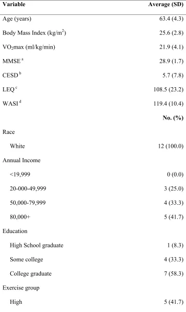
Table 4.1 Baseline demographic characteristics (n=12) ... 69
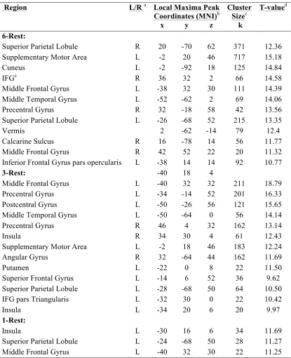
Table 4.2 Brain networks involved during rehearsal of the 1-, 3- and 6-letter sets (1-, 3-, or 6- letter task minus rest) at baseline ... 71
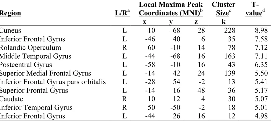
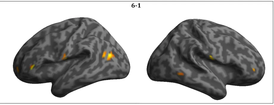
Table 4.3 Brain regions responding more robustly to high- versus low-demand trials (6-1) after exercise training ... 72
Table 5.1 Baseline demographic characteristics (n=35) ... 93
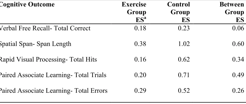
Table 5.2 Change in CANTAB® task performance by group ... 95
Table 5.3 Group differences in change in CANTAB® task performance ... 96
List of Figures
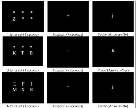
Figure 4.1 Delayed Match to Sample Letter Sternberg fMRI task ... 73 Figure 4.2 Task-related brain regions responding more robustly with greater
cognitive demand (6-1) after exercise training ... 74 Figure 4.3 Change in task-related BOLD signal with exercise training by task condition
Alzheimer’s disease (AD) is becoming a pressing public health concern. In a time when there is no effective treatment for AD, cognitive reserve (CR) theory suggests that a physically and mentally active lifestyle may help older adults stave off AD symptoms (i.e., the dementia) (Stern, 2006). Originally developed to explain the discordance between clinical symptoms of AD and severity of AD pathology upon autopsy, CR theory posits that ‘building’ CR by leading an active lifestyle allows an individuals’ brain to become more efficient, more flexible and/or more capable of recruiting necessary resources. The idea is that neural networks or circuits that are functioning in this manner are more difficult to disrupt, and therefore individuals with more CR should be better able to maintain their cognitive function despite, for example, advancing AD pathology. Importantly, it is believed that individuals can continue to build their CR throughout life; it is never too late to start.
individuals as a function of some proxy variable of mental activity (e.g., years of education).
The within-subjects approach can be used to study the neural implementation of CR in the older adult brain. This approach, in essence, uses increasing cognitive demands to mimic the challenge created by pathology. When using this approach, greater neural network efficiency may be expressed by lesser increases in network activation (within the same network) when switching from low to high-demand
conditions. Alternatively, greater neural network capacity may be expressed as greater increases during this switch, serving as evidence of the brain’s greater ability to recruit neural resources when facing a cognitive challenge. Research suggests that neural reserve in the older adult brain is more likely to manifest as greater efficiency than greater capacity (Scarmeas et al., 2003; Stern et al., 2005). Greater CR in the older adult brain can also be reflected in the ability to use compensatory resources (i.e., different or additional brain regions) in the face of increasing cognitive demands.
incidence of dementia (Physical Activity Guidelines Advisory Committee, 2008), and reduces the risk of AD (Hamer & Chida, 2009). Randomized controlled trials
complement these findings, suggesting that exercise training can improve or maintain cognitive function in the short term (Colcombe & Kramer, 2003). Neuroimaging studies using Stern’s approach could help unveil the mechanisms responsible for AD risk
reduction with higher levels of physical activity and, in doing so, help bolster exercise and physical activity promotion as a non-pharmacological means of addressing the rising incidence of AD.
Scope
This project examined the neural basis of exercise-induced CR in healthy older women using fMRI. The relationship between CR and cognitive performance was examined. In a separate study, the relationship between exercise training and cognitive performance was examined, irrespective of changes in CR.
Aims
1. To determine whether participation in an exercise training program builds CR, evidenced by greater efficiency in a task-related brain network.
2. To determine whether participation in an exercise training program improves cognitive performance.
Hypotheses
Aim 1
program. Changes in task-related activation were expected in premotor, parietal, inferior frontal and middle frontal areas (Zarahn, Rakitin, Abela, Flynn, & Stern, 2007).
1.2. It was hypothesized that greater improvement in fitness would correlate with greater improvement in efficiency in the working memory network. 1.3. It was hypothesized that greater efficiency in the working memory network
would correlate with better performance (i.e., lesser reaction time slope). Aim 2
2.1. It was hypothesized that participation in a 16-week exercise program would improve sustained attention, visuospatial learning, spatial working memory, and verbal free recall, as compared to no exercise; the greatest differential between groups was expected on tasks that challenge executive function capabilities (i.e., spatial span, rapid visual processing).
In the year 2010, 40 million Americans were 65 years or older, representing 13.1% of the population. As the baby boomer generation continues to age that number is projected to balloon to 72.1 million, or 19.3% of the population, by 2030 (U.S.
Department of Health and Human Services, 2011). The astonishing rate at which our population is aging has pushed the prevention and treatment of dementias to the forefront of the public health agenda. Alzheimer’s disease (AD) is by far the most common type of dementia, accounting for 60-80% of cases. It is also deadly; AD is the fifth leading cause of death in older adults. The latest report from the Alzheimer’s Association indicates that 1 in 3 older adults in the United States dies with AD or another dementia (Alzheimer's Association, 2014).
There is currently no effective treatment for AD, making prevention efforts critical. Researchers now know that pathological changes in the brain may begin more than two decades before AD symptoms present (Alzheimer's Association, 2014). With this knowledge, we know that it already too late to prevent the onset of AD pathology in the aging baby boomers. Luckily, although Alzheimer’s disease cannot be stopped, it is still possible that Alzheimer’s dementia can be. The cognitive reserve (CR) theory posits that certain exposures during life, such as mental and physical activity, may improve the brain’s ability to cope with damage. Thus, until scientists find ways to detect the disease in asymptomatic individuals and intervene effectively, prevention efforts with cognitively intact older adults, or even middle-aged adults, might focus on building CR. The tenets of CR theory serve as the basis of the current proposal.
Healthy Cognitive Aging: Changes in Brain & Behavior
decline more sharply after an individual reaches older adulthood (although it is unclear whether these late-life declines are related to disease processes) (Hedden & Gabrieli, 2004). The specificity of these behavioral changes (rather than a global decline in function) suggests that some areas of the brain are more affected by the aging process than others. Below I discuss some of the major structural and functional changes that occur in the ‘healthy’ aging brain that may contribute to changes (or stability) in cognitive performance.
Due a gradual reduction in synapses starting in the third decade of life, the aging brain is smaller in volume than the young brain. In line with observable changes in behavior, brain shrinkage is not uniform across regions (Hedden & Gabrieli, 2004). The prefrontal cortex (largely responsible for executive functioning) shows the largest
function) (Buckner, 2004). With normal aging, volumetric changes in the hippocampus and surrounding areas are less dramatic than those observed in prefrontal cortex; humans typically lose approximately 2-3% of their hippocampal volume per decade (Raz et al., 2004), and 1% annually after age 70 (Jack et al., 1998). In multiple structural MRI studies, hippocampal volume has been shown to predict memory performance after age 60 (Hedden & Gabrieli, 2004). Additionally, volumetric declines are not uniform across regions of the hippocampus and related medial temporal lobe structures; the entorhinal cortex and CA1 region of the hippocampus appear intact with normal aging, but the dentate gyrus and subiculum shrink (Hedden & Gabrieli, 2004).
In multiple fMRI studies with healthy older adults, dampened activity in the left hippocampus during memory task performance is often observed concurrently with alterations in prefrontal activation (usually increases) (Hedden & Gabrieli, 2004).
Conclusions. Even in the healthiest aged brain, structural and functional changes lead to mild decrements in some aspects of cognitive functioning (e.g., working memory, episodic memory). Subtle memory loss in older adulthood may be largely due to
prefrontal cortex-mediated executive function deficits (Buckner, 2004), which is etiologically different than memory loss due to AD (mediated by medial temporal lobe pathology) (Buckner, 2004). Structurally, loss of prefrontal cortex volume is a normal part of aging, while major shrinkage in medial temporal lobe structures tends to be indicative of pathology. There is also a normal decline in frontal white matter integrity, as well as neurotransmitter availability and function (particularly dopamine), with advancing age (Barnhart et al., 2009; Park & Reuter-Lorenz, 2009). Functionally, heightened activity is often found in the prefrontal cortex, while hippocampal activity tends to decline (Hedden & Gabrieli, 2004).
Pathological Cognitive Aging: Changes in Brain & Behavior with AD
Although some subtle changes in cognitive function are normal with advanced age, dementia is defined as disease-related loss of memory and other cognitive functions that interferes with activities of daily living (Jack, 2012). The transitional phase between normal cognitive aging and clinical dementia is known as mild cognitive impairment (MCI) (Jack, 2012). This discussion focuses primarily on AD, the most common cause of dementia; more specifically, the focus is on late onset sporadic AD, which accounts of 95% of AD cases (Alzheimer's Association, 2014).
The most common early symptom of AD is trouble remembering new
completing familiar tasks, confusion with time or place, problems with words in speaking or writing, and trouble understanding visual images or spatial relationships (Alzheimer's Association, 2014). Vascular dementia, caused by microscopic bleeding and vessel blockage, is the second most common type of dementia, and the most likely to co-occur with AD (Alzheimer's Association, 2014). The effects of AD and vascular brain injury appear to be additive (Jack, 2012), and the presence of vascular disease may be the difference between individuals with AD that develop symptoms of dementia and those that do not (Marchant et al., 2013).
AD Pathology. The two pathological hallmarks of AD are amyloid-! (A!) plaques and tau neurofibrillary tangles. The amyloid cascade hypothesis suggests that dysfunction in the A! pathway may be the initial event in AD, or at least one very early in the disease process. Late onset sporadic AD is thought to be a disease of inadequate A! clearance; as A! builds up, neuritic plaques surrounded by inflammatory cells and neuron fragments form in the neocortex (Jack, 2012). Recent evidence suggests that amyloid plaques are necessary but not sufficient for cognitive decline (Jack, 2012); at autopsy, about 30% of cognitively normal individuals have an accumulation of A!
plaques sufficient to meet criteria for AD. Importantly, these individuals usually do not have significant tau tangles (Jack, 2012).
Unlike plaques that form in the extracellular space, neurofibrillary tangles result from the intracellular aggregation of tau protein. In the healthy brain, tau protein binds to and supports the microtubules that form the neuronal cytoskeleton; in AD, tau disengages from the microtubules and binds with other tau threads to cause insoluble tangles.
Department of Health and Human Services, 2008). Tau pathology begins in the
transentorhinal area and progresses to the hippocampus, eventually reaching neocortical association areas, and lastly primary sensorimotor and visual areas (Braak, 1991; Jack, 2012). A closer association has been found between tau tangles and cognitive impairment than amyloid plaques and cognitive impairment, leading some researchers to argue that therapies should target tau rather than amyloid (Jack, 2012).
Conclusions. It can be very difficult to differentiate normal changes in cognitive aging from the slow, gradual onset of AD. Researchers now believe that AD-related changes in the brain may occur more than two decades before the first noticeable
symptoms of AD present themselves (Alzheimer's Association, 2014). The deposition of A! in the cortex is thought to precede the development of intercellular tau tangles; interestingly, cortical amyloid plaques are found in many non-demented individuals at autopsy (Braak, 1991). The trademark memory impairment associated with clinical AD is due largely to cellular pathology and cell loss in medial temporal lobe structures (Buckner, 2004). These structures (especially the entorhinal cortex, which is key in connecting the hippocampus with other brain regions) are the earliest affected by neurofibrillary tau tangles (Braak, 1991). There are many factors that contribute to dementia risk, and although some are out of our control, others are modifiable. As lifestyle choices may contribute substantially to an individuals’ risk profile (and they inherently lend themselves to intervention), they are the focus of the next section.
Lifestyle Choices and Dementia Risk
factors also increase AD risk, including mid-life obesity, diabetes, high cholesterol and midlife hypertension (Alzheimer's Association, 2014). As many lifestyle choices, such as leading an active lifestyle, eating a healthy diet, and not smoking, are known to lower cardiovascular disease risk, it is logical that they may also affect dementia or AD risk. Other factors, such as engagement in mental and social activities, years of education, and level of occupational attainment, have also been studied in relation to dementia risk. Below I highlight some recent studies and review papers focused on these topics. A sub-section focuses exclusively on the role of physical activity in reducing dementia or AD risk. Although cross-sectional studies exist, I review only longitudinal epidemiological studies, as they allow for causal inferences.
In a 2010 review paper, Arab and colleagues discussed Mediterranean diet adherence, supplement intake, and physical activity in relation to dementia or AD risk; although most Mediterranean diet and physical activity studies demonstrated a protective effect, supplement studies were less conclusive (Arab & Sabbagh, 2010). In 2009, Scarmeas et al reported that, when considered simultaneously, both Mediterranean-type diet adherence and physical activity independently reduced the risk of incident AD after an average 5.4 years of follow-up (Scarmeas et al., 2009). In 2012, Norton and colleagues investigated how lifestyle behaviors, specifically diet, exercise, smoking, alcohol
healthy diet, exercised, stayed socially engaged, and didn’t smoke) had a lower risk of AD. Follow-up averaged 6.3 years.
In 2006, Valenzuela and Sachdev conducted a meta-analysis of 22 longitudinal studies examining mental complexity in relation to incident dementia (Valenzuela & Sachdev, 2006). Most studies used years of education as their measure of ‘behavioural brain reserve’, while some used level of occupational attainment, premorbid IQ, or engagement in mental/social activities. After an average 7.1 years of follow-up, higher brain reserve was associated with a significantly lower risk of incident dementia (OR=.54). In 2012, Paillard-Borg and colleagues conducted a study based on 388 incident dementia cases that developed over a 9-year follow-up period, finding that an ‘active lifestyle’, defined as participation in mental, physical, or social activity, delayed the onset of dementia independent of education, medical condition, APoE genotype and other factors. Additionally, the broader the spectrum of activities an individual
participated in, the older the age of dementia onset (Paillard-Borg, Fratiglioni, Xu, Winblad, & Wang, 2012). A 2011 study by James et al focused on the protective effects of social activity, finding that, over an average of 5.2 years of follow-up, the rate of global cognitive decline was reduced 70% in older adults who were frequently socially active compared to those who were infrequently socially active (James, Wilson, Barnes, & Bennett, 2011).
Physical Activity/Exercise and Dementia Risk
and a reduced risk of dementia comes from longitudinal epidemiological studies (Rolland, 2008). In 2008, Rolland and colleagues conducted a systematic review of longitudinal epidemiological studies linking physical activity and risk of cognitive decline, dementia or AD (Rolland, 2008). Relevant studies dated back to 1991, and of the 24 studies reviewed, 20 showed a significant protective effect of physical activity against cognitive decline or dementia. Follow-up periods ranged from 2 to 21 years, and most assessed physical activity as self-reported leisure time physical activity.
Many relevant longitudinal studies have been published since the Rolland review. A 2012 study by Bowen et al found that older adults reporting greater participation in vigorous physical activity (e.g., aerobics, running, heavy housework) in the previous 3-7 years had a lower risk of dementia (Bowen, 2012). In 2012, Buchman and colleagues found that a higher level of total daily physical activity, measured for 10 days with actigraphy, was associated with a reduced risk of AD (average follow-up 4 years)
(Buchman et al., 2012). A 2012 study by Middleton et al found that total activity energy expenditure (measured as total energy expenditure measured using doubly labeled water minus resting metabolic rate measured using indirect calorimetry) was related to risk of cognitive impairment at 2 or 5 year follow-up; older adults in the highest sex-specific tertile of activity energy expenditure had the lower odds of declining at least one standard deviation on the Modified Mini-Mental State Examination compared to those in the lowest tertile (Middleton et al., 2011).
2012, Verdelho et al found that physical activity reduced the risk of MCI and dementia in older adults with vascular cerebral damage (white matter changes), but when dementia criteria were divided further into vascular dementia or AD, only vascular dementia risk was reduced (3 year follow up) (Verdelho et al., 2012).
Conclusions. An independent panel at a 2010 National Institutes of Health State-of-the-Science Conference found that Mediterranean diet, folic acid intake, light to moderate alcohol intake, cognitive activity and physical activity were associated with lower risk of AD (Daviglus et al., 2011). Unfortunately, due to methodological limitations (such as reliance on self-report measures and inconsistent use of proper diagnostic criteria) and poor understanding of the course of AD pathology, the panel deemed the level of evidence to be low and ‘insufficient’ to support the use of lifestyle interventions to prevent AD. In the years since the conference was held, researchers have continued the pursuit and improved the evidence base; specific to physical activity, many recent prospective epidemiological studies have shown a reduced risk of dementia with higher levels of physical activity, even after controlling for relevant demographic, genetic, health, and lifestyle factors (Bowen, 2012; Buchman et al., 2012; Middleton et al., 2011).
Physical Activity/Exercise and Cognitive Function
studies are included in this sub-section, as the use of these technologies in exercise science is relatively new. Plausible mediating and moderating variables of the exercise-cognition relationship are discussed in the final sub-section.
Exercise-cognition research in the 21st century has been heavily informed by the 2003 meta-analysis of randomized controlled trials published by Colcombe and Kramer (Colcombe & Kramer, 2003). This heavily cited landmark paper summarized the evidence of 18 exercise trials conducted with older adults from 1966-2001, verifying a ‘robust but process-specific’ benefit of fitness on cognitive function. An overall effect size of 0.48 was calculated for improvement in all cognitive domains, whereas the effect size was 0.68 for executive control processes. In confirming their previous hypothesis (Kramer et al., 1999) that executive functioning performance benefits most from exercise training, Colcombe and Kramer influenced task selection in many succeeding studies, making executive functioning the most heavily studied cognitive domain in exercise psychology. Other main findings from this landmark meta-analysis include the
following: individuals in dual aerobic/resistance training programs improved more than individuals in aerobic training alone; interventions in which more than half the
Recent studies have confirmed Colcombe and Kramer’s findings in healthy and at-risk populations. In 2008, Lautenschlager and colleagues randomized 170 older adults with MCI or memory complaints to either a 6-month home-based physical activity program (self-directed, mostly walking, 50 min, 3x/wk) or an education and usual care group (Lautenschlager, 2008). After training, older adults in the intervention group had better delayed word recall and performed better on the Alzheimer’s Disease Assessment Scale, which assesses memory, language and praxis; these benefits persisted 12 months after program completion. The authors noted that the observed improvement on the Alzheimer’s Disease Assessment Scale with physical activity compared favorably with the use of donepezil (an acetylcholinesterase inhibitor prescribed for mild to moderate AD) at both 6 and 18 months.
A 2010 study by Baker and colleagues (Baker et al., 2010b) found that a 6-month aerobic exercise program (versus stretching control) improved a number of executive functions (e.g., multitasking, cognitive flexibility, selective attention) but did not improve declarative memory in sedentary older adults with MCI. The executive functioning benefits of exercise were evidenced not only by improvements in the intervention group, but also declines in the control group. In line with findings from the Colcombe and Kramer meta-analysis, many of the effects were greater in women despite similar improvements in cardiorespiratory fitness across genders. In another 2010 study by Baker et al, 28 older adults with glucose intolerance (a risk factor for cognitive impairment and dementia) participated in either 6 months of aerobic exercise or stretching. Again this group found that aerobic exercisers saw improvements in
switching and verbal fluency), but not memory performance compared to the control group (Baker et al., 2010a).
Other studies have attempted to maximize cognitive benefits with joint exercise and cognitive training. In 2013, Barnes et al randomized older adults with memory or thinking complaints to a 3-month aerobic exercise or stretching and toning control program (60 min 3d/wk), as well as a 3-month mental activity training or educational lecture control program (60 min 3d/wk); thus, four groups were formed (Barnes et al., 2013). At the end of the study, all four groups improved on a comprehensive
neuropsychological test battery, but no group improved significantly more than any other. The authors suggested that the amount of activity might be more important than the type in this population. In a 2012 study by Andresen-Haley and colleagues, older adults were randomized to 3 months of cycling either with or without a virtual reality display
(Anderson-Hanley et al., 2012). Participants in the intervention group (the
‘cybercyclists’) experienced 3D tours and competed with a ‘ghost’ rider that rode at the pace of their last best ride; in month 3, they were instructed to outpace on-screen riders. Both groups performed exercise at the same frequency, intensity and duration, but cybercyclists improved or maintained their performance on executive functioning tasks when the control cyclists showed no change or declines. These findings may suggest synergistic effects of mental and physical activity, or they may be due primarily to the unique mental stimulation of virtual reality.
Neuroimaging in Physical Activity Research
goal is to show that these changes mediate the relationship between exercise and
improved cognitive function (or reduced dementia risk). Structural magnetic resonance imaging (MRI) studies have measured changes in gray matter volume, white matter volume, and white matter integrity. Functional MRI (fMRI) studies have focused on changes in task-related and resting-state brain activity. Due to their direct relevance to the current proposal, fMRI studies that have investigated task-related brain activity as a function of exercise training or fitness are summarized below. Structural studies and fMRI studies focused on resting-state brain activity are also discussed briefly.
Functional Neuroimaging Studies. To preface the fMRI discussion, functional changes in the brain can be measured indirectly via tissue perfusion, blood volume, or blood oxygenation levels. The dominant fMRI measure is the latter, measured as the blood-oxygen-level-dependent (BOLD) contrast. The BOLD signal reflects a net decrease in deoxygenated blood in active brain regions. Deoxygenated haemoglobin has different magnetic properties than oxygenated haemoglobin, and therefore when a rush of oxygenated blood arrives at an active brain area, the altered (greater) ratio of oxygenated: deoxygenated haemoglobin is detected as the BOLD signal (Attwell & Iadecola, 2002).
Wolbers, Roder, & Hotting, 2012; Nagamatsu, Handy, Hsu, Voss, & Liu-Ambrose, 2012; Pereira et al., 2007; Smith et al., 2011), although in one study resistance but not aerobic training improved cognitive performance and increased brain activity (Nagamatsu et al., 2012), and in another fitness was related to increased brain activation during spatial memory performance, but only when participants also received spatial training (Holzschneider et al., 2012). Additionally, a cross-sectional positron emission tomography (PET) study found less brain activation during the learning and recall of word pairs in older men with a history of participation in endurance activity (Hollmann, Struder, Tagarakis, & King, 2007). These conflicting findings are hard to explain, especially if they are not mapped to improvements or decrements in cognitive performance. In general, authors that find increases in activity explain that fit older adults have an increased ability to engage task-relevant brain areas, whereas authors that find decreases claim that fit older adults are more efficient in their brain processing and require less compensation (i.e., their brains work more like young brains). Voelcker-Rehage and colleagues explain that both increases and decreases may reflect positive exercise-induced changes in brain function, and that differential findings may be due to differences in cognitive load, amount of practice in completing the task, and sample characteristics, as well as participants’ use of different cognitive strategies (Voelcker-Rehage & Niemann, 2013).
randomized controlled trial, Voss et al found exercise-induced improvements in both default mode and frontal executive network connectivity at rest (Voss et al., 2010b). In both studies, increased connectivity was linked with improved cognitive performance (Voss et al., 2010a; Voss et al., 2010b).
Other Neuroimaging Modalities. Many studies have used structural MRI to evaluate changes in total brain volume or regional gray matter volume with exercise training or differences with high levels of fitness (Colcombe et al., 2003; Colcombe et al., 2006; Erickson et al., 2009; Erickson et al., 2011; Ruscheweyh, 2011; Yuki et al., 2012). These studies, as a whole, have complemented the fMRI literature in finding that exercise seems to delay or reverse age-related shrinkage in frontal and temporal areas (Voelcker-Rehage & Niemann, 2013). Although observed changes in brain volume do not always correlate with or predict improved cognitive performance (and thus their significance remains unclear), multiple studies have shown an association between increased
hippocampal volume and improved spatial memory performance (Erickson et al., 2009; Erickson et al., 2011).
Other structural studies have focused on changes in white matter (which consists mostly of myelinated axons). Most studies show no relationship between physical activity and white matter volume or white matter integrity measured as lesions or microintensities (abnormalities in white matter signals likely resulting from
PET scanning has not yet been used to measure amyloid plaque buildup in
relation to fitness (Johnson et al., 2013), which could potentially be useful in studying the effects of exercise on AD risk. PET could also be used to look at relevant changes in neurotransmission (e.g., dopamine, serotonin) with exercise training (Boecker, 2011). To my knowledge, no studies have used newer modalities such as arterial spin labeling (to determine changes in brain perfusion) or proton magnetic resonance spectroscopy (to look at changes in brain metabolites) in any cross sectional or longitudinal exercise studies.
Conclusions. In summary, the benefits of exercise for cognitive function in both healthy and cognitively impaired older adults have been repeatedly demonstrated. Many domains of cognitive function have been tested in exercise trials (e.g., memory, reaction time, attention), but the most robust effects have been found on executive functioning tasks (Colcombe & Kramer, 2003). Some researchers believe that this ‘selective effect’ may be even more specific to executive functioning tasks that are timed and especially demanding (Smiley-Oyen, Lowry, Francois, Kohut, & Ekkekakis, 2008). In many studies, the benefits have been larger in women (Baker et al., 2010b; Colcombe &
Kramer, 2003). The majority of exercise trials have focused on aerobic exercise, perhaps due to the predominance of the cardiovascular fitness hypothesis, although recent studies have begun to study the cognitive benefits of resistance training (Cassilhas, 2007; Liu-Ambrose & Donaldson, 2008; Liu-Liu-Ambrose et al., 2012) and Tai Chi Chuan (Chang, Nien, Tsai, & Etnier, 2010).
paper (Voelcker-Rehage & Niemann, 2013), a total of 12 studies (6 interventions) have used fMRI to investigate the relationship between exercise/fitness and brain function in adults; 9 of these studies (5 interventions) have focused specifically on older adults. Although the quality of neuroimaging studies varies (e.g., cross-sectional versus intervention, self-reported physical activity versus fitness measured via VO2max) and
many fail to show a link between brain level changes and behavioral improvement, the majority of the ‘exercise cognitive-neuroscience’ research has provided exciting
preliminary evidence that fitness may be neuroprotective, and that exercise training can induce adaptive changes in the brain over even short periods of time.
Mediators and Moderators of the Exercise-Cognition Relationship
Neuroimaging studies are mechanistic in that they attempt to explain what is happening in the brain at the systems level with exercise training, but they do not (for the most part) provide direct insight into cellular and molecular changes responsible for cognitive improvement. The biological mechanisms linking exercise and cognition are complex and poorly understood, but some highly plausible key players have been identified, both centrally and in the periphery. Although some studies suggest that exercise has a direct effect on AD pathology (e.g., reduced amyloid deposition in cortex) (Rolland, 2008), most mechanistic discussions focus on enhanced neuroplasticity and lower cardiovascular and metabolic risk factors with exercise. Studies have also
provided insight into moderators of the exercise-cognition relationship (i.e., variables that predict which individuals may benefit most from exercise training).
dendritic length, dendritic complexity, and spine density. With these structural changes there are exercise-induced increases in synaptic proteins, glutamate receptors, and growth factors, all which promote synaptic plasticity, and, therefore, learning. In addition to priming pre-established neurons for learning, hippocampus neurogenesis is one of the most replicated cellular-level changes found in exercise studies. Although the behavioral significance of new neurons is unclear, they do become functionally integrated in the hippocampus, and have been linked with improved learning and memory. Increases in synaptic plasticity and neurogenesis come with increased energy demands, and therefore exercise also leads to a widespread growth of blood vessels for the delivery of oxygen and nutrients (Cotman, Berchtold, & Christie, 2007).
Three molecules thought to be highly responsible for exercise-induced
neuroplasticity are brain-derived neurotrophic factor (BDNF), insulin-like growth factor 1 (IGF-1) and vascular endothelial-derived growth factor (VEGF). Upregulation of BDNF and IGF-1 in the hippocampus is thought to be key to enhanced hippocampal-dependent learning with exercise. Both of these growth factors also appear to have anti-depressant effects, which may overlap with or partially explain their effects on cognition. Peripheral increases in IGF-1 and VEGF are directly linked with exercise-induced
neurogenesis and angiogenesis (Cotman et al., 2007). Although these molecules are clearly involved, the mechanisms of these mechanisms (i.e., the downstream effects) are largely unknown.
hypertension and glucose intolerance (Cotman et al., 2007; Rolland, 2008). Most peripheral risk factors that affect cognitive function involve systemic inflammation; Cotman and colleagues suggest that reducing peripheral inflammation is another means by which exercise increases the levels of key growth factors (as inflammation inhibits their function) (Cotman et al., 2007). There is also a direct effect of vascular health on risk of cognitive decline and dementia. Hypertension is one of the greatest risk factors for white matter damage, which has been associated with decrements in executive functioning and memory (Buckner, 2004). Lange-Asschenfeldt and Kodja advocate the concept of using exercise to build ‘vascular reserve’ to offset the vascular oxidative stress and reduced cerebral blood flow caused by early AD pathology (Lange-Asschenfeldt & Kojda, 2008).
in a cascade of physiological events and may simply not be a sensitive enough measure of mediation. It may also be the case that psychological or physiological events unrelated to aerobic fitness are the true mediators of the exercise-cognition relationship in humans.
Gender and genotype are often discussed as important moderators of the physical activity-cognition relationship. Many studies have found greater neuroprotective effects in women (Colcombe & Kramer, 2003), possibly due to an interaction between the cognitive-enhancing effects of estrogens and physical activity (Erickson et al., 2007; Rolland, 2008). Alternatively, women may not benefit more than men, but smaller samples of men may have precluded the detection of cognitive improvements. There is also debate over the effect of ApoE genotype on response to exercise training; some studies show a greater effect in individuals with risky genotypes (i.e., ApoE-"4 carriers) (Etnier et al., 2007) and other show a lesser effect (Kramer, Erickson, & Colcombe, 2006; Rolland, 2008).
2006), and that gender and ApoE genotype appear to influence the effectiveness of physical activity in preserving brain and cognitive function.
Cognitive Reserve (CR) Theory
The last section of this literature review discusses the theoretical basis of the current proposal. As mentioned previously, CR theory was originally developed to explain the discordance between clinical symptoms of AD and severity of AD pathology upon autopsy (i.e., amyloid plaques, neurofibrillary tangles, widespread atrophy). The theory posits that engaging in mental and physical activities may protect against cognitive decline in the face of brain insult (e.g., normal aging, neurodegenerative conditions, traumatic brain injury). When a healthy individual ‘builds’ CR, the neural processing underlying his or her cognitive performance becomes more efficient, more flexible and/or more capable of recruiting necessary resources. The idea is that neural networks or circuits that are functioning in this manner are more difficult to disrupt. Importantly, it is believed that individuals can continue to build their CR throughout life; it is never too late to start.
Stern suggests that the mechanisms of CR may be divided into two
subcomponents of neural processing in the brain: neural reserve and neural compensation. Neural reserve refers to inter-individual variation in the efficiency, capacity or flexibility of brain networks subserving cognitive functions in the healthy brain. A brain that is more efficient (i.e., requires fewer neural resources), capable (i.e., can recruit more neural resources) and/or flexible (i.e., can use multiple networks to accomplish the same task) is thought to be less susceptible to disruption when met with a challenge. Alternatively, neural compensation refers to the ability to use alternative brain networks when original networks are damaged (Stern, 2009). Neural reserve and neural compensation are considered active models for maintaining cognitive function, as opposed to the related passive model of brain reserve. Brain reserve simply implies that pathology will translate to behavioral impairment when a critical atrophy threshold is reached (i.e., bigger brains are better) without considering neural processing (Stern, 2009). Although this
terminology is not universal (for example, some researchers use ‘neurological brain reserve’ rather than ‘brain reserve’, and ‘behavioral brain reserve’ rather than ‘cognitive reserve’ (Valenzuela & Sachdev, 2006), I use Stern’s terminology and definitions throughout this review.
of task-related activation can be assessed across groups of individuals (e.g., young versus old) or within a group of individuals as a function of some proxy of mental activity. Neural network efficiency and capacity are more straightforward to study using fMRI than neural network flexibility or compensation (Stern, 2009), and thus Stern’s approach focuses on the former. It should be noted that although Stern and colleagues use verbal and non-verbal working memory tasks to test CR theory, CR likely mediates
advantageous neural processing during the performance of a variety of cognitive tasks, and may do so via a non-task-specific assistive network (Stern, 2009).
additional brain regions; this would be evidence of neural compensation rather than neural reserve.
Using this theoretical framework, Habeck et al found that healthy young adults with higher IQs showed greater neural efficiency (identified as a smaller increase in network activation with increasing task difficulty) during the retention phase of a verbal memory task performance (Habeck et al., 2005). Greater efficiency also correlated with lower reaction time. Another study by Habeck et al showed that young adults with higher IQs displayed greater neural efficiency during the encoding and recognition phase of a shape recognition task (Habeck et al., 2003). In a study comparing the brain
Other studies have compared not just young and older adults, but also young and older adults with varying levels of mental activity engagement. In a 2003 PET study, Scarmeas et al found that changes in brain activation with increasing task demand (more shapes to remember) correlated with a composite variable of education and IQ measures (Scarmeas et al., 2003). The major finding from this study was that young adults with high education/IQ showed relatively large increases in activity in some brain regions where older adults with high education/IQ showed relatively small increases; this suggests that CR may present differently in the face of age-related neural changes. A second PET study using the same shape memory task and composite variable found similar results when using different a different analytic approach (looking at covariance networks rather than separate brain regions); older adults with high education/IQ showed an opposite pattern of activation from young adults with high education/IQ (Stern et al., 2005).
Conclusions. CR theory is exciting because it suggests that, through our lifestyle choices, we have a degree of control over how our brain’s age and whether or not our cognitive function is preserved. Stern and colleagues have shown that differential brain activation as a function IQ (a proxy variable of mental activity) can be observed even in young adults (Habeck et al., 2003; Habeck et al., 2005). Using the young brain as the ‘gold standard’, they have also found differences in efficiency and capacity in the young versus older adult brain during memory task performance, as well as the use of
young, high-CR brain is ideal, reliance solely on these networks might not be attainable even in the most advantageously functioning older adult brain. For intervention purposes, the goal may therefore not be to make an old brain look young, but to make an old brain look like a healthier old brain.
The current studies were performed in collaboration with an on-going study in the exercise science department led by Dr. Xuewen Wang. For the WeWalk study, older women (n=72) are randomized to either a higher (14 kcal/kg body weight, weekly) or lower-dose (8 kcal/kg body weight, weekly) 16-week aerobic exercise program.
Supervised exercise is performed at a moderate intensity (60-65% VO2max), 3 d/wk, and
primarily consists of walking and jogging on an inclined motor-driven treadmill. The difference between the higher and lower-dose programs is one of session duration; women in the higher dose program walk, on average, 55-60 minutes per session, while women in the lower dose program walk, on average 30-35 minutes per session. Prior to and at the completion of the program, participants undergo a graded exercise test to determine maximum oxygen consumption (VO2max).
Study 1
Purpose
differencein task-related neural network activation during low versus
high-demand working memory trials after completion of the 16-week exercise program. Although the task-related network was expected to encompass a wide range of brain areas, changes in task-related activation with exercise training were
expected in premotor, parietal, inferior frontal and middle frontal areas (Zarahn et al., 2007).
1.2. It was hypothesized that greater improvement in fitness would correlate with greater improvement in efficiency in the working memory network. 1.3. It was hypothesized that greater efficiency in the working memory network
would correlate with better performance (i.e. reaction time slope).
Design
The experiment was a quasi-experimental pilot study (pre-post, no control group).
Participants
A sub-set of healthy older women participating in WeWalk (n=17) participated in this study. After meeting the eligibility criteria for WeWalk (e.g., age 60-75 yrs,
sedentary for previous 3 mo), additional inclusion criteria for my study were: 1) visual acuity of at least 20/40 (with or without contacts) and 2) right-handedness. Additional or overlapping exclusion criteria were: 1) mild cognitive impairment or dementia, 2),
implanted medical devices, joint replacements, and history of certain medical conditions or claustrophobia.
Recruitment
At the first of six WeWalk baseline sessions (session B1), I was given the opportunity to talk to WeWalk participants about additional participation in my study (session B1 is held approximately 4-5 weeks before WeWalk baseline testing is complete and exercise training begins). Individuals who indicated an interest in additionally participating in my study were contacted by phone, and baseline session 1 was scheduled for those still interested.
Procedures
Baseline Session 1. Prior to beginning the 16-week exercise program, participants came to the McCausland Center for Brain Imaging (MCBI) at Palmetto Richland Hospital for their first baseline session (120 min). Participants were asked not to participate in any moderate or vigorous physical activity 48 hours prior to their arrival to avoid any acute effects of exercise on cognitive performance. Upon arrival,
participants reviewed and signed a consent form approved by the USC IRB. I summarized and orally restated the major sections of the consent form and gave
participants the opportunity to ask questions. Consenting participants completed a series of computerized, touch-screen cognitive tasks to assess performance in different
participants completed 5 questions from the Profile of Mood States (McNair, Lorr, & Droppleman, 1992) relating to nervousness/anxiety prior to all cognitive testing and MRI scanning to verify that pre-post changes were not the result of greater familiarity and lower stress at post-testing.
Baseline Session 2.Participants returned to the MCBI for their second baseline session (90 min total) prior to beginning their exercise training program. Once again, participants were asked not to participate in any moderate or vigorous physical activity 48 hours prior to their MRI scanning appointment. Prior to scanning, contraindications for MRI scanning were discussed again, informed consent was obtained, and participants changed into MRI safe attire (if necessary). Participants also practiced the task on a computer monitor outside of the scanner until they were comfortable (usually 5-10 minutes). For each participant, we acquired a structural image and functional images during the performance of a working memory task. Participants underwent 55 total minutes of MRI scanning.
Follow Up Sessions 1 & 2.Within the final week or up to 1 week after the
follow-up scanning session, each participant also received a CD with a picture of her brain (i.e., her structural MRI scan).
Measures
The following are assessed as part of WeWalk screening and data collection: Sociodemographics. Participants provide their date of birth, race, highest grade/level of school completed, total yearly household/family income, work/retirement status, and marital/relationship status.
Medical History. Participants provide general self-rated health information, as well as information regarding specific health conditions past and present (e.g.,
hypertension, heart conditions, diabetes). Participants also indicate whether or not they smoke and what medications they are currently taking.
Cognitive Impairment. The Mini Mental Status Exam (MMSE) (Folstein, Folstein, & McHugh, 1975)) is used to screen for dementia. Participants scoring lower than 24 are excluded from participation in WeWalk.
Depression. The Center for Epidemiologic Studies Depression Scale (Radloff, 1977) is used to screen for depression. Participants scoring higher than 16 are excluded from participation in WeWalk.
Aerobic Fitness. Agraded exercise test (modified Astrand-Saltin protocol (Hawkins, Raven, Snell, Stray-Gundersen, & Levine, 2007; Levine & Stray-Gundersen,
1997) determines fitness, measured as maximum oxygen consumption (VO2max).
Before starting the test, a trained exercise physiologist measures heart rate and blood pressure and conducts a standard 12-lead ECG; ECG, heart rate, blood pressure, rating of
protocol. Participants perform the test at a constant, self-selected walking speed. The
treadmill grade is increased 2% every 2 minutes until volitional exhaustion (usually
occurring 8-12 minutes after the start of the test). The test may be terminated early by
study staff if exercise blood pressure exceeds 250/115 mmHg or if abnormal ECG
tracings are observed. All tests are performed under the supervision of the WeWalk
medical director.
The following were additionally assessed in the current study:
Demographics/Health History. Three demographic/health history questions not addressed as part of WeWalk were asked: 1) ‘Have you ever taken hormone replacement therapy? If so, for how long?’, 2) ‘Have you ever experienced a traumatic brain injury?’, and 3) ‘Have you ever had an alcohol or other drug addiction?’.
MRI Safety. The MCBI’s MRI Participant Screening Document was used to ensure that all participants could safely undergo MRI scanning. The document asks about metal in or on the body, past surgical procedures, relevant medical conditions, and claustrophobia.
Handedness. An abbreviated version of the Edinburgh Handedness Inventory determined handedness (Oldfield, 1971; Veale, 2013).
History of Leisure Activity. A modified version of the Lifetime Experiences Questionnaire (LEQ) (Valenzuela & Sachdev, 2007) assessed complex mental activity throughout the lifespan. This questionnaire assesses educational, occupational and leisure activities in young adulthood (13-30y), midlife (30-60y) and late life (60y+).
Corporation, 1999). This abbreviated measure can be issued in 15 minutes, and includes a vocabulary test and a matrix-reasoning test. The vocabulary test assesses word
knowledge, verbal concept formation, and fund of knowledge; as responses are subjective, I transcribed the participants’ responses verbatim and Dr. Wilcox later assisted in scoring. The matrix-reasoning test assesses visual information processing and abstract reasoning skills. For this test, participants select the missing portion of a pattern from five possible choices (responses are objective [correct or incorrect]). A combined score from the two tests was used as the measure of IQ.
Cognitive Function. The Cambridge Neuropsychological Test Automated Battery (CANTAB®) is a series of computerized, touch-screen tasks used to assess performance in different cognitive domains. Performance in the domains tested in this study have been shown to predict rates of cognitive decline (Blacker et al., 2007; Collie, Maruff, & Currie, 2002; De Jager, Blackwell, Budge, & Sahakian, 2005; De Jager & Budge, 2005) and/or have been shown to improve with exercise training (Colcombe & Kramer, 2003). Prior to completing the chosen battery, the Motor Screening Task was used to introduce the participant to the touch screen; the task screens for visual,
movement or comprehension difficulties and takes approximately 2 minutes.
Visuospatial learning. The Paired Associates Learning task is a challenging test of visual memory and new learning. During this task, boxes around the perimeter of the screen are opened in random order. One or more of them will contain a pattern. The patterns are then displayed in the middle of the screen, and the participant must touch the box where each pattern was originally seen. Outcome measures include number of errors made, number of trials required to locate the pattern(s) correctly, memory scores and stages completed. The task takes about 10 minutes.
Working memory. Spatial Span was used to assess working memory capacity. For this task, a series of boxes (starting at two and increasing to nine) change color in
sequence and the participant must touch the boxes in the same order that they changed. Outcome measures include span length achieved and errors. The task takes about 5 minutes.
Verbal memory. The Verbal Recognition Memory task tested free recall and recognition memory. To complete this task, participants are shown a list of 12 words and asked to: produce as many words as possible immediately after presentation, recognize the original words from a list of 24 (12 original words, 12 distractors), recognize the original word list from another list of 24 following a 20 minute delay. Outcome measures include correct and incorrect responses. This task takes about 7 minutes.
not the probe letter was part of the previous set. This letter memory task (Habeck et al., 2005; Steffener et al., 2012; Stern et al., 2008; Zarahn et al., 2007), as well as similar working memory tasks using abstract shapes (Habeck et al., 2003; Holtzer et al., 2009; Scarmeas et al., 2003; Stern et al., 2005; Stern et al., 2008; Stern et al., 2003) , has been utilized in multiple MRI investigations of CR mechanisms because the internal
manipulation of difficulty (i.e., changes in memory load) is ideal for studying how neural processing changes in the face of a challenge. Behaviorally, reaction time slope with increasing set size is an indicator of working memory scanning speed (Stern, 2009).
Statistical Analyses
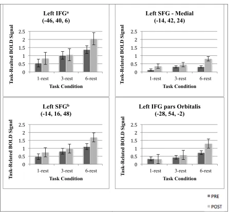
MRI data were analyzed using SPM8, a MATLAB based software package specifically designed to analyze brain imaging data (www.fil.ion.ucl.ac.uk/spm/). All data were preprocessed using standard SPM protocols (realignment, coregistration, normalization and smoothing). Brain areas involved in the performance of each task condition (i.e., 6-, 3-, and 1-letter trials) were identified using baseline data. Next we modeled the following events in a first-level analysis: high-demand trials (6-letter trials) and low-demand trials (1-letter trials). For each individual participant we calculated a statistical parametric map that subtracted brain activation during low-demand trial performance from activation during high-demand trial performance to identify the areas where the BOLD signal changed with increasing cognitive demand. These first-level analyses were conducted on both baseline and post-exercise scans, and focused
pre scan (post minus pre). These maps were entered into a second-level random-effects (group) analysis, and parameter estimates for changes in BOLD activation with exercise training were extracted.
For reaction time analyses, reaction times corresponding with response errors were removed, and means and standard deviations for 1-, 3-, and 6-letter sets (across all 3 runs) were calculated. Reaction times #2 standard deviations from their respective means were then also removed. Reaction time slope with increasing set size (1 to 3 to 6 letters) was calculated using the 1-, 3- and 6- letter mean reaction time values for both baseline and post-exercise scanning data.
Basic descriptive statistics were run in SAS, and included frequencies and means of key variables (age, health status, IQ, lifetime experience, baseline fitness, group randomization). PROC UNIVARIATE was used to test the distribution and skewness of these variables. Residualized changes scores were created to determine change in fitness and change in reaction time slope and used in place of simple change scores in all
analyses.
Hypothesis 1.2. Pearson’s correlation coefficient determined whether change in aerobic fitness was associated with change in neural network efficiency, measured as the difference in task-related network activation during low versus high-demand trials. Pearson’s correlations were also run between neural network efficiency and four other variables (age, health status, lifetime experience score, and baseline fitness) to verify that the observed correlation between change in aerobic fitness and change in efficiency was unique.
Hypothesis 1.3. Pearson’s correlation coefficient determined whether change in neural network efficiency was associated with change in task performance, measured a change in mean reaction time slope (between low and high-demand trials).
to a stretching and toning control group that improved only 2.9%) (Colcombe et al., 2004).
Study 2
Purpose
This study addressed Aim 2: To determine whether participation in an exercise training program improves cognitive performance.
Hypotheses
2.1. It was hypothesized that participation in a 16-week exercise program would improve sustained attention, visuospatial learning, spatial working memory, verbal free recall, and verbal recognition memory (immediate and delayed), as compared to no exercise.
2.2 It was hypothesized that greater improvement in cognitive performance would correlate with greater improvement in fitness.
Design
The proposed research was a quasi-experimental pilot study (non-randomized, controlled).
Participants
The sub-sample of women from WeWalk who participated in Study 1 (n=17) also participated in Study 2. Additional WeWalk participants were part of Study 2, only (i.e., no MRI; n=5). Study 2 also included a non- randomized, no exercise control group (n=19). Inclusion/exclusion criteria for the control group were the same as the for the
having a physical limitation that would preclude exercise training. A total of 41 women participated in Study 2.
Recruitment
Recruitment efforts for participants in the experimental (exercise group) were described in Study 1. Additional women from the community were recruited through a newspaper advertisement and word of mouth to serve as the non-exercise control group. Recruitment efforts for the control group advertised a study relating to memory and thinking skills. Individuals interested in the memory and thinking study also completed an initial screening with the WeWalk study coordinator.
Procedures
For intervention group data collection, procedures for Study 2 were identical to Study 1, except that the additional WeWalk participants recruited for Study 2, only, did not undergo MRI scanning. Thus, these participants (n=5) partook in Baseline Session 1 and Follow-up Session 1, only.
For control group data collection, we followed near identical cognitive testing procedures, asking participants (n=19) to complete their two sessions approximately 16 weeks apart. Prior to their baseline session we mailed a packet of questionnaires
Compensation. Intervention participants in Study 2, only, and control group participants received $20 at the completion of each session, for a total of $40 if they completed the study in its entirety.
Measures
For the intervention (exercise) group, Study 2 measures were identical to those used in Study 1 except that the MRI safety questionnaire and CR MRI task were not administered. Additional measures collected with control participants were height, weight, and blood pressure. Height was measured with a Seca mobile stadiometer to the nearest quarter inch. Weight was measured with a Seca scale to the nearest tenth of a pound. Blood pressure was measured using an Omron Automatic Blood Pressure Monitor (Model HEM-780). We asked control group participants not to begin a structured exercise program in the 16 weeks between sessions.
Statistical Analyses
We performed analyses using SAS, version 9.3 (SAS Institute, Inc., Cary, NC). Basic descriptive statistics included frequencies and means of key variables. PROC UNIVARIATE tested the distribution and skewness of these variables. Chi-square and t-tests assessed differences between groups at baseline. We created residualized change scores for change variables (e.g., changes in fitness, cognitive performance) and used them instead of simple change scores in all ANOVA and correlation analyses.
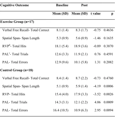
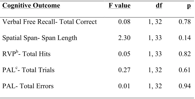
exercise. Separate paired t-tests verified the relationship between exercise training and change in performance on each cognitive task. Due to the small sample size, we also calculated effect sizes to assess the magnitude of change in cognitive performance within and between groups. We divided the exercise group by randomization assignment (high or low exercise group), and conducted an ANOVA to determine whether there were differential changes in cognitive performance based on exercise dose (high, low, no exercise [control]). The exercise group was also divided based on age, baseline fitness, IQ score, lifetime experience (median splits), and family history of AD (yes/no) to look for trends relating to changes in cognitive performance (paired t-tests).
Hypothesis 2.2. For each cognitive task, Pearson’s correlation determined
whether change in aerobic fitness was associated with change in cognitive performance in intervention participants.
!!!!!!!!!!!!!!!!!!!!!!!!!!!!!!!!!!!!!!!!!!!!!!!!!!!!!!!!
Abstract
Cognitive reserve theory suggests that physical activity may protect individuals from cognitive decline. At the brain level, cognitive reserve may manifest as neural network efficiency. Our purpose was to determine 1) whether participation in a 16-week walking program increased brain efficiency, and 2) whether change in fitness and
cognitive performance correlated with increased brain efficiency. Twelve participants underwent fMRI scanning before and after exercise training. During scanning,
participants completed the Sternberg delayed-match-to-sample letter task. Brain activation during the low-demand task condition was subtracted from brain activation during the high-demand condition. We expected this difference to become lesser with exercise training. Within our sample (100% female; mean age 63), the difference became greater in the following brain regions with exercise training: left inferior frontal gyrus, left cuneus, right rolandic operculum, left middle temporal gyrus, left postcentral gyrus, left superior med frontal, left superior frontal gyrus, right caudate, right inferior temporal gyrus (ps < 0.001). No task-related brain regions were utilized more efficiently after exercise training (ps > 0.001). These findings suggest that exercise-induced cognitive reserve may present as a greater ability to recruit neural resources, rather than greater brain efficiency, in this sample. As there were no significant correlations between change in task-related brain activation and change in performance (reaction time slope) with exercise training (r values < 0.49), these findings should be interpreted with caution.
Exercise and Cognitive Reserve: An fMRI Investigation in Healthy Older Women An estimated 5.2 million Americans will have dementia due to Alzheimer’s disease (AD) in 2014 (Alzheimer's Association, 2014). A projected 7.1 Americans will have the disease by 2025, and this number is expected to swell to 13.8-16 million by 2050 (Alzheimer's Association, 2014). Although AD is officially listed as the 6th leading cause of death in the US, a recent report suggests that AD-related deaths are vastly underreported (James et al., 2014). Although current pharmacological treatments (namely cholinesterase inhibitors, including donepezil, galantamine, and rivastigmine) may help with some cognitive and behavioral symptoms of AD, they cannot stop or reverse AD progression (U.S. Department of Health and Human Services, 2008). Importantly, pathological changes in the brain may begin more than two decades before AD symptoms present (Alzheimer's Association, 2014), meaning it is already too late to prevent the onset of pathology in much of the aging baby boomer generation (i.e.,
individuals born between 1946 and 1964). Although the disease is already developing in many Americans aged 40+, it may still be possible to effectively intervene to prevent the dementia. Interventions aimed at building cognitive reserve may help prevent AD pathology from manifesting as dementia.