Drug Design, Development and Therapy 2017:11 3511–3517
Drug Design, Development and Therapy
Dove
press
submit your manuscript | www.dovepress.com 3511
R e v i e w
open access to scientific and medical research
Open Access Full Text Article
Regulation profile of the intestinal peptide
transporter 1 (PepT1)
Chun-Yang wang1,2
Shu Liu1,2
Xiao-Nv Xie1,2
Zhi-Rong Tan1,2
1Department of Clinical Pharmacology,
Xiangya Hospital, Xiangya School of Medicine, Central South University, 2Hunan Key Laboratory
of Pharmacogenetics, Institute of Clinical Pharmacology, Central South University, Changsha, Hunan, People’s Republic of China
Abstract: The intestinal peptide transporter 1 (PepT1) was first identified in 1994. It plays a crucial role in the absorption of small peptides including not only .400 different dipeptides and 8,000 tripeptides digested from dietary proteins but also a repertoire of structurally related compounds and drugs. Owing to its critical role in the bioavailability of peptide-like drugs, such as the anti-cancer agents and anti-virus drug, PepT1 is increasingly becoming a striking prodrug-designing target. Therefore, the understanding of PepT1 gene regulation is of great importance both for dietary adaptation and for clinical drug treatment. After decades of research, it has been recognized that PepT1 could be regulated at the transcriptional and post-transcriptional levels by numerous factors. Therefore, the present review intends to summarize the progress made in the regulation of PepT1 and provide insights into the PepT1’s potential in clinical aspects of nutritional and drug therapies.
Keywords: PepT1, dietary, regulation, transport activity, absorption, bioavailability
Introduction
The uptake and digestion of dietary proteins in the intestinal lumen are mainly in the form of free amino acids and small peptides. These free amino acids and small peptides absorbed are delivered to various tissues through the blood as resources for protein synthesis and energy metabolites.1 While the free amino acids could be absorbed via
several kinds of amino acid transporters located in the intestinal apical membrane, the di- and tripeptides could solely be transported by the oligopeptide transporter PepT1.2
PepT1 was first identified in 1994 as a small peptide transporter. The comple-mentary DNA (cDNA) encoding the human PepT1 is 2,263 base pairs in length and has an open reading frame of 2,127 base pairs encoding 708 amino acids. However, the 3D structures of the proton-dependent peptide transporter remain elusive. Crystal structure analysis revealed that the high-capacity/low-affinity peptide transporter pro-tein contains 12 putative membrane-spanning domains (TMD)3 and a large extracellular
domain (ECD) that is highly conserved within the mammalian homology.4,5 The ECD
of PepT1 consists of two immunoglobulin-like domains connected in tandem, which provides structural insight into the transport of mammalian peptides.4
PepT1, a prototypical member of the SLC15 family,6 is mainly located on apical
membranes of small intestinal epithelial cells. However, there are still controversies regarding PepT1’s location in other tissues for a long while. Some studies reported that PepT1 can be expressed in tissues such as liver, pancreas and colon, while others proposed that the expression is only found in intestine.7–13 In our opinion, these
controversies may originate from the detection of only mRNA or protein level and lack of detailed region-specific analysis.
Correspondence: Zhi-Rong Tan Department of Clinical Pharmacology, Xiangya Hospital, Xiangya School of Medicine, Central South University, 445, 110 Xiangya Road, Changsha, Hunan 410078, People’s Republic of China Tel +86 731 8480 5380
Fax +86 731 8235 4476 email [email protected]
Journal name: Drug Design, Development and Therapy Article Designation: Review
Year: 2017 Volume: 11
Running head verso: Wang et al
Running head recto: Regulation profile of PepT1 DOI: http://dx.doi.org/10.2147/DDDT.S151725
Drug Design, Development and Therapy downloaded from https://www.dovepress.com/ by 118.70.13.36 on 21-Aug-2020
For personal use only.
Dovepress
wang et al
Multiple assays revealed that PepT1 has an enormous range of substrates, including .400 different dipeptides and 8,000 tripeptides produced from the digestion of dietary or body proteins and a repertoire of structurally closely related compounds and drugs.5,12,14 The influx of small peptides
into intestinal epithelial cells is via inwardly directed proton electrochemical gradient, which is mediated by PepT1.7,15
Considering its critical role in the absorption and bioavail-ability of numerous peptide-like drugs, such as the anticancer agents, bestatin, β-lactamantibiotics,16 angiotensin-converting
enzyme (ACE) inhibitors for hypertension,17 anti-virus drug
valacyclovir and L-DOPA-L-Phe,18,19 PepT1 is increasingly
becoming into a prodrug-designing target. Especially, some poorly absorbed drugs can be modified as peptidomimetic prodrugs targeting PepT1, so as to improve the membrane permeability. Therefore, detailed knowledge about the regu-lation of PepT1 may enhance its potential in clinical aspects of nutritional and drug therapies. The present review aimed to give an updated overview about the elementary knowledge on the factors modulating PepT1’s expression and function. This information may be a useful application in pharmacological and clinical treatments.
Regulation by substrates
It was well accepted that one of the most possible regulators of function and/or expression of any transporter is its own substrates. This is true in case of PepT1; multiple studies have investigated this possibility and provided firm evidences as well. A study revealed that PepT1 could be upregulated in the transcriptional level by certain dipeptides produced from the dietary via directly stimulating the transcriptional activity of the PepT1 gene promoter.20 Consistently, a subsequent study
demonstrated an upregulation of PepT1 gene expression in human colon adenocarcinoma (Caco-2) cells after treatment with dipeptides such as Gly-Sar and Gly-Phe (chemically synthesized dipeptide).21 In addition, Gly-Gln (a natural
dipeptide) was also reported to be able to cause a threefold increase in the PepT1 mRNA level in the apical membrane of Caco-2 cells,14 which is consistent with the research of Walker
et al22 that the V
max of Gly-Sar uptake increased ~1.64-fold
after Caco-2 cells were treated with a peptide-rich medium. As Walker et al showed, the magnitude of functional increase in response to Gly-Gln may fully attribute to the increased expression of PepT1’s mRNA and protein. Furthermore, the increase in the mRNA level was probably due to the enhanced PepT1 mRNA stability and the incremental transcription. Above all, the substrates have been firmly confirmed as regulators of PepT1 in the transcriptional level.
Regulation by proteins
It is well known that PepT1 cannot work alone under physi-ological conditions. The expression and activity may vary in response to several factors. In 2008, the post-transcriptional regulation of PepT1 caused by the postsynaptic density-95/ disk-large/ZO-1 [PDZ] domain-containing protein (PDZK1) was first identified.23 In this context, a significant decrease
in the PepT1’s location at the apical membrane of small intestinal epithelial cells was found in PDZK1 gene knockout mice, consistently, in Hek293/PDZK1 cells; both the PepT1 protein expression and the transport activity increased to ~1.7-fold without any change in the PepT1 mRNA level, which further clarified the post-transcriptional regulation by PDZK1. In 2012, Park et al suggested a strong inhibition effect of the aflavins on the transport of small peptides across Caco-2 cell monolayer via suppressing the AMPK-mediated PepT1 expression, which was consistent with previous reports that the apical PepT1 transport systems of Caco-2 cells can be regulated by AMP-activated protein kinase.24
In addition, the AICAR (AMPK activator) has been shown to alter the peptide uptake in the small intestine model cell line Caco-2 cells.25 Meanwhile, Coon et al26 also investigated
the effect of GIP on PepT1 and revealed that PepT1-mediated Gly-Sar absorption can be activated directly by GIP through the cAMP-dependent signaling pathway,27,28 as far as the
interaction between the AMP-activated protein kinase and PepT1 is clarified; further researches are still needed to iden-tify the underlying molecular mechanisms. Of course, such post-transcriptional stimulation may also provide functional coupling of PepT1, just as proposed in the case of PepT1 and Na+/H+ exchanger (NHE3). It was reported that NHE3
located on apical membranes acted as PepT1’s driving force via changing the H+ gradient. In addition, the acid-loading
activity of PepT1 and the requirement of the apical NHE3 activity for the recovery from the acid load have been clearly demonstrated by the use of a fluorescent reporter to monitor the intracellular pH changes. Furthermore, the functional coupling of PepT1 is evolutionarily conserved; the essential action of NHE3 in the mammalian enterocytes has already been established in the intestine of Caenorhabditis elegans by its ortholog NHX-2.29–31
In 2000, Fei et al described the cDNA structure and the promoter region of the mouse-Pept1; they proposed that the promoter region possesses three GC boxes that can bind to the transcription activator SP1.32 Subsequently, Shimakura
et al33 cloned the human Pept1 promoter region and
high-lighted the direct interaction of Sp1 with the GC boxes in mammalian Pept1 expression. One year later, the same group
Drug Design, Development and Therapy downloaded from https://www.dovepress.com/ by 118.70.13.36 on 21-Aug-2020
Dovepress Regulation profile of PepT1
reported that Cdx2 can also regulate PepT1 transcription in concert with SP1 in mammals.34 Another transcriptional
regulation of PepT1 was reported in 2014; as reported, the transcriptional activity of PepT1 gene promoter can be stimulated directly via elevating Nrf2 binding with PepT1-ARE1 (a biologically active Nrf2 binding site within the PepT1 promoter),35 and the endogenous PepT1 protein
and mRNA abundance as well as transport activity are all enhanced via stimulating the Nrf2 pathway. All abovemen-tioned explanation may provide new ideas in PepT1’s protein regulations, and we can change the intake of nutrition and structurally related compounds via regulating these above proteins’ level.
Regulation by hormones
Studies concerning the regulation of PepT1 by hormones are sparse. Evidences indicate that both insulin and epi-dermal growth factor (EGF) can be involved in the regula-tion of PepT1 even with different molecular mechanisms. Thamotharan et al hardly ever discovered changes in the PepT1 mRNA level in Caco-2 cells incubating with insulin, while the protein level of PepT1 in the apical membrane was increased. Following this, they proposed that insulin may regulate PepT1 via increasing the protein recruitment of the plasma membrane from the preformed cytoplasm pool. That is to say, although insulin cannot be found in the gut lumen, the peptide transport activity still can be regulated by binding to its receptor located in the basolateral membrane of the intestinal mucosal cells.36
Few years later, another study was performed to evalu-ate the role of EGF in PepT1’s regulation; it proposed that a long-term incubation of EGF resulted in a cut down of PepT1’s expression in both protein and mRNA levels in Caco-2 cells.7,14 However, a following study demonstrated
that short-term incubation with EGF may upregulate the PepT1’s mRNA expression in enterocyte-like cells that express the specific small peptide transporter PepT1.37 In other
words, the effect of EGF appears paradoxical; to resolve this paradox, additional studies including different incubating times and diverse incubating doses need to be carried out.
Sun et al also evaluated the potential effect of recombi-nant human growth hormone (rhGH) on the human PepT1 expression. As a result of Caco-2 cells treated with rhGH, the dipeptide transport of PepT1 was upregulated greatly. In addition, it demonstrated that the remarkable increase in the PepT1 gene expression mostly attributed to the stimula-tion of PepT1 gene transcripstimula-tional activity.38 In words, all
of these three hormones act by combining with the tyrosine
kinase receptor; the EGF and rhGH work via regulating the PepT1 gene expression, yet insulin acts by regulating protein recruitment. PepT1 can also be regulated by ghrelin (an orexigenic hormone). Liu et al39 proposed that the uptake
function of PepT1 can be upregulated by ghrelin, the small intestine epithelium of rats, and the expression of PepT1 both in the protein and mRNA levels was higher in ghrelin-treated group rats. This may be a transcriptional regulation even though the molecular mechanisms are still unclear.
Regulation by diseases
Intestinal diseases such as ulcerative colitis, Crohn’s disease, diabetes and some cancer types may have effects on PepT1’s expression. As already mentioned, insulin could regulate the protein expression of PepT1. Studies were also performed to investigate the expression of PepT1 in diabetes; the results showed that the protein expression of PepT1 was increased in uncontrolled diabetes without any change in gene transcription;36 however, whether the abnormal
expres-sion of PepT1 in diabetes was caused by insulin or not still needs further research. Unlike diabetes, controversial findings exist in inflammatory bowel disease (IBD). To assess the association between PepT1 and IBD, Wuensch et al built IBD mouse models to analyze PepT1’s expression during intestinal inflammation and studied the susceptibility of PepT1-deficient mice to experimental colitis. Eventually, they proposed that PepT1’s expression level was decreased in the descending colon in both IBD patients and IBD mouse models.9 However, this report contradicts with previous
studies that proposed no expression of PepT1 in healthy colon but significant expression in inflamed colonic tissues in patients with IBD.7,9,10,14,40,41 As has been mentioned, further
investigation of PepT1’s expression in patients with IBD is warranted. PepT1’s abnormal expressions were also reported in tumors. As PepT1’s critical role in intestinal inflammation, a recent study was performed to research if PepT1 could be involved in colitis-associated cancer development; it showed that PepT1 was highly expressed in colorectal tumor biopsy specimens, and mice with PepT1 overexpression had larger tumor sizes and increased tumor burdens compared with wild-type (WT) mice and PepT1-KO mice.10 In addition, studies
show that increasing peptide transport activity of PepT1 exists in the human fibrosarcoma cell HT1080, human pancreatic carcinoma cell AsPC-1, Capan-2, human colon cancer cell Caco-2, prostatic cancer cell PC-3, lung cancer cell A549, gastric cancer cell MKN45 and ovarian adenocarcinoma cell C13.42–45 As is known to all, cancer cells are easy to be killed,
but it is difficult to kill cancer cells without impairing the
Drug Design, Development and Therapy downloaded from https://www.dovepress.com/ by 118.70.13.36 on 21-Aug-2020
Dovepress
wang et al
normal physiological functions. If PepT1 can be a specific biomarker for carcinogenesis and tumor growth, it may have many useful applications in clinical treatments.
Regulation by pharmacological
agents
Through decades of research, it has been recognized that PepT1 could be regulated by many pharmacological agents, such as floxuridine, gemcitabine and 5-fluorouracil. Studies revealed that when treated with 5-fluorouracil, both protein and mRNA levels for amino acid and glucose transporters were profoundly depressed, yet PepT1 conversely increased. It was proposed that the increasing of PepT1’s expression and transport activity may be caused by stimulating the transcrip-tional activity of PepT1 directly rather than by changing the kinetic properties.13 In our opinion, PepT1’s high expression
may affect the uptake of small peptides and then facilitate the proliferation of tumor and increase tumor burdens or even result in chemotherapy resistance. Further studies need to be carried out to confirm our hypothesis.
Likewise, another experiment was performed to investi-gate the effects of enrofloxacin and xylanase on PepT1, and the results showed that either treatment with enrofloxacin or
xylanase leads to a significantly high expression of PepT1.46,47
There are also studies that revealed that the transport activity of PepT1 can be altered directly by α2-adrenergic agonist clonidine via increasing the translocation of preformed cytoplasmic PepT1 to the apical membrane of Caco-2 cells.48 Moreover, pentazocine, a selective σ1 ligand, was
reported as a regulator of PepT1’s expression in Caco-2 cells. Pentazocine can upregulate the mRNA level of PepT1, which leads to the increased protein level at the cell membrane, causing an increase in peptide transport activity eventually.45
Simultaneously, an experiment has been performed to inves-tigate if PepT1 can be affected by lipopolysaccharide (LPS), and in this context, the authors proposed that a significantly decrease in the abundance both in mRNA and protein levels was found after injecting rats with LPS.49,50 Above all, even
many pharmacological agents were mentioned; the molecu-lar processes modifying PepT1’s expression and transport property still need further discussion.
Summary
Within the past 2 decades, several factors that directly or indirectly influence the PepT1 on either the transcription level or the intracellular trafficking have been investigated.
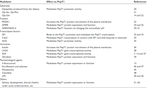
Table 1 Overview of selected factors modulating the gene and/or protein expression and function of the intestinal peptide transporter
PepT1
Modulator Effect on PepT1 References
Substrates
Dipeptides produced from the dietary Modulates PepT1 promoter activity 20
Gly-Sar, Gly-Phe 21
Gly-Gln 14 and 22
Proteins
PDZK1 Increases the PepT1 protein recruitment of the plasma membrane 23
AMPK Modulates PepT1 protein expression and function 24 and 25
NHE3/NHX-2 Modulates PepT1 function via changing the intracellular pH 29–31 Transcription factors
SP1 Binds to the PepT1 promoter and modulates the PepT1 transcription 32 and 33
Cdx2 Modulates PepT1 transcription in concert with SP1 and with butyrate in mammals 34
Nrf2 Modulates PepT1 promoter activity 35
Hormones
insulin Increases the PepT1 protein recruitment of the plasma membrane 36
rhGH Modulates PepT1 gene transcriptional activity 38
eGF Modulates PepT1 gene transcriptional activity 7, 14 and 37
Ghrelinin Modulates PepT1 protein expression and function 39
Pharmacological agents
5-fluorouracil Modulates PepT1 protein expression or function 13
Enrofloxacin and xylanase 46 and 47
Pentazocine 45
Clonidine 48
LPS 49 and 50
Others
Salinity, development, diurnal rhythm, under acute undernutrition, etc.
Modulates PepT1 protein expression or function 51–56
Abbreviations: PepT1, peptide transporter 1; PDZK1, PDZ domain-containing protein; AMPK, adenosine 5′-monophosphate (AMP)-activated protein kinase; NHE3/ NHX-2, Na+/H+ exchanger; rhGH, recombinant human growth hormone; EGF, epidermal growth factor; LPS, lipopolysaccharide.
Drug Design, Development and Therapy downloaded from https://www.dovepress.com/ by 118.70.13.36 on 21-Aug-2020
Dovepress Regulation profile of PepT1
For example, multiple studies revealed that the expression of PepT1 was increased under acute undernutrition on nutrient transport,51,52 similar to results from a previous morphological
study of the rat intestinal lining in relation to depletion of body stores during fasting and after refeeding. In this study, Habold et al proposed the major mechanisms of intestinal adaptation to fasting and refeeding and showed a remarkable increase in the apical PepT1 expression during both metabolic fasting phases. Additionally, they also revealed an upregula-tion of transporter expression along with the restoraupregula-tion of the villus structure and function when refeeding after both fasting phases.53 Furthermore, there are also studies demonstrating
that high-fat diet may upregulate the expression of PepT1.21,54
Meanwhile, it was also reported that salinity, development and diurnal rhythm55–57 could have effects on the function and/
or expression of PepT1, although the pathways for the trans-mission of these signals is still unclear. These factors regulate PepT1 mainly via the following three ways: first, modifying PepT1’s driving force via changing the H+ gradient; second,
regulating PepT1’s amount via increasing the protein recruit-ment of the plasma membrane from the preformed cytoplasm pool and last, altering the transcription and stability of PepT1. Interestingly, the recent reporting of PepT1’s 3D structure has opened up a fertile ground for exploring the molecular mechanisms of the regulation of PepT1.
To our knowledge, mammals absorb nitrogen via the uptake of small peptides in the gastrointestinal tract through PepT1. As far as clinical application is concerned, the knowl-edge of regulation of PepT1 has implications in studies of enteral nutrition and drug therapy. In this review, we tried to summarize the factors modulating the intestinal peptide transporter PepT1’s expression and function so as to provide an exhaustive knowledge about the regulation of PepT1 (Table 1). As is well known to us, even the study of the proton-dependent peptide transporter PepT1 has achieved initial success; copious studies remain to be done. In-depth study of the 3D structure and factors affecting PepT1 activity and mechanisms of action, including gene promoter stimula-tion and post-transcripstimula-tional and translastimula-tional mechanisms, can better utilize PepT1’s unique biological property, so as to be applied in pharmacological and clinical treatments.
Acknowledgments
This study was supported by The Research of Major Disease Prevention and Control of Precision Medical Research of Hunan Province Education Department (number 2016JC2069) and The National Science and Technology Major Projects for Major New Drugs Innovation and Development (numbers 2017zx09306012 and 2017zx09304014).
Author contributions
All authors contributed toward data analysis, drafting and critically revising the paper and agree to be accountable for all aspects of the work.
Disclosure
The authors report no conflicts of interest in this work.
References
1. Bröer S. Amino acid transport across mammalian intestinal and renal epithelia. Physiol Rev. 2008;88(1):249–286.
2. Spanier B. Transcriptional and functional regulation of the intestinal peptide transporter PEPT1. J Physiol. 2014;592(5):871–879. 3. Bolger MB, Haworth IS, Yeung AK, et al. Structure, function, and
molecular modeling approaches to the study of the intestinal dipeptide transporter PepT1. J Pharm Sci. 1998;87(11):1286–1291.
4. Beale John H, Parker Joanne L, Samsudin F, et al. Crystal structures of the extracellular domain from PepT1 and PepT2 provide novel insights into mammalian peptide transport. Structure. 2015;23(10):1889–1899. 5. Newstead S. Recent advances in understanding proton coupled peptide
transport via the POT family. Curr Opin Struct Biol. 2016;45:17–24. 6. Daniel H, Kottra G. The proton oligopeptide cotransporter family
SLC15 in physiology and pharmacology. Pflugers Arch. 2004;447(5): 610–618.
7. Ingersoll SA, Ayyadurai S, Charania MA, Laroui H, Yan Y, Merlin D. The role and pathophysiological relevance of membrane transporter PepT1 in intestinal inflammation and inflammatory bowel disease.
Am J Physiol Gastrointest Liver Physiol. 2012;302(5):G484.
8. Wuensch T, Schulz S, Ullrich S, et al. The peptide transporter PEPT1 is expressed in distal colon in rodents and humans and contributes to water absorption. Am J Physiol Gastrointest Liver Physiol. 2013;305(1):G66. 9. Wuensch T, Ullrich S, Schulz S, et al. Colonic expression of the peptide
transporter PEPT1 is downregulated during intestinal inflammation and is not required for NOD2-dependent immune activation. Inflamm Bowel
Dis. 2014;20(4):671–684.
10. Viennois E, Ingersoll SA, Ayyadurai S, et al. Critical role of PepT1 in promoting colitis-associated cancer and therapeutic benefits of the anti-inflammatory PepT1-mediated tripeptide KPV in a murine model.
Cell Mol Gastroenterol Hepatol. 2016;2(3):340–357.
11. Terada T, Shimada Y, Pan X, et al. Expression profiles of various transporters for oligopeptides, amino acids and organic ions along the human digestive tract. Biochem Pharmacol. 2005;70(12):1756–1763. 12. Jappar D, Wu SP, Hu Y, Smith DE. Significance and regional depen-dency of peptide transporter (PEPT) 1 in the intestinal permeability of glycylsarcosine: in situ single-pass perfusion studies in wild-type and Pept1 knockout mice. Drug Metab Dispos. 2010;38(10):1740–1746. 13. Inoue M, Terada T, Okuda M, Inui K. Regulation of human peptide
transporter 1 (PEPT1) in gastric cancer cells by anticancer drugs. Cancer
Lett. 2005;230(1):72–80.
14. Adibi SA. Regulation of expression of the intestinal oligopeptide transporter (Pept-1) in health and disease. Am J Physiol Gastrointest
Liver Physiol. 2003;285(5):G779–G788.
15. Newstead S. Molecular insights into proton coupled peptide transport in the PTR family of oligopeptide transporters. Biochim Biophys Acta. 2015;1850(3):488–499.
16. Tamai I, Nakanishi T, Hayashi K, et al. The predominant contribu-tion of oligopeptide transporter PepT1 to intestinal absorpcontribu-tion of beta-lactam antibiotics in the rat small intestine. J Pharm Pharmacol. 1997;49(8):796–801.
17. Zhang Y, Sun J, Sun Y, Wang Y, He Z. Prodrug design targeting intestinal PepT1 for improved oral absorption: design and performance.
Curr Drug Metab. 2013;14(6):675–687.
18. Nakanishi T, Tamai I, Takaki A, Tsuji A. Cancer cell-targeted drug delivery utilizing oligopeptide transport activity. Int J Cancer. 2000; 88(2):274.
Drug Design, Development and Therapy downloaded from https://www.dovepress.com/ by 118.70.13.36 on 21-Aug-2020
Dovepress
wang et al
19. Brandsch M. Drug transport via the intestinal peptide transporter PepT1.
Curr Opin Pharmacol. 2013;13(6):881–887.
20. Shiraga T, Miyamoto K, Tanaka H, et al. Cellular and molecular mecha-nisms of dietary regulation on rat intestinal H+/Peptide transporter PepT1. Gastroenterology. 1999;116(2):354.
21. Wisniewski JR, Friedrich A, Keller T, Mann M, Koepsell H. The impact of high-fat diet on metabolism and immune defense in small intestine mucosa. J Proteome Res. 2015;14(1):353–365.
22. Walker D, Thwaites DT, Simmons NL, Gilbert HJ, Hirst BH. Substrate upregulation of the human small intestinal peptide transporter, hPepT1.
J Physiol. 1998;507(3):697.
23. Sugiura T, Kato Y, Wakayama T, et al. PDZK1 regulates two intestinal solute carriers (Slc15a1 and Slc22a5) in mice. Drug Metab Dispos. 2008;36(6):1181–1188.
24. Park HY, Kunitake Y, Matsui T. Benzotropolone moiety in theaflavins is responsible for inhibiting peptide-transport and activating AMP-activated protein kinase in Caco-2 cells. Functional Foods in Health
& Disease. 2013;3(5).
25. Pieri M, Christian HC, Wilkins RJ, Boyd CA, Meredith D. The apical (hPepT1) and basolateral peptide transport systems of Caco-2 cells are regulated by AMP-activated protein kinase. Am J Physiol Gastrointest
Liver Physiol. 2010;299(1):G136–G143.
26. Coon SD, Schwartz JH, Rajendran VM, Jepeal L, Singh SK. Glucose-dependent insulinotropic polypeptide regulates dipeptide absorption in mouse jejunum. Am J Physiol Gastrointest Liver Physiol. 2013; 305(10):G678.
27. Myrtani P, Christian HC, Wilkins RJ, Boyd CAR, David M. The apical (hPepT1) and basolateral peptide transport systems of Caco-2 cells are regulated by AMP-activated protein kinase. Am J Physiol Gastrointest
Liver Physiol. 2010;299(1):136–143.
28. Lang F, Foller M. Regulation of ion channels and transporters by AMP-activated kinase (AMPK). Channels. 2014;8(1):20–28. 29. Romano A, Barca A, Storelli C, Verri T. Teleost fish models in
mem-brane transport research: the PEPT1(SLC15A1) H+-oligopeptide transporter as a case study. J Physiol. 2014;592(5):881–897. 30. Yu Q, Liu X, Liu Y, et al. Defective small intestinal anion secretion,
dipeptide absorption, and intestinal failure in suckling NBCe1-deficient mice. Pflugers Arch. 2016;468(8):1419–1432.
31. Watanabe C, Kato Y, Ito S, Kubo Y, Sai Y, Tsuji A. Na+/H+ exchanger 3 affects transport property of H+/oligopeptide transporter 1. Drug
Metab Pharmacokinet. 2005;20(6):443.
32. Fei YJ, Sugawara M, Liu JC, et al. cDNA structure, genomic organiza-tion, and promoter analysis of the mouse intestinal peptide transporter PEPT1. Biochim Biophys Acta. 2000;1492(1):145–154.
33. Shimakura J, Terada T, Katsura T, Inui K. Characterization of the human peptide transporter PEPT1 promoter: Sp1 functions as a basal transcriptional regulator of human PEPT1. Am J Physiol Gastrointest
Liver Physiol. 2005;289(3):G471.
34. Shimakura J, Terada T, Shimada Y, Katsura T, Inui K. The transcrip-tion factor Cdx2 regulates the intestine-specific expression of human peptide transporter 1 through functional interaction with Sp1. Biochem
Pharmacol. 2006;71(11):1581–1588.
35. Geillinger KE, Kipp AP, Schink K, Roder PV, Spanier B, Daniel H. Nrf2 regulates the expression of the peptide transporter PEPT1 in the human colon carcinoma cell line Caco-2. Biochim Biophys Acta. 2014; 1840(6):1747–1754.
36. Thamotharan M, Bawani SZ, Zhou X, Adibi SA. Hormonal regula-tion of oligopeptide transporter pept-1 in a human intestinal cell line.
American Journal of Physiology. 1999;276(4 Pt 1):C821.
37. Iwao T, Toyota M, Miyagawa Y, et al. Differentiation of human induced pluripotent stem cells into functional enterocyte-like cells using a simple method. Drug Metab Pharmacokinet. 2014;29(1):44–51.
38. Sun BW, Zhao XC, Wang GJ, Li N, Li JS. Hormonal regulation of dipeptide transporter (PepT1) in Caco-2 cells with normal and anoxia/ reoxygenation management. World J Gastroenterol. 2003;9(4): 808–812.
39. Liu J, Shi B, Shi K, et al. Ghrelin upregulates PepT1 activity in the small intestine epithelium of rats with sepsis. Biomed Pharmacother. 2016;86:669–676.
40. Merlin D, Si-Tahar M, Sitaraman SV, et al. Colonic epithelial hPepT1 expression occurs in inflammatory bowel disease: transport of bac-terial peptides influences expression of MHC class 1 molecules.
Gastroenterology. 2001;120(7):1666–1679.
41. Sato T, Mushiake S, Kato Y, et al. The Rab8 GTPase regulates apical pro-tein localization in intestinal cells. Nature. 2007;448(7151):366–369. 42. Tai W, Chen Z, Cheng K. Expression profile and functional activity
of peptide transporters in prostate cancer cells. Mol Pharm. 2013; 10(2):477–487.
43. Fang L, Wang M, Gou S, Liu X, Zhang H, Cao F. Combination of amino acid/dipeptide with nitric oxide donating oleanolic acid derivatives as PepT1 targeting antitumor prodrugs. J Med Chem. 2014; 57(3):1116–1120.
44. Ai Y, Kang F, Huang Z, et al. Synthesis of CDDO-amino acid-nitric oxide donor trihybrids as potential antitumor agents against both drug-sensitive and drug-resistant colon cancer. J Med Chem. 2015;58(5): 2452–2464.
45. Kouodom MN, Ronconi L, Celegato M, et al. Toward the selective delivery of chemotherapeutics into tumor cells by targeting peptide transporters: tailored gold-based anticancer peptidomimetics. J Med
Chem. 2012;55(5):2212–2226.
46. Pavlova I, Milanova A, Danova S, Fink-Gremmels J. Enrofloxacin and probiotic lactobacilli influence PepT1 and LEAP-2 mRNA expression in poultry. Probiotics Antimicrob Proteins. 2016;8(4):215–220. 47. Guo S, Liu D, Zhao X, Li C, Guo Y. Xylanase supplementation of a
wheat-based diet improved nutrient digestion and mRNA expression of intestinal nutrient transporters in broiler chickens infected with Clostridium perfringens. Poult Sci. 2014;93(1):94.
48. Berlioz F, Maoret JJ, Paris H, Laburthe M, Farinotti R, Roze C. Alpha(2)-adrenergic receptors stimulate oligopeptide transport in a human intes-tinal cell line. J Pharmacol Exp Ther. 2000;294(2):466–472. 49. Shu HJ, Takeda H, Shinzawa H, Takahashi T, Kawata S. Effect of
lipopolysaccharide on peptide transporter 1 expression in rat small intestine and its attenuation by dexamethasone. Digestion. 2002;65(1): 21–29.
50. Motohashi H, Katsura T, Saito H, Inui K. Effects of tacrolimus and cyclosporin A on peptide transporter PEPT1 in Caco-2 cells. Pharm Res. 2001;18(5):713–717.
51. Sampaio IC, Medeiros PH, Rodrigues FA, et al. Impact of acute undernutrition on growth, ileal morphology and nutrient transport in a murine model. Braz J Med Biol Res. 2016;49(10):e5340.
52. Qiu K, Qin CF, Luo M, et al. Protein restriction with amino acid-balanced diets shrinks circulating pool size of amino acid by decreasing expression of specific transporters in the small intestine. PLoS One. 2016;11(9):e0162475.
53. Habold C, Reichardt F, Foltzer-Jourdainne C, Lignot JH. Morphologi-cal changes of the rat intestinal lining in relation to body stores deple-tion during fasting and after refeeding. Pflugers Arch. 2007;455(2): 323–332.
54. Boudry G, Rome V, Perrier C, Jamin A, Savary G, Le Huerou-Luron I. A high-protein formula increases colonic peptide transporter 1 activity during neonatal life in low-birth-weight piglets and disturbs barrier function later in life. Br J Nutr. 2014;112(7):1073–1080.
55. Con P, Nitzan T, Cnaani A. Salinity-dependent shift in the localization of three peptide transporters along the intestine of the Mozambique tilapia (Oreochromis mossambicus). Front Physiol. 2017;8:8. 56. Geillinger KE, Kuhlmann K, Eisenacher M, et al. Intestinal amino acid
availability via PEPT-1 affects TORC1/2 signaling and the unfolded protein response. J Proteome Res. 2014;13(8):3685–3692.
57. Hussain MM, Pan X. Circadian regulation of macronutrient absorption.
J Biol Rhythms. 2015;30(6):459–469.
Drug Design, Development and Therapy downloaded from https://www.dovepress.com/ by 118.70.13.36 on 21-Aug-2020
Drug Design, Development and Therapy
Publish your work in this journal
Submit your manuscript here: http://www.dovepress.com/drug-design-development-and-therapy-journal
Drug Design, Development and Therapy is an international, peer-reviewed open-access journal that spans the spectrum of drug design and development through to clinical applications. Clinical outcomes, patient safety, and programs for the development and effective, safe, and sustained use of medicines are the features of the journal, which
has also been accepted for indexing on PubMed Central. The manu-script management system is completely online and includes a very quick and fair peer-review system, which is all easy to use. Visit http://www.dovepress.com/testimonials.php to read real quotes from published authors.
Dovepress
Dove
press
Regulation profile of PepT1
Drug Design, Development and Therapy downloaded from https://www.dovepress.com/ by 118.70.13.36 on 21-Aug-2020